Rim JS et al. (JAN 2011)
Methods in molecular biology (Clifton,N.J.) 702 299--316
Screening for Epigenetic Target Genes that Enhance Reprogramming Using Lentiviral-Delivered shRNA
Small molecules will need to be identified and/or developed that target protein classes limiting reprogramming efficiency. A specific class of proteins includes epigenetic regulators that silence,or minimize expression,of pluripotency genes in differentiated cells. To better understand the role of specific epigenetic modulators in reprogramming,we have used shRNA delivered by lentivirus to assess the significance of individual epi-proteins in reprogramming pluripotent gene expression.
View Publication
产品类型:
产品号#:
85850
85857
产品名:
mTeSR™1
mTeSR™1
Ikeda Y et al. (MAR 2015)
Gene therapy 23 November 2015 256--262
A novel intranuclear RNA vector system for long-term stem cell modification.
Genetically modified stem and progenitor cells have emerged as a promising regenerative platform in the treatment of genetic and degenerative disorders,highlighted by their successful therapeutic use in inherent immunodeficiencies. However,biosafety concerns over insertional mutagenesis resulting from integrating recombinant viral vectors have overshadowed the widespread clinical applications of genetically modified stem cells. Here,we report an RNA-based episomal vector system,amenable for long-term transgene expression in stem cells. Specifically,we used a unique intranuclear RNA virus,Borna disease virus (BDV),as the gene transfer vehicle,capable of persistent infections in various cell types. BDV-based vectors allowed for long-term transgene expression in mesenchymal stem cells (MSCs) without affecting cellular morphology,cell surface CD105 expression,or the adipogenicity of MSCs. Similarly,replication-defective BDV vectors achieved long-term transduction of human induced pluripotent stem cells (iPSCs),while maintaining the ability to differentiate into three embryonic germ layers. Thus,the BDV-based vectors offer a genomic modification-free,episomal RNA delivery system for sustained stem cell transduction.Gene Therapy accepted article preview online,03 December 2015. doi:10.1038/gt.2015.108.
View Publication
产品类型:
产品号#:
85850
85857
产品名:
mTeSR™1
mTeSR™1
Kumar S et al. ( 2016)
Stem Cells International 2016 1--20
Utility of Lymphoblastoid Cell Lines for Induced Pluripotent Stem Cell Generation
A large number of EBV immortalized LCLs have been generated and maintained in genetic/epidemiological studies as a perpetual source of DNA and as a surrogate in vitro cell model. Recent successes in reprograming LCLs into iPSCs have paved the way for generating more relevant in vitro disease models using this existing bioresource. However,the overall reprogramming efficiency and success rate remain poor and very little is known about the mechanistic changes that take place at the transcriptome and cellular functional level during LCL-to-iPSC reprogramming. Here,we report a new optimized LCL-to-iPSC reprogramming protocol using episomal plasmids encoding pluripotency transcription factors and mouse p53DD (p53 carboxy-terminal dominant-negative fragment) and commercially available reprogramming media. We achieved a consistently high reprogramming efficiency and 100% success rate using this optimized protocol. Further,we investigated the transcriptional changes in mRNA and miRNA levels,using FC-abs ≥ 2.0 and FDR ≤ 0.05 cutoffs; 5,228 mRNAs and 77 miRNAs were differentially expressed during LCL-to-iPSC reprogramming. The functional enrichment analysis of the upregulated genes and activation of human pluripotency pathways in the reprogrammed iPSCs showed that the generated iPSCs possess transcriptional and functional profiles very similar to those of human ESCs.
View Publication
A viral strategy for targeting and manipulating interneurons across vertebrate species.
A fundamental impediment to understanding the brain is the availability of inexpensive and robust methods for targeting and manipulating specific neuronal populations. The need to overcome this barrier is pressing because there are considerable anatomical,physiological,cognitive and behavioral differences between mice and higher mammalian species in which it is difficult to specifically target and manipulate genetically defined functional cell types. In particular,it is unclear the degree to which insights from mouse models can shed light on the neural mechanisms that mediate cognitive functions in higher species,including humans. Here we describe a novel recombinant adeno-associated virus that restricts gene expression to GABAergic interneurons within the telencephalon. We demonstrate that the viral expression is specific and robust,allowing for morphological visualization,activity monitoring and functional manipulation of interneurons in both mice and non-genetically tractable species,thus opening the possibility to study GABAergic function in virtually any vertebrate species.
View Publication
产品类型:
产品号#:
05790
05792
05793
05794
05795
85850
85857
产品名:
BrainPhys™神经元培养基
BrainPhys™神经元培养基和SM1试剂盒
BrainPhys™ 神经元培养基N2-A和SM1试剂盒
BrainPhys™原代神经元试剂盒
BrainPhys™ hPSC 神经元试剂盒
mTeSR™1
mTeSR™1
M. R. Hildebrandt et al. (dec 2019)
Stem cell reports 13 6 1126--1141
Precision Health Resource of Control iPSC Lines for Versatile Multilineage Differentiation.
Induced pluripotent stem cells (iPSC) derived from healthy individuals are important controls for disease-modeling studies. Here we apply precision health to create a high-quality resource of control iPSCs. Footprint-free lines were reprogrammed from four volunteers of the Personal Genome Project Canada (PGPC). Multilineage-directed differentiation efficiently produced functional cortical neurons,cardiomyocytes and hepatocytes. Pilot users demonstrated versatility by generating kidney organoids,T lymphocytes,and sensory neurons. A frameshift knockout was introduced into MYBPC3 and these cardiomyocytes exhibited the expected hypertrophic phenotype. Whole-genome sequencing-based annotation of PGPC lines revealed on average 20 coding variants. Importantly,nearly all annotated PGPC and HipSci lines harbored at least one pre-existing or acquired variant with cardiac,neurological,or other disease associations. Overall,PGPC lines were efficiently differentiated by multiple users into cells from six tissues for disease modeling,and variant-preferred healthy control lines were identified for specific disease settings.
View Publication
产品类型:
产品号#:
05835
05839
05010
85850
85857
产品名:
STEMdiff™ 神经诱导培养基
STEMdiff™ 神经诱导培养基
STEMdiff™ 心肌细胞分化培养基试剂盒
mTeSR™1
mTeSR™1
Xia L et al. (NOV 2004)
Blood 104 10 3091--6
Surface fucosylation of human cord blood cells augments binding to P-selectin and E-selectin and enhances engraftment in bone marrow.
Murine hematopoietic stem and progenitor cells (HSPCs) home to bone marrow in part by rolling on P-selectin and E-selectin expressed on endothelial cells. Human adult CD34(+) cells,which are enriched in HSPCs,roll on endothelial selectins in bone marrow vessels of nonobese diabetic/severe combined immune deficiency (NOD/SCID) mice. Many human umbilical cord blood (CB) CD34(+) cells do not roll in these vessels,in part because of an uncharacterized defect in binding to P-selectin. Selectin ligands must be alpha1-3 fucosylated to form glycan determinants such as sialyl Lewis x (sLe(x)). We found that inadequate alpha1-3 fucosylation of CB CD34(+) cells,particularly CD34(+)CD38(-/low) cells that are highly enriched in HSPCs,caused them to bind poorly to E-selectin as well as to P-selectin. Treatment of CB CD34(+) cells with guanosine diphosphate (GDP) fucose and exogenous alpha1-3 fucosyltransferase VI increased cell-surface sLe(x) determinants,augmented binding to fluid-phase P- and E-selectin,and improved cell rolling on P- and E-selectin under flow. Similar treatment of CB mononuclear cells enhanced engraftment of human hematopoietic cells in bone marrows of irradiated NOD/SCID mice. These observations suggest that alpha1-3 fucosylation of CB cells might be a simple and effective method to improve hematopoietic cell homing to and engraftment in bone marrows of patients receiving CB transplants.
View Publication
产品类型:
产品号#:
产品名:
Li H et al. (SEP 2016)
In vitro cellular & developmental biology. Animal 52 8 885--893
Directed differentiation of human embryonic stem cells into keratinocyte progenitors in vitro: an attempt with promise of clinical use.
Human embryonic stem cells (hESCs) can differentiate into all somatic lineages including stratified squamous epithelia. Thus,efficient methods are required to direct hESC differentiation to obtain a pure subpopulation for tissue engineering. The study aimed to assess the effects of retinoic acid (RA),bone morphogenetic protein-4 (BMP4),and ascorbic acid (AA) on the differentiation of hESCs into keratinocyte progenitors in vitro. The first media contained AA and BMP4; the second contained RA,AA,and BMP4; the third was commercial-defined keratinocyte serum-free medium,which was used to differentiate H9 hESCs (direct approach) or embryoid bodies (EBs) (indirect approach) into keratinocyte progenitors. Real-time RT-PCR,immunofluorescence,and flow-cytometry were used to characterize the differentiated cells. Cells induced by AA + BMP4 + RA showed the typical epithelial morphology,while cells induced by AA + BMP4 showed multiple appearances. CK14 and p63 messenger RNA (mRNA) expressions in the AA + BMP4 + RA-treated cells were higher than those of the AA + BMP4-treated cells (CK14: 22.4-fold; p63: 84.7-fold). Epithelial marker CK18 mRNA expressions at 14 d of differentiation and keratinocyte marker CK14 and transcription factor p63 mRNA expressions at 35 d of differentiation were higher in cells differentiated from hESCs compared with those differentiated from EBs (CK18 10.51 ± 3.26 vs. 6.67 ± 1.28; CK14 9.27 ± 3.61 vs. 5.32 ± 1.86; p63 0.73 ± 0.06 vs. 0.44 ± 0.12,all P textless 0.05) After hESC induction by AA+BMP4+RA,CK14 mRNA expression was upregulated after day 21,peaking by 35 d of differentiation. Combined RA,BMP4,and AA could effectively induce differentiation of hESCs into keratinocyte progenitors in vitro. These keratinocytes could be used for oral mucosa and skin tissue engineering.
View Publication
产品类型:
产品号#:
07923
85850
85857
产品名:
Dispase (1 U/mL)
mTeSR™1
mTeSR™1
Daga A et al. (MAY 2000)
Experimental hematology 28 5 569--74
The retroviral transduction of HOXC4 into human CD34(+) cells induces an in vitro expansion of clonogenic and early progenitors.
OBJECTIVE: +HOX genes are expressed in the hematopoietic system and increasing data point to their involvement in the control of proliferation and/or differentiation. Genes belonging to the C cluster are preferentially expressed in developing and differentiated lymphoid lineages. However,recent studies demonstrated,by RT-PCR,that the HOXC4 gene is also actively transcribed in the most undifferentiated hematopoietic cells (CD34(+)38(low)) and in more mature myeloid and erythroid progenitors. We evaluated the expression of HOXC4 protein on human CD34(+) cells and the in vitro effect of its overexpression on proliferation and differentiation. MATERIALS AND METHODS: We assessed the expression of HOXC4 on human CD34(+) cells using a polyclonal antibody raised against the C-terminal portion of the protein expressed using the baculovirus system. Overexpression of HOXC4 in human CD34(+) cells was obtained by retroviral gene transfer; its effect on clonogenic (CFU-GM,BFU-E,and CFU-GEMM) and early progenitors (LTC-IC) was evaluated. RESULTS: The HOXC4 protein is indeed expressed in human CD34(+) cells,and its overexpression in human CD34(+) cells increases the proliferation potential of clonogenic and early progenitors. CFU-GM showed a median threefold expansion (range: 1.1-19.4; p textless 0.002) compared with control transduced with the vector alone. The increment of BFU-E was higher (median ninefold,range 2.5-35; p textless 0. 0009) and erythroid colonies presented a larger size with normal morphology. An even more marked effect was observed on LTC-IC (median 13,onefold; range 4.1-102.1,p textless 0.0001). CONCLUSION: We demonstrate that HOXC4 is expressed in CD34(+) cells and that its overexpression induces an in vitro expansion of committed as well as very early hematopoietic progenitors. The most striking effect was obtained on LTC-IC with an expansion of 13.1-fold. The enforced expression of HOXC4 induced a significant increase (p textless 0.009) in the number of erythroid colonies compared with CFU-GM,although without perturbing,at least in vitro,the maturation program of the cells. On the other hand,the effect of the gene overexpression did not induce any skewing in the colony types derived from the myeloid lineage.
View Publication
Wang H et al. (APR 2016)
The Journal of biological chemistry 291 16 8644--8652
Germ Cell Nuclear Factor (GCNF) Represses Oct4 Expression and Globally Modulates Gene Expression in Human Embryonic Stem (hES) Cells.
Oct4 is considered a key transcription factor for pluripotent stem cell self-renewal. It binds to specific regions within target genes to regulate their expression and is downregulated upon induction of differentiation of pluripotent stem cells; however,the mechanisms that regulate the levels of human Oct4 expression remain poorly understood. Here we show that expression of human Oct4 is directly repressed by germ cell nuclear factor (GCNF),an orphan nuclear receptor,in hES cells. Knockdown of GCNF by siRNA resulted in maintenance of Oct4 expression during RA-induced hES cell differentiation. While overexpression of GCNF promoted repression of Oct4 expression in both undifferentiated and differentiated hES cells. The level of Oct4 repression was dependent on the level of GCNF expression in a dose-dependent manner. mRNA microarray analysis demonstrated that overexpression of GCNF globally regulates gene expression in undifferentiated and differentiated hES cells. Within the group of altered genes,GCNF down-regulated 36% of the genes,and up-regulated 64% in undifferentiated hES cells. In addition,GCNF also showed a regulatory gene pattern that is different from RA treatment during hES cell differentiation. These findings increase our understanding of the mechanisms that maintain hES cell pluripotency and regulate gene expression during the differentiation process.
View Publication
产品类型:
产品号#:
85850
85857
产品名:
mTeSR™1
mTeSR™1
Kim Y et al. (OCT 2016)
Scientific reports 6 35145
Islet-like organoids derived from human pluripotent stem cells efficiently function in the glucose responsiveness in vitro and in vivo.
Insulin secretion is elaborately modulated in pancreatic ß cells within islets of three-dimensional (3D) structures. Using human pluripotent stem cells (hPSCs) to develop islet-like structures with insulin-producing ß cells for the treatment of diabetes is challenging. Here,we report that pancreatic islet-like clusters derived from hESCs are functionally capable of glucose-responsive insulin secretion as well as therapeutic effects. Pancreatic hormone-expressing endocrine cells (ECs) were differentiated from hESCs using a step-wise protocol. The hESC-derived ECs expressed pancreatic endocrine hormones,such as insulin,somatostatin,and pancreatic polypeptide. Notably,dissociated ECs autonomously aggregated to form islet-like,3D structures of consistent sizes (100-150 μm in diameter). These EC clusters (ECCs) enhanced insulin secretion in response to glucose stimulus and potassium channel inhibition in vitro. Furthermore,ß cell-deficient mice transplanted with ECCs survived for more than 40 d while retaining a normal blood glucose level to some extent. The expression of pancreatic endocrine hormones was observed in tissues transplanted with ECCs. In addition,ECCs could be generated from human induced pluripotent stem cells. These results suggest that hPSC-derived,islet-like clusters may be alternative therapeutic cell sources for treating diabetes.
View Publication
产品类型:
产品号#:
85850
85857
产品名:
mTeSR™1
mTeSR™1
Lim MN et al. (MAY 2012)
Molecular vision 18 1289--300
Ex vivo expanded SSEA-4+ human limbal stromal cells are multipotent and do not express other embryonic stem cell markers.
PURPOSE: The presence of multipotent human limbal stromal cells resembling mesenchymal stromal cells (MSC) provides new insights to the characteristic of these cells and its therapeutic potential. However,little is known about the expression of stage-specific embryonic antigen 4 (SSEA-4) and the embryonic stem cell (ESC)-like properties of these cells. We studied the expression of SSEA-4 surface protein and the various ESC and MSC markers in the ex vivo cultured limbal stromal cells. The phenotypes and multipotent differentiation potential of these cells were also evaluated.backslashnbackslashnMETHODS: Limbal stromal cells were derived from corneoscleral rims. The SSEA-4(+) and SSEA-4(-) limbal stromal cells were sorted by fluorescence-activated cells sorting (FACS). Isolated cells were expanded and reanalyzed for their expression of SSEA-4. Expression of MSC and ESC markers on these cells were also analyzed by FACS. In addition,expression of limbal epithelial and corneal stromal proteins such as ATP-binding cassette sub-family G member 2 (ABCG2),tumour protein p63 (p63),paired box 6 (Pax6),cytokeratin 3 (AE5),cytokeratin 10,and keratocan sulfate were evaluated either by immunofluorecence staining or reverse transcription polymerase chain reaction. Appropriate induction medium was used to differentiate these cells into adipocytes,osteocytes,and chondrocytes.backslashnbackslashnRESULTS: Expanded limbal stromal cells expressed the majority of mesenchymal markers. These cells were negative for ABCG2,p63,Pax6,AE-5,and keratocan sulfate. After passaged,a subpopulation of these cells showed low expression of SSEA-4 but were negative for other important ESC surface markers such as Tra-1-60,Tra-1-81,and transcription factors like octamer-binding transcription factor 4 (Oct4),SRY(sex determining region Y)-box 2 (Sox2),and Nanog. Early passaged cells when induced were able to differentiate into adipocytes,osteocytes and chondrocytes.backslashnbackslashnCONCLUSIONS: The expanded limbal stromal cells showed features of multipotent MSC. Our study confirmed the expression of SSEA-4 by a subpopulation of cultured limbal stromal cells. However,despite the expression of SSEA-4,these cells did not express any other markers of ESC. Therefore,we conclude that the cells did not show properties of ESC.
View Publication

 EasySep™小鼠TIL(CD45)正选试剂盒


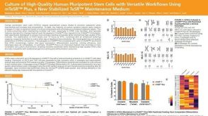 科学海报Culture of High-Quality Human Pluripotent Stem Cells with Versatile Workflows Using mTeSR™ Plus, a New Stabilized TeSR™ Maintenance Medium
 沪公网安备31010102008431号