Requirement for Dot1l in murine postnatal hematopoiesis and leukemogenesis by MLL translocation.
Disruptor of telomeric silencing 1-like (Dot1l) is a histone 3 lysine 79 methyltransferase. Studies of constitutive Dot1l knockout mice show that Dot1l is essential for embryonic development and prenatal hematopoiesis. DOT1L also interacts with translocation partners of Mixed Lineage Leukemia (MLL) gene,which is commonly translocated in human leukemia. However,the requirement of Dot1l in postnatal hematopoiesis and leukemogenesis of MLL translocation proteins has not been conclusively shown. With a conditional Dot1l knockout mouse model,we examined the consequences of Dot1l loss in postnatal hematopoiesis and MLL translocation leukemia. Deletion of Dot1l led to pancytopenia and failure of hematopoietic homeostasis,and Dot1l-deficient cells minimally reconstituted recipient bone marrow in competitive transplantation experiments. In addition,MLL-AF9 cells required Dot1l for oncogenic transformation,whereas cells with other leukemic oncogenes,such as Hoxa9/Meis1 and E2A-HLF,did not. These findings illustrate a crucial role of Dot1l in normal hematopoiesis and leukemogenesis of specific oncogenes.
View Publication
产品类型:
产品号#:
03234
产品名:
MethoCult™M3234
Volpe DA and Warren MK (JUN 2003)
Toxicology in vitro : an international journal published in association with BIBRA 17 3 271--7
Myeloid clonogenic assays for comparison of the in vitro toxicity of alkylating agents.
A battery of clonal assays for myeloid progenitor cells (HPP-CFC,CFU-gemm,CFU-gm,CFU-g) was utilized to evaluate the myelotoxicity of a series of alkylating agents representing the spectrum of clinical times to nadir. Bone marrow aspirates from normal volunteers were incubated with mechlorethamine,busulfan,melphalan,carmustine or lomustine for 1 h and then cultured in methylcellulose with 30% serum and cytokines. There was a concentration-dependent inhibition of colony formation and often a differential toxicity to the myeloid progenitors with the alkylators tested. On a molar basis,mechlorethamine and melphalan were the most toxic of the alkylator drugs to the myeloid precursors. The most sensitive progenitor was CFU-gemm with the lowest inhibitory concentration IC(70) concentrations for mechlorethamine,melphalan,carmustine and lomustine. Generally,there was great similarity for drug effects between CFU-g and CFU-gm with overlapping inhibition curves. HPP-CFC proved to be the least sensitive of the progenitors to the toxic actions of the drugs. While there was no correlation between the time to clinical neutropenic nadir and the most sensitive progenitor in the clonal assays,the CFU-gm assay remains a suitable method for determining the myelotoxic potential of cytotoxic agents.
View Publication
产品类型:
产品号#:
04535
04545
产品名:
MethoCult™H4535富集无EPO
MethoCult™ H4535 Enriched,不含EPO
An MC et al. ( 2014)
PLoS currents 6 1--19
Polyglutamine Disease Modeling: Epitope Based Screen for Homologous Recombination using CRISPR/Cas9 System.
We have previously reported the genetic correction of Huntington's disease (HD) patient-derived induced pluripotent stem cells using traditional homologous recombination (HR) approaches. To extend this work,we have adopted a CRISPR-based genome editing approach to improve the efficiency of recombination in order to generate allelic isogenic HD models in human cells. Incorporation of a rapid antibody-based screening approach to measure recombination provides a powerful method to determine relative efficiency of genome editing for modeling polyglutamine diseases or understanding factors that modulate CRISPR/Cas9 HR.
View Publication
产品类型:
产品号#:
85850
85857
产品名:
mTeSR™1
mTeSR™1
Gorman BR et al. (DEC 2014)
PLoS ONE 9 12 e116037
Multi-scale imaging and informatics pipeline for in situ pluripotent stem cell analysis
Human pluripotent stem (hPS) cells are a potential source of cells for medical therapy and an ideal system to study fate decisions in early development. However,hPS cells cultured in vitro exhibit a high degree of heterogeneity,presenting an obstacle to clinical translation. hPS cells grow in spatially patterned colony structures,necessitating quantitative single-cell image analysis. We offer a tool for analyzing the spatial population context of hPS cells that integrates automated fluorescent microscopy with an analysis pipeline. It enables high-throughput detection of colonies at low resolution,with single-cellular and sub-cellular analysis at high resolutions,generating seamless in situ maps of single-cellular data organized by colony. We demonstrate the tool's utility by analyzing inter- and intra-colony heterogeneity of hPS cell cycle regulation and pluripotency marker expression. We measured the heterogeneity within individual colonies by analyzing cell cycle as a function of distance. Cells loosely associated with the outside of the colony are more likely to be in G1,reflecting a less pluripotent state,while cells within the first pluripotent layer are more likely to be in G2,possibly reflecting a G2/M block. Our multi-scale analysis tool groups colony regions into density classes,and cells belonging to those classes have distinct distributions of pluripotency markers and respond differently to DNA damage induction. Lastly,we demonstrate that our pipeline can robustly handle high-content,high-resolution single molecular mRNA FISH data by using novel image processing techniques. Overall,the imaging informatics pipeline presented offers a novel approach to the analysis of hPS cells that includes not only single cell features but also colony wide,and more generally,multi-scale spatial configuration.
View Publication
Expression of pluripotency-associated genes in the surviving fraction of cultured human embryonic stem cells is not significantly affected by ionizing radiation.
Human embryonic stem cells (hESC) are capable to give rise to all cell types in the human body during the normal course of development. Therefore,these cells hold a great promise in regenerative cell replacement based therapeutical approaches. However,some controversy exists in literature concerning the ultimate fate of hESC after exposure to genotoxic agents,in particular,regarding the effect of DNA damaging insults on pluripotency of hESC. To comprehensively address this issue,we performed an analysis of the expression of marker genes,associated with pluripotent state of hESC,such as Oct-4,Nanog,Sox-2,SSEA-4,TERT,TRA-1-60 and TRA-1-81 up to 65h after exposure to ionizing radiation (IR) using flow cytometry,immunocytochemistry and quantitative real-time polymerase chain reaction techniques. We show that irradiation with relatively low doses of gamma-radiation (0.2Gy and 1Gy) does not lead to loss of expression of the pluripotency-associated markers in the surviving hESC. While changes in the levels of expression of some of the pluripotency markers were observed at different time points after IR exposure,these alterations were not persistent,and,in most cases,the expression of the pluripotency-associated markers remained significantly higher than that observed in fully differentiated human fibroblasts,and in hESCs differentiated into definitive endodermal lineage. Our data suggest that exposure of hESC to relatively low doses of IR as a model genotoxic agent does not significantly affect pluripotency of the surviving fraction of hESC.
View Publication
产品类型:
产品号#:
85850
85857
产品名:
mTeSR™1
mTeSR™1
Kadari A et al. ( 2014)
Stem cell research & therapy 5 2 47
Excision of viral reprogramming cassettes by Cre protein transduction enables rapid, robust and efficient derivation of transgene-free human induced pluripotent stem cells.
Integrating viruses represent robust tools for cellular reprogramming; however,the presence of viral transgenes in induced pluripotent stem cells (iPSCs) is deleterious because it holds the risk of insertional mutagenesis leading to malignant transformation. Here,we combine the robustness of lentiviral reprogramming with the efficacy of Cre recombinase protein transduction to derive iPSCs devoid of transgenes. By genome-wide analysis and targeted differentiation towards the cardiomyocyte lineage,we show that transgene-free iPSCs are superior to iPSCs before Cre transduction. Our study provides a simple,rapid and robust protocol for the generation of clinical-grade iPSCs suitable for disease modeling,tissue engineering and cell replacement therapies.
View Publication
产品类型:
产品号#:
85850
85857
产品名:
mTeSR™1
mTeSR™1
Czysz K et al. (FEB 2015)
PLoS ONE 10 2 e0117689
Dmso efficiently down regulates pluripotency genes in human embryonic stem cells during definitive endoderm derivation and increases the proficiency of hepatic differentiation
BACKGROUND Definitive endoderm (DE) is one of the three germ layers which during in vivo vertebrate development gives rise to a variety of organs including liver,lungs,thyroid and pancreas; consequently efficient in vitro initiation of stem cell differentiation to DE cells is a prerequisite for successful cellular specification to subsequent DE-derived cell types [1,2]. In this study we present a novel approach to rapidly and efficiently down regulate pluripotency genes during initiation of differentiation to DE cells by addition of dimethyl sulfoxide (DMSO) to Activin A-based culture medium and report its effects on the downstream differentiation to hepatocyte-like cells. MATERIALS AND METHODS Human embryonic stem cells (hESC) were differentiated to DE using standard methods in medium supplemented with 100ng/ml of Activin A and compared to cultures where DE specification was additionally enhanced with different concentrations of DMSO. DE cells were subsequently primed to generate hepatic-like cells to investigate whether the addition of DMSO during formation of DE improved subsequent expression of hepatic markers. A combination of flow cytometry,real-time quantitative reverse PCR and immunofluorescence was applied throughout the differentiation process to monitor expression of pluripotency (POUF5/OCT4 & NANOG),definitive endoderm (SOX17,CXCR4 & GATA4) and hepatic (AFP & ALB) genes to generate differentiation stage-specific signatures. RESULTS Addition of DMSO to the Activin A-based medium during DE specification resulted in rapid down regulation of the pluripotency genes OCT4 and NANOG,accompanied by an increase expression of the DE genes SOX17,CXCR4 and GATA4. Importantly,the expression level of ALB in DMSO-treated cells was also higher than in cells which were differentiated to the DE stage via standard Activin A treatment.
View Publication
Critical role for PI 3-kinase in the control of erythropoietin-induced erythroid progenitor proliferation.
The production of red blood cells is tightly regulated by erythropoietin (Epo). The phosphoinositide 3-kinase (PI 3-kinase) pathway was previously shown to be activated in response to Epo. We studied the role of this pathway in the control of Epo-induced survival and proliferation of primary human erythroid progenitors. We show that phosphoinositide 3 (PI 3)-kinase associates with 4 tyrosine-phosphorylated proteins in primary human erythroid progenitors,namely insulin receptor substrate-2 (IRS2),Src homology 2 domain-containing inositol 5'-phosphatase (SHIP),Grb2-associated binder-1 (Gab1),and the Epo receptor (EpoR). Using different in vitro systems,we demonstrate that 3 alternative pathways independently lead to Epo-induced activation of PI 3-kinase and phosphorylation of its downstream effectors,Akt,FKHRL1,and P70S6 kinase: through direct association of PI 3-kinase with the last tyrosine residue (Tyr479) of the Epo receptor (EpoR),through recruitment and phosphorylation of Gab proteins via either Tyr343 or Tyr401 of the EpoR,or through phosphorylation of IRS2 adaptor protein. The mitogen-activated protein (MAP) kinase pathway was also activated by Epo in erythroid progenitors,but we found that this process is independent of PI 3-kinase activation. In erythroid progenitors,the functional role of PI 3-kinase was both to prevent apoptosis and to stimulate cell proliferation in response to Epo stimulation. Finally,our results show that PI 3-kinase-mediated proliferation of erythroid progenitors in response to Epo occurs mainly through modulation of the E3 ligase SCF(SKP2),which,in turn,down-regulates p27(Kip1) cyclin-dependent kinase (CDK) inhibitor via proteasome degradation.
View Publication

 EasySep™小鼠TIL(CD45)正选试剂盒


 20:24
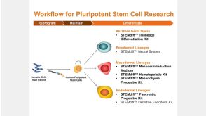
线上讲座STEMdiff™ Kits for Robust and Efficient Differentiation of hPSCs to Multiple Cell Types发布日期: 06/24/2016
20:24
线上讲座STEMdiff™ Kits for Robust and Efficient Differentiation of hPSCs to Multiple Cell Types发布日期: 06/24/2016

 沪公网安备31010102008431号