Hematopoietic cells regulate the angiogenic switch during tumorigenesis.
Hematopoietic cells (HCs) promote blood vessel formation by producing various proangiogenic cytokines and chemokines and matrix metalloproteinases. We injected mouse colon26 colon cancer cells or human PC3 prostate adenocarcinoma cells into mice and studied the localization of HCs during tumor development. HCs were distributed in the inner tumor mass in all of the tumor tissues examined; however,the localization of HCs in the tumor tissue differed depending on the tumor cell type. In the case of colon26 tumors,as the tumor grew,many mature HCs migrated into the tumor mass before fine capillary formation was observed. On the other hand,although very few HCs migrated into PC3 tumor tissue,c-Kit+ hematopoietic stem/progenitor cells accumulated around the edge of the tumor. Bone marrow suppression induced by injection of anti-c-Kit neutralizing antibody suppressed tumor angiogenesis by different mechanisms according to the tumor cell type: bone marrow suppression inhibited the initiation of sprouting angiogenesis in colon26 tumors,while it suppressed an increase in the caliber of newly developed blood vessels at the tumor edge in PC3 tumors. Our findings suggest that HCs are involved in tumor angiogenesis and regulate the angiogenic switch during tumorigenesis.
View Publication
产品类型:
产品号#:
03434
03444
产品名:
MethoCult™ GF M3434
MethoCult™ GF M3434
Wognum AW et al. ( )
Archives of medical research 34 6 461--75
Identification and isolation of hematopoietic stem cells.
Hematopoietic stem cells (HSCs) are defined by their ability to repopulate all of the hematopoietic lineages in vivo and sustain the production of these cells for the life span of the individual. In the absence of reliable direct markers for HSCs,their identification and enumeration depends on functional long-term,multilineage,in vivo repopulation assays. The extremely low frequency of HSCs in any tissue and the absence of a specific HSC phenotype have made their purification and characterization a highly challenging goal. HSCs and primitive hematopoietic cells can be distinguished from mature blood cells by their lack of lineage-specific markers and presence of certain other cell-surface antigens,such as CD133 (for human cells) and c-kit and Sca-1 (for murine cells). Functional analyses of purified subpopulations of primitive hematopoietic cells have led to the development of several procedures for isolating cell populations that are highly enriched in cells with in vivo stem cell activity. Simplified methods for obtaining these cells at high yield have been important to the practical exploitation of such advances. This article reviews recent progress in identifying human and mouse HSCs and current techniques for their purification.
View Publication
产品类型:
产品号#:
18056
18056RF
产品名:
Zhang J et al. (FEB 2007)
The Journal of clinical investigation 117 2 473--81
Primitive hematopoietic cells resist HIV-1 infection via p21.
Hematopoietic stem cells are resistant to HIV-1 infection. Here,we report a novel mechanism by which the cyclin-dependent kinase inhibitor (CKI) p21(Waf1/Cip1/Sdi1) (p21),a known regulator of stem cell pool size,restricts HIV-1 infection of primitive hematopoietic cells. Modifying p21 expression altered HIV-1 infection prior to changes in cell cycling and was selective for p21 since silencing the related CKIs,p27(Kip1) and p18(INK4C),had no effect on HIV-1. We show that p21 blocked viral infection by complexing with HIV-1 integrase and aborting chromosomal integration. A closely related lentivirus with a distinct integrase,SIVmac-251,and the other cell-intrinsic inhibitors of HIV-1,Trim5alpha,PML,Murr1,and IFN-alpha,were unaffected by p21. Therefore,p21 is an endogenous cellular component in stem cells that provides a unique molecular barrier to HIV-1 infection and may explain how these cells remain an uninfected sanctuary" in HIV disease."
View Publication
Rebel VI et al. (NOV 2002)
Proceedings of the National Academy of Sciences of the United States of America 99 23 14789--94
Distinct roles for CREB-binding protein and p300 in hematopoietic stem cell self-renewal.
Hematopoietic stem cells (HSC) are tightly regulated through,as yet,undefined mechanisms that balance self-renewal and differentiation. We have identified a role for the transcriptional coactivators CREB-binding protein (CBP) and p300 in such HSC fate decisions. A full dose of CBP,but not p300,is crucial for HSC self-renewal. Conversely,p300,but not CBP,is essential for proper hematopoietic differentiation. Furthermore,in chimeric mice,hematologic malignancies emerged from both CBP(-/-) and p300(-/-) cell populations. Thus,CBP and p300 play essential but distinct roles in maintaining normal hematopoiesis,and,in mice,both are required for preventing hematologic tumorigenesis.
View Publication
产品类型:
产品号#:
06902
06952
00321
00322
00323
00324
00325
产品名:
Qin J et al. (NOV 2016)
Scientific reports 6 37388
Connexin 32-mediated cell-cell communication is essential for hepatic differentiation from human embryonic stem cells.
Gap junction-mediated cell-cell interactions are highly conserved and play essential roles in cell survival,proliferation,differentiation and patterning. We report that Connexin 32 (Cx32)-mediated gap junctional intercellular communication (GJIC) is necessary for human embryonic stem cell-derived hepatocytes (hESC-Heps) during step-wise hepatic lineage restriction and maturation. Vitamin K2,previously shown to promote Cx32 expression in mature hepatocytes,up-regulated Cx32 expression and GJIC activation during hepatic differentiation and maturation,resulting in significant increases of hepatic markers expression and hepatocyte functions. In contrast,negative Cx32 regulator 2-aminoethoxydiphenyl borate blocked hESC-to-hepatocyte maturation and muted hepatocyte functions through disruption of GJIC activities. Dynamic gap junction organization and internalization are phosphorylation-dependent and the p38 mitogen-activated protein kinases pathway (MAPK) can negatively regulate Cxs through phosphorylation-dependent degradation of Cxs. We found that p38 MAPK inhibitor SB203580 improved maturation of hESC-Heps correlating with up-regulation of Cx32; by contrast,the p38 MAPK activator,anisomycin,blocked hESC-Heps maturation correlating with down-regulation of Cx32. These results suggested that Cx32 is essential for cell-cell interactions that facilitate driving hESCs through hepatic-lineage maturation. Regulators of both Cx32 and other members of its pathways maybe used as a promising approach on regulating hepatic lineage restriction of pluripotent stem cells and optimizing their functional maturation.
View Publication
产品类型:
产品号#:
05850
05857
05870
05875
85850
85857
85870
85875
产品名:
mTeSR™1
mTeSR™1
Sugii S et al. (FEB 2010)
Proceedings of the National Academy of Sciences of the United States of America 107 8 3558--63
Human and mouse adipose-derived cells support feeder-independent induction of pluripotent stem cells.
Although adipose tissue is an expandable and readily attainable source of proliferating,multipotent stem cells,its potential for use in regenerative medicine has not been extensively explored. Here we report that adult human and mouse adipose-derived stem cells can be reprogrammed to induced pluripotent stem (iPS) cells with substantially higher efficiencies than those reported for human and mouse fibroblasts. Unexpectedly,both human and mouse iPS cells can be obtained in feeder-free conditions. We discovered that adipose-derived stem cells intrinsically express high levels of pluripotency factors such as basic FGF,TGFbeta,fibronectin,and vitronectin and can serve as feeders for both autologous and heterologous pluripotent cells. These results demonstrate a great potential for adipose-derived cells in regenerative therapeutics and as a model for studying the molecular mechanisms of feeder-free iPS generation and maintenance.
View Publication
产品类型:
产品号#:
05850
05857
05870
05875
85850
85857
85870
85875
产品名:
mTeSR™1
mTeSR™1
Prowse ABJ et al. (NOV 2010)
Biomaterials 31 32 8281--8288
Long term culture of human embryonic stem cells on recombinant vitronectin in ascorbate free media.
Human embryonic stem cells (hESC) are expected to provide revolutionary therapeutic applications and drug discovery technologies. In order for this to be achieved a reproducible,defined animal component free culture system is required for the scale-up production of undifferentiated hESC. In this work we have investigated the applicability of a recombinantly produced domain of human vitronectin as an extracellular matrix alternative to the common standards Geltrex or Matrigel. In addition we have validated an ascorbate free media capable of supporting CD30(low) populations of hESC through a multi-factorial analysis of bFGF and Activin A. The recombinant vitronectin domain combined with the ascorbate free media were capable of supporting 3 cell lines,MEL1,MEL2 and hES3 for 10 or more passages while maintaining hESC pluripotency markers and differentiation capacity. The culture method outlined here provides a platform for future investigation into growth factor and extracellular matrix effects on hESC maintenance prior to bioreactor scale-up.
View Publication
Lidonnici MR et al. (MAY 2008)
Blood 111 9 4771--9
Requirement of c-Myb for p210(BCR/ABL)-dependent transformation of hematopoietic progenitors and leukemogenesis.
The c-Myb gene encodes a transcription factor required for proliferation and survival of normal myeloid progenitors and leukemic blast cells. Targeting of c-Myb by antisense oligodeoxynucleotides has suggested that myeloid leukemia blasts (including chronic myelogenous leukemia [CML]-blast crisis cells) rely on c-Myb expression more than normal progenitors,but a genetic approach to assess the requirement of c-Myb by p210(BCR/ABL)-transformed hematopoietic progenitors has not been taken. We show here that loss of a c-Myb allele had modest effects (20%-28% decrease) on colony formation of nontransduced progenitors,while the effect on p210(BCR/ABL)-expressing Lin(-) Sca-1(+) and Lin(-) Sca-1(+)Kit(+) cells was more pronounced (50%-80% decrease). Using a model of CML-blast crisis,mice (n = 14) injected with p210(BCR/ABL)-transduced p53(-/-)c-Myb(w/w) marrow cells developed leukemia rapidly and had a median survival of 26 days,while only 67% of mice (n = 12) injected with p210(BCR/ABL)-transduced p53(-/-)c-Myb(w/d) marrow cells died of leukemia with a median survival of 96 days. p210(BCR/ABL)-transduced c-Myb(w/w) and c-Myb(w/d) marrow progenitors expressed similar levels of the c-Myb-regulated genes c-Myc and cyclin B1,while those of Bcl-2 were reduced. However,ectopic Bcl-2 expression did not enhance colony formation of p210(BCR/ABL)-transduced c-Myb(w/d) Lin(-)Sca-1(+)Kit(+) cells. Together,these studies support the requirement of c-Myb for p210(BCR/ABL)-dependent leukemogenesis.
View Publication
产品类型:
产品号#:
04230
产品名:
MethoCult™ H4230
Eckardt S et al. (FEB 2007)
Genes & development 21 4 409--19
Hematopoietic reconstitution with androgenetic and gynogenetic stem cells.
Parthenogenetic embryonic stem (ES) cells with two oocyte-derived genomes (uniparental) have been proposed as a source of autologous tissue for transplantation. The therapeutic applicability of any uniparental cell type is uncertain due to the consequences of genomic imprinting that in mammalian uniparental tissues causes unbalanced expression of imprinted genes. We transplanted uniparental fetal liver cells into lethally irradiated adult mice to test their capacity to replace adult hematopoietic tissue. Both maternal (gynogenetic) and paternal (androgenetic) derived cells conveyed long-term,multilineage reconstitution of hematopoiesis in recipients,with no associated pathologies. We also establish that uniparental ES cells can differentiate into transplantable hematopoietic progenitors in vitro that contribute to long-term hematopoiesis in recipients. Hematopoietic tissue in recipients maintained fidelity of parent-of-origin methylation marks at the Igf2/H19 locus; however,variability occurred in the maintenance of parental-specific methylation marks at other loci. In summary,despite genomic imprinting and its consequences on development that are particularly evident in the androgenetic phenotype,uniparental cells of both parental origins can form adult-transplantable stem cells and can repopulate an adult organ.
View Publication
产品类型:
产品号#:
03434
03444
产品名:
MethoCult™ GF M3434
MethoCult™ GF M3434
Rathjen J and Rathjen PD (OCT 2001)
Current opinion in genetics & development 11 5 587--94
Mouse ES cells: experimental exploitation of pluripotent differentiation potential.
Pluripotent ES cells can be used to generate a wide variety of cell populations in vitro in a manner resembling embryonic development. Recent advances in controlling ES cell differentiation,combined with the power of genetic and biochemical manipulation,are providing insights into cell biology and the determination of cell fate.
View Publication

 EasySep™小鼠TIL(CD45)正选试剂盒


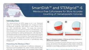 产品手册SmartDish™ and STEMgrid™-6 Meniscus-Free Cultureware for More Accurate Counting of Hematopoietic Colonies

 沪公网安备31010102008431号