A novel I-branching beta-1,6-N-acetylglucosaminyltransferase involved in human blood group I antigen expression.
The human blood group i and I antigens are determined by linear and branched poly-N-acetyllactosamine structures,respectively. In erythrocytes,the fetal i antigen is converted to the adult I antigen by I-branching beta-1,6-N-acetylglucosaminyltransferase (IGnT) during development. Dysfunction of the I-branching enzyme may result in the adult i phenotype in erythrocytes. However,the I gene responsible for blood group I antigen has not been fully confirmed. We report here a novel human I-branching enzyme,designated IGnT3. The genes for IGnT1 (reported in 1993),IGnT2 (also presented in this study),and IGnT3 consist of 3 exons and share the second and third exons. Bone marrow cells preferentially expressed IGnT3 transcript. During erythroid differentiation using CD34(+) cells,IGnT3 was markedly up-regulated with concomitant decrease in IGnT1/2. Moreover,reticulocytes expressed the IGnT3 transcript,but IGnT1/2 was below detectable levels. By molecular genetic analyses of an adult i pedigree,individuals with the adult i phenotype were revealed to have heterozygous alleles with mutations in exon 2 (1006GtextgreaterA; Gly336Arg) and exon 3 (1049GtextgreaterA; Gly350Glu),respectively,of the IGnT3 gene. Chinese hamster ovary (CHO) cells transfected with each mutated IGnT3 cDNA failed to express I antigen. These findings indicate that the expression of the blood group I antigen in erythrocytes is determined by a novel IGnT3,not by IGnT1 or IGnT2.
View Publication
产品类型:
产品号#:
09500
09600
09650
产品名:
BIT 9500血清替代物
StemSpan™ SFEM
StemSpan™ SFEM
Finkbeiner SR et al. (NOV 2015)
Biology open 4 11 bio.013235--
Generation of tissue-engineered small intestine using embryonic stem cell-derived human intestinal organoids.
Short bowel syndrome (SBS) is characterized by poor nutrient absorption due to a deficit of healthy intestine. Current treatment practices rely on providing supportive medical therapy with parenteral nutrition; while life saving,such interventions are not curative and are still associated with significant co-morbidities. As approaches to lengthen remaining intestinal tissue have been met with only limited success and intestinal transplants have poor survival outcomes,new approaches to treating SBS are necessary. Human intestine derived from embryonic stem cells (hESCs) or induced pluripotent stem cells (iPSCs),called human intestinal organoids (HIOs),have the potential to offer a personalized and scalable source of intestine for regenerative therapies. However,given that HIOs are small three-dimensional structures grown in vitro,methods to generate usable HIO-derived constructs are needed. We investigated the ability of hESCs or HIOs to populate acellular porcine intestinal matrices and artificial polyglycolic/poly L lactic acid (PGA/PLLA) scaffolds,and examined the ability of matrix/scaffolds to thrive when transplanted in vivo. Our results demonstrate that the acellular matrix alone is not sufficient to instruct hESC differentiation towards an endodermal or intestinal fate. We observed that while HIOs reseed acellular porcine matrices in vitro,the HIO-reseeded matrices do not thrive when transplanted in vivo. In contrast,HIO-seeded PGA/PLLA scaffolds thrive in vivo and develop into tissue that looks nearly identical to adult human intestinal tissue. Our results suggest that HIO-seeded PGA/PLLA scaffolds are a promising avenue for developing the mucosal component of tissue engineered human small intestine,which need to be explored further to develop them into fully functional tissue.
View Publication
产品类型:
产品号#:
85850
85857
产品名:
mTeSR™1
mTeSR™1
Ang Y-S et al. (DEC 2016)
Cell 167 7 1734--1749.e22
Disease Model of GATA4 Mutation Reveals Transcription Factor Cooperativity in Human Cardiogenesis.
Mutation of highly conserved residues in transcription factors may affect protein-protein or protein-DNA interactions,leading to gene network dysregulation and human disease. Human mutations in GATA4,a cardiogenic transcription factor,cause cardiac septal defects and cardiomyopathy. Here,iPS-derived cardiomyocytes from subjects with a heterozygous GATA4-G296S missense mutation showed impaired contractility,calcium handling,and metabolic activity. In human cardiomyocytes,GATA4 broadly co-occupied cardiac enhancers with TBX5,another transcription factor that causes septal defects when mutated. The GATA4-G296S mutation disrupted TBX5 recruitment,particularly to cardiac super-enhancers,concomitant with dysregulation of genes related to the phenotypic abnormalities,including cardiac septation. Conversely,the GATA4-G296S mutation led to failure of GATA4 and TBX5-mediated repression at non-cardiac genes and enhanced open chromatin states at endothelial/endocardial promoters. These results reveal how disease-causing missense mutations can disrupt transcriptional cooperativity,leading to aberrant chromatin states and cellular dysfunction,including those related to morphogenetic defects.
View Publication
NANOG Is a Direct Target of TGF$\$/Activin-Mediated SMAD Signaling in Human ESCs
Self-renewal of human embryonic stem cells (ESCs) is promoted by FGF and TGFbeta/Activin signaling,and differentiation is promoted by BMP signaling,but how these signals regulate genes critical to the maintenance of pluripotency has been unclear. Using a defined medium,we show here that both TGFbeta and FGF signals synergize to inhibit BMP signaling; sustain expression of pluripotency-associated genes such as NANOG,OCT4,and SOX2; and promote long-term undifferentiated proliferation of human ESCs. We also show that both TGFbeta- and BMP-responsive SMADs can bind with the NANOG proximal promoter. NANOG promoter activity is enhanced by TGFbeta/Activin and FGF signaling and is decreased by BMP signaling. Mutation of putative SMAD binding elements reduces NANOG promoter activity to basal levels and makes NANOG unresponsive to BMP and TGFbeta signaling. These results suggest that direct binding of TGFbeta/Activin-responsive SMADs to the NANOG promoter plays an essential role in sustaining human ESC self-renewal.
View Publication
产品类型:
产品号#:
85850
85857
产品名:
mTeSR™1
mTeSR™1
Malik J et al. (NOV 2013)
Haematologica 98 11 1778--1787
Erythropoietin critically regulates the terminal maturation of murine and human primitive erythroblasts
Primitive erythroid cells,the first red blood cells produced in the mammalian embryo,are necessary for embryonic survival. Erythropoietin and its receptor EpoR,are absolutely required for survival of late-stage definitive erythroid progenitors in the fetal liver and adult bone marrow. Epo- and Epor-null mice die at E13.5 with a lack of definitive erythrocytes. However,the persistence of circulating primitive erythroblasts raises questions about the role of erythropoietin/EpoR in primitive erythropoiesis. Using Epor-null mice and a novel primitive erythroid 2-step culture we found that erythropoietin is not necessary for specification of primitive erythroid progenitors. However,Epor-null embryos develop a progressive,profound anemia by E12.5 as primitive erythroblasts mature as a synchronous cohort. This anemia results from reduced primitive erythroblast proliferation associated with increased p27 expression,from advanced cellular maturation,and from markedly elevated rates of apoptosis associated with an imbalance in pro- and anti-apoptotic gene expression. Both mouse and human primitive erythroblasts cultured without erythropoietin also undergo accelerated maturation and apoptosis at later stages of maturation. We conclude that erythropoietin plays an evolutionarily conserved role in promoting the proliferation,survival,and appropriate timing of terminal maturation of primitive erythroid precursors.
View Publication
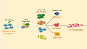
High-throughput fingerprinting of human pluripotent stem cell fate responses and lineage bias.
Populations of cells create local environments that lead to emergent heterogeneity. This is particularly evident with human pluripotent stem cells (hPSCs): microenvironmental heterogeneity limits hPSC cell fate control. We developed a high-throughput platform to screen hPSCs in configurable microenvironments in which we optimized colony size,cell density and other parameters to achieve rapid and robust cell fate responses to exogenous cues. We used this platform to perform single-cell protein expression profiling,revealing that Oct4 and Sox2 costaining discriminates pluripotent,neuroectoderm,primitive streak and extraembryonic cell fates. We applied this Oct4-Sox2 code to analyze dose responses of 27 developmental factors to obtain lineage-specific concentration optima and to quantify cell line–specific endogenous signaling pathway activation and differentiation bias. We demonstrated that short-term responses predict definitive endoderm induction efficiency and can be used to rescue differentiation of cell lines reticent to cardiac induction. This platform will facilitate high-throughput hPSC-based screening and quantification of lineage-induction bias.
View Publication
产品类型:
产品号#:
85850
85857
产品名:
mTeSR™1
mTeSR™1
Kharas MG et al. (JAN 2007)
Blood 109 2 747--55
KLF4 suppresses transformation of pre-B cells by ABL oncogenes.
Genes that are strongly repressed after B-cell activation are candidates for being inactivated,mutated,or repressed in B-cell malignancies. Krüppel-like factor 4 (Klf4),a gene down-regulated in activated murine B cells,is expressed at low levels in several types of human B-cell lineage lymphomas and leukemias. The human KLF4 gene has been identified as a tumor suppressor gene in colon and gastric cancer; in concordance with this,overexpression of KLF4 can suppress proliferation in several epithelial cell types. Here we investigate the effects of KLF4 on pro/pre-B-cell transformation by v-Abl and BCR-ABL,oncogenes that cause leukemia in mice and humans. We show that overexpression of KLF4 induces arrest and apoptosis in the G1 phase of the cell cycle. KLF4-mediated death,but not cell-cycle arrest,can be rescued by Bcl-XL overexpression. Transformed pro/pre-B cells expressing KLF4 display increased expression of p21CIP and decreased expression of c-Myc and cyclin D2. Tetracycline-inducible expression of KLF4 in B-cell progenitors of transgenic mice blocks transformation by BCR-ABL and depletes leukemic pre-B cells in vivo. Collectively,our work identifies KLF4 as a putative tumor suppressor in B-cell malignancies.
View Publication
产品类型:
产品号#:
03630
产品名:
MethoCult™M3630
West FD et al. ( 2015)
1330 153--167
Generation of Chimeras from Porcine Induced Pluripotent Stem Cells
Pig induced pluripotent stem cells (piPSCs) offer a great opportunity and a number of advantages in the generation of transgenic animals. These immortalized cells can undergo multiple rounds of genetic modifications (e.g.,gene knock-in,knockout) and selection leading to animals that have optimized traits of biomedical or agricultural interests. In this chapter we describe the production and characterization of piPSCs,microinjection of piPSCs into embryos,embryo transfer and production of chimeric animals based on successful protocols.
View Publication
产品类型:
产品号#:
85850
85857
产品名:
mTeSR™1
mTeSR™1
Moore JC (JAN 2013)
997 35--43
Generation of Human-Induced Pluripotent Stem Cells by Lentiviral Transduction
Human somatic cells can be reprogrammed to the pluripotent state to become human-induced pluripotent stem cells (hiPSC). This reprogramming is achieved by activating signaling pathways that are expressed during early development. These pathways can be induced by ectopic expression of four transcription factors—Oct4,Sox2,Klf4,and c-Myc. Although there are many ways to deliver these transcription factors into the somatic cells,this chapter will provide protocols that can be used to generate hiPSC from lentiviruses.
View Publication
Chagraoui J et al. (APR 2003)
Blood 101 8 2973--82
Fetal liver stroma consists of cells in epithelial-to-mesenchymal transition.
Liver becomes the predominant site of hematopoiesis by 11.5 dpc (days after coitus) in the mouse and 15 gestational weeks in humans and stays so until the end of gestation. The reason the liver is the major hematopoietic site during fetal life is not clear. In this work,we tried to define which of the fetal liver microenvironmental cell populations would be associated with the development of hematopoiesis and found that a population of cells with mixed endodermal and mesodermal features corresponded to hematopoietic-supportive fetal liver stroma. Stromal cells generated from primary cultures or stromal lines from mouse or human fetal liver in the hematopoietic florid phase expressed both mesenchymal markers (vimentin,osteopontin,collagen I,alpha smooth muscle actin,thrombospondin-1,EDa fibronectin,calponin,Stro-1 antigens,myocyte-enhancer factor 2C) and epithelial (alpha-fetoprotein,cytokeratins 8 and 18,albumin,E-cadherin,hepatocyte nuclear factor 3 alpha) markers. Such a cell population fits with the description of cells in epithelial-to-mesenchymal transition (EMT),often observed during development,including that of the liver. The hematopoietic supportive capacity of EMT cells was lost after hepatocytic maturation,induced by oncostatin M in the cell line AFT024. EMT cells were observed in the fetal liver microenvironment during the hematopoietic phase but not in nonhematopoietic liver by the end of gestation and in the adult. EMT cells represent a novel stromal cell type that may be generated from hepatic endodermal or mesenchymal stem cells or even from circulating hematopoietic stem cells (HSCs) seeding the liver rudiment.
View Publication

 EasySep™小鼠TIL(CD45)正选试剂盒


 沪公网安备31010102008431号