Musah S et al. (SEP 2014)
Proceedings of the National Academy of Sciences of the United States of America 111 38 13805--10
Substratum-induced differentiation of human pluripotent stem cells reveals the coactivator YAP is a potent regulator of neuronal specification.
Physical stimuli can act in either a synergistic or antagonistic manner to regulate cell fate decisions,but it is less clear whether insoluble signals alone can direct human pluripotent stem (hPS) cell differentiation into specialized cell types. We previously reported that stiff materials promote nuclear localization of the Yes-associated protein (YAP) transcriptional coactivator and support long-term self-renewal of hPS cells. Here,we show that even in the presence of soluble pluripotency factors,compliant substrata inhibit the nuclear localization of YAP and promote highly efficient differentiation of hPS cells into postmitotic neurons. In the absence of neurogenic factors,the effective substrata produce neurons rapidly (2 wk) and more efficiently (textgreater75%) than conventional differentiation methods. The neurons derived from substrate induction express mature markers and possess action potentials. The hPS differentiation observed on compliant surfaces could be recapitulated on stiff surfaces by adding small-molecule inhibitors of F-actin polymerization or by depleting YAP. These studies reveal that the matrix alone can mediate differentiation of hPS cells into a mature cell type,independent of soluble inductive factors. That mechanical cues can override soluble signals suggests that their contributions to early tissue development and lineage commitment are profound.
View Publication
产品类型:
产品号#:
85850
85857
产品名:
mTeSR™1
mTeSR™1
Bogomazova AN et al. (JAN 2015)
Scientific reports 5 7749
No DNA damage response and negligible genome-wide transcriptional changes in human embryonic stem cells exposed to terahertz radiation.
Terahertz (THz) radiation was proposed recently for use in various applications,including medical imaging and security scanners. However,there are concerns regarding the possible biological effects of non-ionising electromagnetic radiation in the THz range on cells. Human embryonic stem cells (hESCs) are extremely sensitive to environmental stimuli,and we therefore utilised this cell model to investigate the non-thermal effects of THz irradiation. We studied DNA damage and transcriptome responses in hESCs exposed to narrow-band THz radiation (2.3 THz) under strict temperature control. The transcription of approximately 1% of genes was subtly increased following THz irradiation. Functional annotation enrichment analysis of differentially expressed genes revealed 15 functional classes,which were mostly related to mitochondria. Terahertz irradiation did not induce the formation of γH2AX foci or structural chromosomal aberrations in hESCs. We did not observe any effect on the mitotic index or morphology of the hESCs following THz exposure.
View Publication
产品类型:
产品号#:
85850
85857
产品名:
mTeSR™1
mTeSR™1
Xu X et al. ( 2010)
Biotechnology Progress 26 3 827--837
The roles of apoptotic pathways in the low recovery rate after cryopreservation of dissociated human embryonic stem cells
Human embryonic stem (hES) cells have enormous potential for clinical applications. However,one major challenge is to achieve high cell recovery rate after cryopreservation. Understanding how the conventional cryopreservation protocol fails to protect the cells is a prerequisite for developing efficient and successful cryopreservation methods for hES cell lines and banks. We investigated how the stimuli from cryopreservation result in apoptosis,which causes the low cell recovery rate after cryopreservation. The level of reactive oxygen species (ROS) is significantly increased,F-actin content and distribution is altered,and caspase-8 and caspase-9 are activated after cryopreservation. p53 is also activated and translocated into nucleus. During cryopreservation apoptosis is induced by activation of both caspase-8 through the extrinsic pathway and caspase-9 through the intrinsic pathway. However,exactly how the extrinsic pathway is activated is still unclear and deserves further investigation.
View Publication
产品类型:
产品号#:
85850
85857
产品名:
mTeSR™1
mTeSR™1
Ghule PN et al. (MAY 2011)
Journal of cellular physiology 226 5 1149--56
Reprogramming the pluripotent cell cycle: restoration of an abbreviated G1 phase in human induced pluripotent stem (iPS) cells.
Induced pluripotent stem (iPS) cells derived from terminally differentiated human fibroblasts are reprogrammed to possess stem cell like properties. However,the extent to which iPS cells exhibit unique properties of the human embryonic stem (hES) cell cycle remains to be established. hES cells are characterized by an abbreviated G1 phase (∼ 2.5 h) and accelerated organization of subnuclear domains that mediate the assembly of regulatory machinery for histone gene expression [i.e.,histone locus bodies (HLBs)]. We therefore examined cell cycle parameters of iPS cells in comparison to hES cells. Analysis of DNA synthesis [5-bromo-2'-deoxy-uridine (BrdU) incorporation],cell cycle distribution (FACS analysis and Ki67 staining) and subnuclear organization of HLBs [immunofluorescence microscopy and fluorescence in situ hybridization (FISH)] revealed that human iPS cells have a short G1 phase (∼ 2.5 h) and an abbreviated cell cycle (16-18 h). Furthermore,HLBs are formed and reorganized rapidly after mitosis (within 1.5-2 h). Thus,reprogrammed iPS cells have cell cycle kinetics and dynamic subnuclear organization of regulatory machinery that are principal properties of pluripotent hES cells. Our findings support the concept that the abbreviated cell cycle of hES and iPS cells is functionally linked to pluripotency.
View Publication
产品类型:
产品号#:
85850
85857
产品名:
mTeSR™1
mTeSR™1
Inamdar AA et al. (JAN 2012)
Mycopathologia 173 1 13--20
A Model to Evaluate the Cytotoxicity of the Fungal Volatile Organic Compound 1-octen-3-ol in Human Embryonic Stem Cells
Microbial growth in damp indoor environments has been correlated with risks to human health. This study was aimed to determine the cytotoxicity of 1-octen-3-ol (mushroom alcohol")�
View Publication
Kunishima S et al. (MAR 2008)
Blood 111 6 3015--23
Differential expression of wild-type and mutant NMMHC-IIA polypeptides in blood cells suggests cell-specific regulation mechanisms in MYH9 disorders.
MYH9 disorders such as May-Hegglin anomaly are characterized by macrothrombocytopenia and cytoplasmic granulocyte inclusion bodies that result from mutations in MYH9,the gene for nonmuscle myosin heavy chain-IIA (NMMHC-IIA). We examined the expression of mutant NMMHC-IIA polypeptide in peripheral blood cells from patients with MYH9 5770delG and 5818delG mutations. A specific antibody to mutant NMMHC-IIA (NT629) was raised against the abnormal carboxyl-terminal residues generated by 5818delG. NT629 reacted to recombinant 5818delG NMMHC-IIA but not to wild-type NMMHC-IIA,and did not recognize any cellular components of normal peripheral blood cells. Immunofluorescence and immunoblotting revealed that mutant NMMHC-IIA was present and sequestrated only in inclusion bodies within neutrophils,diffusely distributed throughout lymphocyte cytoplasm,sparsely localized on a diffuse cytoplasmic background in monocytes,and uniformly distributed at diminished levels only in large platelets. Mutant NMMHC-IIA did not translocate to lamellipodia in surface activated platelets. Wild-type NMMHC-IIA was homogeneously distributed among megakaryocytes derived from the peripheral blood CD34(+) cells of patients,but coarse mutant NMMHC-IIA was heterogeneously scattered without abnormal aggregates in the cytoplasm. We show the differential expression of mutant NMMHC-IIA and postulate that cell-specific regulation mechanisms function in MYH9 disorders.
View Publication
产品类型:
产品号#:
09600
09650
产品名:
StemSpan™ SFEM
StemSpan™ SFEM
Porayette P et al. (AUG 2009)
The Journal of Biological Chemistry 284 35 23806--17
Differential Processing of Amyloid-β Precursor Protein Directs Human Embryonic Stem Cell Proliferation and Differentiation into Neuronal Precursor Cells
The amyloid-beta precursor protein (AbetaPP) is a ubiquitously expressed transmembrane protein whose cleavage product,the amyloid-beta (Abeta) protein,is deposited in amyloid plaques in neurodegenerative conditions such as Alzheimer disease,Down syndrome,and head injury. We recently reported that this protein,normally associated with neurodegenerative conditions,is expressed by human embryonic stem cells (hESCs). We now report that the differential processing of AbetaPP via secretase enzymes regulates the proliferation and differentiation of hESCs. hESCs endogenously produce amyloid-beta,which when added exogenously in soluble and fibrillar forms but not oligomeric forms markedly increased hESC proliferation. The inhibition of AbetaPP cleavage by beta-secretase inhibitors significantly suppressed hESC proliferation and promoted nestin expression,an early marker of neural precursor cell (NPC) formation. The induction of NPC differentiation via the non-amyloidogenic pathway was confirmed by the addition of secreted AbetaPPalpha,which suppressed hESC proliferation and promoted the formation of NPCs. Together these data suggest that differential processing of AbetaPP is normally required for embryonic neurogenesis.
View Publication
产品类型:
产品号#:
85850
85857
产品名:
mTeSR™1
mTeSR™1
Jä et al. (SEP 2010)
Proceedings of the National Academy of Sciences of the United States of America 107 37 16280--5
Isolation and killing of candidate chronic myeloid leukemia stem cells by antibody targeting of IL-1 receptor accessory protein.
Chronic myeloid leukemia (CML) is genetically characterized by the Philadelphia (Ph) chromosome,formed through a reciprocal translocation between chromosomes 9 and 22 and giving rise to the constitutively active tyrosine kinase P210 BCR/ABL1. Therapeutic strategies aiming for a cure of CML will require full eradication of Ph chromosome-positive (Ph(+)) CML stem cells. Here we used gene-expression profiling to identify IL-1 receptor accessory protein (IL1RAP) as up-regulated in CML CD34(+) cells and also in cord blood CD34(+) cells as a consequence of retroviral BCR/ABL1 expression. To test whether IL1RAP expression distinguishes normal (Ph(-)) and leukemic (Ph(+)) cells within the CML CD34(+)CD38(-) cell compartment,we established a unique protocol for conducting FISH on small numbers of sorted cells. By using this method,we sorted cells directly into drops on slides to investigate their Ph-chromosome status. Interestingly,we found that the CML CD34(+)CD38(-)IL1RAP(+) cells were Ph(+),whereas CML CD34(+)CD38(-)IL1RAP(-) cells were almost exclusively Ph(-). By performing long-term culture-initiating cell assays on the two cell populations,we found that Ph(+) and Ph(-) candidate CML stem cells could be prospectively separated. In addition,by generating an anti-IL1RAP antibody,we provide proof of concept that IL1RAP can be used as a target on CML CD34(+)CD38(-) cells to induce antibody-dependent cell-mediated cytotoxicity. This study thus identifies IL1RAP as a unique cell surface biomarker distinguishing Ph(+) from Ph(-) candidate CML stem cells and opens up a previously unexplored avenue for therapy of CML.
View Publication
产品类型:
产品号#:
09600
09650
04435
04445
产品名:
StemSpan™ SFEM
StemSpan™ SFEM
MethoCult™H4435富集
MethoCult™H4435富集
Zhang L-Z et al. (JUN 2010)
Zhonghua xue ye xue za zhi = Zhonghua xueyexue zazhi 31 6 398--402
[In vitro effects of anti-CD44 monoclonal antibody on the adhesion and migration of chronic myeloid leukemia stem cells.]
OBJECTIVE: To explore the effects of anti-CD44 monoclonal antibody-IM7 on the in vitro adhesion and migration of chronic myeloid leukemia stem cell (CML-LSC) and its mechanism. METHODS: CD34(+)CD38(-)CD123(+) leukemic stem cells (LSC) from 20 newly-diagnosed chronic myeloid leukemia (CML) patients BM cells and CD34(+)CD38(-) hematopoietic stem cells (HSC) from 20 full-term newborn cord blood cells were isolated with EasySep(TM) magnet beads. The CD44 expression of the LSC and HSC was detected by flow cytometry (FCM),and the adhesion and migration ability of the LSC and HSC pre- and post-incubated with IM7 in vitro by MTT assay and transendothelial migration assay,respectively. RESULTS: (1) After incubated with IM7,the LSC and HSC CD44 expression rates were (86.60 ± 2.10)% vs. (25.40 ± 1.70)% (P textless 0.05),respectively. (2) The adhesive ability of the LSC to endothelial cells was decreased markedly after incubated with IM7,the OD value (A(570)) changing from pre-incubation of (0.62 ± 0.11) to post-incubation of (0.34 ± 0.07),while there was little change of A(570) in the HSC group. (3) The migration ability of the LSC group was inhibited evidently after incubated with IM7,the inhibition rate being 46% ∼ 63%,while little change of that in HSC group was detected. (4) The adhesive ability of the LSC group to marrow stromal cells was decreased markedly after incubated with IM7,while little change was found in that of HSC group. CONCLUSION: The anti-CD44 monoclonal antibody-IM7 can effectively inhibit the adhesion and migration abilities of the LSC in vitro,which might provide a theoretical evidence for targeting therapy.
View Publication
产品类型:
产品号#:
产品名:
Lian R-L et al. (FEB 2016)
Molecular and cellular biochemistry 413 1-2 69--85
Effects of induced pluripotent stem cells-derived conditioned medium on the proliferation and anti-apoptosis of human adipose-derived stem cells.
Human adipose-derived stem cells (hASCs) become an appealing source for regenerative medicine. However,with the multi-passage or cryopreservation for large-scale growth procedures in terms of preclinical and clinical purposes,hASCs often reveal defective cell viability,which is a major obstacle for cell therapy. In our study,the effects of induced pluripotent stem cells-derived conditioned medium (iPS-CM) on the proliferation and anti-apoptosis in hASCs were investigated. hASCs at passage 1 were identified by the analysis of typical surface antigens with flow cytometry assay and adipogenic and osteogenic differentiation. The effect of iPS-CM on the proliferation in hASCs was analyzed by cell cycle assay and Ki67/P27 quantitative polymerase chain reaction analysis. The effect of iPS-CM on the anti-apoptosis of hASCs irradiated by 468 J/m(2) of ultraviolet C was investigated by annexin v/propidium iodide analysis,mitochondrial membrane potential assay,intracellular reactive oxygen species assay,Western blotting and caspase activity assays. The effect of iPS-CM on the surface antigen expressions of hASCs was analyzed using flow cytometry assay. The levels of Activin A and bFGF in culture supernatant of hASCs with different treatments were also detected by enzyme-linked immunosorbent assay. iPS-CM promoted proliferation and inhibited apoptosis of hASCs. This discovery demonstrates that iPS-CM might be used as one of the available means to overcome the propagation obstacle for hASCs and make for large-scale growth procedures in terms of preclinical and clinical purposes.
View Publication
产品类型:
产品号#:
85850
85857
产品名:
mTeSR™1
mTeSR™1
Nong K et al. (AUG 2016)
Cytotherapy
Hepatoprotective effect of exosomes from human-induced pluripotent stem cell-derived mesenchymal stromal cells against hepatic ischemia-reperfusion injury in rats.
BACKGROUND This study aimed to evaluate the effect of exosomes produced by human-induced pluripotent stem cell-derived mesenchymal stromal cells (hiPSC-MSCs-Exo) on hepatic ischemia-reperfusion (I/R) injury. METHODS Exosomes were isolated and concentrated from conditioned medium using ultracentrifugation and ultrafiltration. hiPSC-MSCs-Exo were injected systemically via the inferior vena cava in a rat model of 70% warm hepatic I/R injury,and the therapeutic effect was evaluated. The serum levels of transaminases (aspartate aminotransferase [AST] and alanine aminotransferase [ALT]) were measured using an automatic analyzer. The expression of inflammatory factors was measured using enzyme-linked immunosorbent assay (ELISA). Histological changes indicated changes in pathology and inflammatory infiltration in liver tissue. Apoptosis of hepatic cells in liver tissue was measured using terminal-deoxynucleoitidyl transferase mediated nick end labeling (TUNEL) staining along with apoptotic markers. RESULTS hiPSCs were efficiently induced into hiPSC-MSCs with typical MSC characteristics. hiPSC-MSCs-Exo had diameters ranging from 50 to 60 nm and expressed exosomal markers (CD9,CD63 and CD81). Hepatocyte necrosis and sinusoidal congestion were markedly suppressed with a lower Suzuki score after hiPSC-MSCs-Exo administration. The levels of the hepatocyte injury markers AST and ALT were significantly lower in the treated group than in the control group. Inflammatory markers,such as tumor necrosis factor (TNF)-α,interleukin (IL)-6 and high mobility group box 1 (HMGB1),were significantly reduced after administration of hiPSC-MSCs-Exo,which suggests that the exosomes have a role in suppressing the inflammatory response. Additionally,in liver tissues from the experimental group,the levels of apoptotic markers,such as caspase-3 and bax,were significantly lower and the levels of oxidative markers,such as glutathione (GSH),glutathione peroxidase (GSH-Px) and superoxide dismutase (SOD),were significantly higher than in the control group. These data point to an anti-apoptotic,anti-oxidative stress response role for hiPSC-MSCs-Exo. CONCLUSIONS Our results demonstrated that hiPSC-MSCs-Exo alleviate hepatic I/R injury,possibly via suppression of inflammatory responses,attenuation of the oxidative stress response and inhibition of apoptosis.
View Publication

 EasySep™小鼠TIL(CD45)正选试剂盒


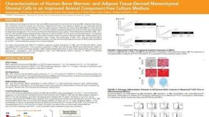 科学海报Characterization of Human Bone Marrow- and Adipose Tissue-Derived Mesenchymal Stromal Cells in an Improved Animal Component-Free Culture Medium
 沪公网安备31010102008431号