Li H et al. (SEP 2016)
In vitro cellular & developmental biology. Animal 52 8 885--893
Directed differentiation of human embryonic stem cells into keratinocyte progenitors in vitro: an attempt with promise of clinical use.
Human embryonic stem cells (hESCs) can differentiate into all somatic lineages including stratified squamous epithelia. Thus,efficient methods are required to direct hESC differentiation to obtain a pure subpopulation for tissue engineering. The study aimed to assess the effects of retinoic acid (RA),bone morphogenetic protein-4 (BMP4),and ascorbic acid (AA) on the differentiation of hESCs into keratinocyte progenitors in vitro. The first media contained AA and BMP4; the second contained RA,AA,and BMP4; the third was commercial-defined keratinocyte serum-free medium,which was used to differentiate H9 hESCs (direct approach) or embryoid bodies (EBs) (indirect approach) into keratinocyte progenitors. Real-time RT-PCR,immunofluorescence,and flow-cytometry were used to characterize the differentiated cells. Cells induced by AA + BMP4 + RA showed the typical epithelial morphology,while cells induced by AA + BMP4 showed multiple appearances. CK14 and p63 messenger RNA (mRNA) expressions in the AA + BMP4 + RA-treated cells were higher than those of the AA + BMP4-treated cells (CK14: 22.4-fold; p63: 84.7-fold). Epithelial marker CK18 mRNA expressions at 14 d of differentiation and keratinocyte marker CK14 and transcription factor p63 mRNA expressions at 35 d of differentiation were higher in cells differentiated from hESCs compared with those differentiated from EBs (CK18 10.51 ± 3.26 vs. 6.67 ± 1.28; CK14 9.27 ± 3.61 vs. 5.32 ± 1.86; p63 0.73 ± 0.06 vs. 0.44 ± 0.12,all P textless 0.05) After hESC induction by AA+BMP4+RA,CK14 mRNA expression was upregulated after day 21,peaking by 35 d of differentiation. Combined RA,BMP4,and AA could effectively induce differentiation of hESCs into keratinocyte progenitors in vitro. These keratinocytes could be used for oral mucosa and skin tissue engineering.
View Publication
产品类型:
产品号#:
07923
85850
85857
产品名:
Dispase (1 U/mL)
mTeSR™1
mTeSR™1
Daga A et al. (MAY 2000)
Experimental hematology 28 5 569--74
The retroviral transduction of HOXC4 into human CD34(+) cells induces an in vitro expansion of clonogenic and early progenitors.
OBJECTIVE: +HOX genes are expressed in the hematopoietic system and increasing data point to their involvement in the control of proliferation and/or differentiation. Genes belonging to the C cluster are preferentially expressed in developing and differentiated lymphoid lineages. However,recent studies demonstrated,by RT-PCR,that the HOXC4 gene is also actively transcribed in the most undifferentiated hematopoietic cells (CD34(+)38(low)) and in more mature myeloid and erythroid progenitors. We evaluated the expression of HOXC4 protein on human CD34(+) cells and the in vitro effect of its overexpression on proliferation and differentiation. MATERIALS AND METHODS: We assessed the expression of HOXC4 on human CD34(+) cells using a polyclonal antibody raised against the C-terminal portion of the protein expressed using the baculovirus system. Overexpression of HOXC4 in human CD34(+) cells was obtained by retroviral gene transfer; its effect on clonogenic (CFU-GM,BFU-E,and CFU-GEMM) and early progenitors (LTC-IC) was evaluated. RESULTS: The HOXC4 protein is indeed expressed in human CD34(+) cells,and its overexpression in human CD34(+) cells increases the proliferation potential of clonogenic and early progenitors. CFU-GM showed a median threefold expansion (range: 1.1-19.4; p textless 0.002) compared with control transduced with the vector alone. The increment of BFU-E was higher (median ninefold,range 2.5-35; p textless 0. 0009) and erythroid colonies presented a larger size with normal morphology. An even more marked effect was observed on LTC-IC (median 13,onefold; range 4.1-102.1,p textless 0.0001). CONCLUSION: We demonstrate that HOXC4 is expressed in CD34(+) cells and that its overexpression induces an in vitro expansion of committed as well as very early hematopoietic progenitors. The most striking effect was obtained on LTC-IC with an expansion of 13.1-fold. The enforced expression of HOXC4 induced a significant increase (p textless 0.009) in the number of erythroid colonies compared with CFU-GM,although without perturbing,at least in vitro,the maturation program of the cells. On the other hand,the effect of the gene overexpression did not induce any skewing in the colony types derived from the myeloid lineage.
View Publication
Wang H et al. (APR 2016)
The Journal of biological chemistry 291 16 8644--8652
Germ Cell Nuclear Factor (GCNF) Represses Oct4 Expression and Globally Modulates Gene Expression in Human Embryonic Stem (hES) Cells.
Oct4 is considered a key transcription factor for pluripotent stem cell self-renewal. It binds to specific regions within target genes to regulate their expression and is downregulated upon induction of differentiation of pluripotent stem cells; however,the mechanisms that regulate the levels of human Oct4 expression remain poorly understood. Here we show that expression of human Oct4 is directly repressed by germ cell nuclear factor (GCNF),an orphan nuclear receptor,in hES cells. Knockdown of GCNF by siRNA resulted in maintenance of Oct4 expression during RA-induced hES cell differentiation. While overexpression of GCNF promoted repression of Oct4 expression in both undifferentiated and differentiated hES cells. The level of Oct4 repression was dependent on the level of GCNF expression in a dose-dependent manner. mRNA microarray analysis demonstrated that overexpression of GCNF globally regulates gene expression in undifferentiated and differentiated hES cells. Within the group of altered genes,GCNF down-regulated 36% of the genes,and up-regulated 64% in undifferentiated hES cells. In addition,GCNF also showed a regulatory gene pattern that is different from RA treatment during hES cell differentiation. These findings increase our understanding of the mechanisms that maintain hES cell pluripotency and regulate gene expression during the differentiation process.
View Publication
产品类型:
产品号#:
85850
85857
产品名:
mTeSR™1
mTeSR™1
Galat V et al. (MAY 2016)
Stem cells and development 25 14 1060--1072
Transgene Reactivation in Induced Pluripotent Stem Cell Derivatives and Reversion to Pluripotency of Induced Pluripotent Stem Cell-Derived Mesenchymal Cells.
Induced pluripotent stem cells (iPSCs) have enormous potential in regenerative medicine and disease modeling. It is now felt that clinical trials should be performed with iPSCs derived with non-integrative constructs. Numerous studies,however,including those describing disease models,are still being published using cells derived from iPSCs generated with integrative constructs. Our experimental work presents the first evidence of spontaneous transgene reactivation in vitro in several cellular types. Our results show that the transgenes were predominantly silent in parent iPSCs,but in mesenchymal and endothelial iPSC derivatives,the transgenes experienced random up-regulation of Nanog and c-Myc. Additionally,we provide evidence of spontaneous secondary reprogramming and reversion to pluripotency in mesenchymal stem cells derived from iPSCs. These findings strongly suggest that the studies,which utilize cellular products derived from iPSCs generated with retro- or lentiviruses,should be evaluated with consideration of the possibility of transgene reactivation. The in vitro model described here provides insight into the earliest events of culture transformation and suggests the hypothesis that reversion to pluripotency may be responsible for the development of tumors in cell replacement experiments. The main goal of this work,however,is to communicate the possibility of transgene reactivation in retro- or lenti- iPSC derivatives and the associated loss of cellular fidelity in vitro,which may impact the outcomes of disease modeling and related experimentation.
View Publication
产品类型:
产品号#:
85850
85857
产品名:
mTeSR™1
mTeSR™1
Kim Y et al. (OCT 2016)
Scientific reports 6 35145
Islet-like organoids derived from human pluripotent stem cells efficiently function in the glucose responsiveness in vitro and in vivo.
Insulin secretion is elaborately modulated in pancreatic ß cells within islets of three-dimensional (3D) structures. Using human pluripotent stem cells (hPSCs) to develop islet-like structures with insulin-producing ß cells for the treatment of diabetes is challenging. Here,we report that pancreatic islet-like clusters derived from hESCs are functionally capable of glucose-responsive insulin secretion as well as therapeutic effects. Pancreatic hormone-expressing endocrine cells (ECs) were differentiated from hESCs using a step-wise protocol. The hESC-derived ECs expressed pancreatic endocrine hormones,such as insulin,somatostatin,and pancreatic polypeptide. Notably,dissociated ECs autonomously aggregated to form islet-like,3D structures of consistent sizes (100-150 μm in diameter). These EC clusters (ECCs) enhanced insulin secretion in response to glucose stimulus and potassium channel inhibition in vitro. Furthermore,ß cell-deficient mice transplanted with ECCs survived for more than 40 d while retaining a normal blood glucose level to some extent. The expression of pancreatic endocrine hormones was observed in tissues transplanted with ECCs. In addition,ECCs could be generated from human induced pluripotent stem cells. These results suggest that hPSC-derived,islet-like clusters may be alternative therapeutic cell sources for treating diabetes.
View Publication
产品类型:
产品号#:
85850
85857
产品名:
mTeSR™1
mTeSR™1
Lim MN et al. (MAY 2012)
Molecular vision 18 1289--300
Ex vivo expanded SSEA-4+ human limbal stromal cells are multipotent and do not express other embryonic stem cell markers.
PURPOSE: The presence of multipotent human limbal stromal cells resembling mesenchymal stromal cells (MSC) provides new insights to the characteristic of these cells and its therapeutic potential. However,little is known about the expression of stage-specific embryonic antigen 4 (SSEA-4) and the embryonic stem cell (ESC)-like properties of these cells. We studied the expression of SSEA-4 surface protein and the various ESC and MSC markers in the ex vivo cultured limbal stromal cells. The phenotypes and multipotent differentiation potential of these cells were also evaluated.backslashnbackslashnMETHODS: Limbal stromal cells were derived from corneoscleral rims. The SSEA-4(+) and SSEA-4(-) limbal stromal cells were sorted by fluorescence-activated cells sorting (FACS). Isolated cells were expanded and reanalyzed for their expression of SSEA-4. Expression of MSC and ESC markers on these cells were also analyzed by FACS. In addition,expression of limbal epithelial and corneal stromal proteins such as ATP-binding cassette sub-family G member 2 (ABCG2),tumour protein p63 (p63),paired box 6 (Pax6),cytokeratin 3 (AE5),cytokeratin 10,and keratocan sulfate were evaluated either by immunofluorecence staining or reverse transcription polymerase chain reaction. Appropriate induction medium was used to differentiate these cells into adipocytes,osteocytes,and chondrocytes.backslashnbackslashnRESULTS: Expanded limbal stromal cells expressed the majority of mesenchymal markers. These cells were negative for ABCG2,p63,Pax6,AE-5,and keratocan sulfate. After passaged,a subpopulation of these cells showed low expression of SSEA-4 but were negative for other important ESC surface markers such as Tra-1-60,Tra-1-81,and transcription factors like octamer-binding transcription factor 4 (Oct4),SRY(sex determining region Y)-box 2 (Sox2),and Nanog. Early passaged cells when induced were able to differentiate into adipocytes,osteocytes and chondrocytes.backslashnbackslashnCONCLUSIONS: The expanded limbal stromal cells showed features of multipotent MSC. Our study confirmed the expression of SSEA-4 by a subpopulation of cultured limbal stromal cells. However,despite the expression of SSEA-4,these cells did not express any other markers of ESC. Therefore,we conclude that the cells did not show properties of ESC.
View Publication
产品类型:
产品号#:
85850
85857
产品名:
mTeSR™1
mTeSR™1
Mohamad O et al. (MAY 2013)
PLoS ONE 8 5 e64160
Vector-Free and Transgene-Free Human iPS Cells Differentiate into Functional Neurons and Enhance Functional Recovery after Ischemic Stroke in Mice
Stroke is a leading cause of human death and disability in the adult population in the United States and around the world. While stroke treatment is limited,stem cell transplantation has emerged as a promising regenerative therapy to replace or repair damaged tissues and enhance functional recovery after stroke. Recently,the creation of induced pluripotent stem (iPS) cells through reprogramming of somatic cells has revolutionized cell therapy by providing an unlimited source of autologous cells for transplantation. In addition,the creation of vector-free and transgene-free human iPS (hiPS) cells provides a new generation of stem cells with a reduced risk of tumor formation that was associated with the random integration of viral vectors seen with previous techniques. However,the potential use of these cells in the treatment of ischemic stroke has not been explored. In the present investigation,we examined the neuronal differentiation of vector-free and transgene-free hiPS cells and the transplantation of hiPS cell-derived neural progenitor cells (hiPS-NPCs) in an ischemic stroke model in mice. Vector-free hiPS cells were maintained in feeder-free and serum-free conditions and differentiated into functional neurons in vitro using a newly developed differentiation protocol. Twenty eight days after transplantation in stroke mice,hiPS-NPCs showed mature neuronal markers in vivo. No tumor formation was seen up to 12 months after transplantation. Transplantation of hiPS-NPCs restored neurovascular coupling,increased trophic support and promoted behavioral recovery after stroke. These data suggest that using vector-free and transgene-free hiPS cells in stem cell therapy are safe and efficacious in enhancing recovery after focal ischemic stroke in mice.
View Publication
产品类型:
产品号#:
85850
85857
产品名:
mTeSR™1
mTeSR™1
Neves H et al. (MAY 2006)
Stem cells (Dayton,Ohio) 24 5 1328--37
Effects of Delta1 and Jagged1 on early human hematopoiesis: correlation with expression of notch signaling-related genes in CD34+ cells.
It has been shown that Notch signaling mediated by ligands of both Jagged and Delta families expands the hematopoietic stem cell compartment while blocking or delaying terminal myeloid differentiation. Here we show that Delta1- and Jagged1-expressing stromal cells have distinct effects on the clonogenic and differentiation capacities of human CD34(+) CD38(+) cells. Jagged1 increases the number of bipotent colony-forming unit-granulocyte macrophage (CFU-GM) and unipotent progenitors (CFU-granulocytes and CFU-macrophages),without quantitatively affecting terminal cell differentiation,whereas Delta1 reduces the number of CFU-GM and differentiated monocytic cells. Expression analysis of genes coding for Notch receptors,Notch targets,and Notch signaling modulators in supernatant CD34(+) cells arising upon contact with Jagged1 and Delta1 shows dynamic and differential gene expression profiles over time. At early time points,modest upregulation of Notch1,Notch3,and Hes1 was observed in Jagged1-CD34(+) cells,whereas those in contact with Delta1 strikingly upregulated Notch3 and Hes1. Later,myeloid progenitors with strong clonogenic potential emerging upon contact with Jagged1 upregulated Notch1 and Deltex and downregulated Notch signaling modulators,whereas T/NK progenitors originated by Delta1 strikingly upregulated Notch3 and Deltex and,to a lesser extent,Hes1,Lunatic Fringe,and Numb. Together,the data unravel previously unrecognized expression patterns of Notch signaling-related genes in CD34(+) CD38(+) cells as they develop in Jagged1- or Delta1-stromal cell environments,which appear to reflect sequential maturational stages of CD34(+) cells into distinct cell lineages.
View Publication
产品类型:
产品号#:
04435
04445
产品名:
MethoCult™H4435富集
MethoCult™H4435富集
Bruserud &O et al. (MAR 2007)
Haematologica 92 3 332--41
Subclassification of patients with acute myelogenous leukemia based on chemokine responsiveness and constitutive chemokine release by their leukemic cells.
BACKGROUND AND OBJECTIVES: Chemokines are soluble mediators involved in angiogenesis,cellular growth control and immunomodulation. In the present study we investigated the effects of various chemokines on proliferation of acute myelogenous leukemia (AML) cells and constitutive chemokine release by primary AML cells. DESIGN AND METHODS: Native human AML cells derived from 68 consecutive patients were cultured in vitro. We investigated AML cell proliferation (3H-thymidine incorporation,colony formation),chemokine receptor expression,constitutive chemokine release and chemotaxis of normal peripheral blood mononuclear cells. RESULTS: Exogenous chemokines usually did not have any effect on AML blast proliferation in the absence of hematopoietic growth factors,but when investigating growth factor-dependent (interleukin 3 + granulocyte-macrophage colony-stimulating factor + stem cell factor) proliferation in suspension cultures the following patient subsets were identified: (i) patients whose cells showed chemokine-induced growth enhancement (8 patients); (ii) divergent effects on proliferation (15 patients); and (iii) no effect (most patients). These patient subsets did not differ in chemokine receptor expression,but,compared to CD34- AML cells,CD34+ cells showed higher expression of several receptors. Chemokines also increased the proliferation of clonogenic AML cells from the first subset of patients. Furthermore,a broad constitutive chemokine release profile was detected for most patients,and the following chemokine clusters could be identified: CCL2-4/CXCL1/8,CCL5/CXCL9-11 (possibly also CCL23) and CCL13/17/22/24/CXCL5 (possibly also CXCL6). Only the CCL2-4/CXCL1/8 cluster showed significant correlations between corresponding mRNA levels and NFkB levels/activation. The chemotaxis of normal immunocompetent cells for patients without constitutive chemokine release was observed to be decreased. INTERPRETATION AND CONCLUSIONS: Differences in chemokine responsiveness as well as chemokine release contribute to patient heterogeneity in AML. Patients with AML can be classified into distinct subsets according to their chemokine responsiveness and chemokine release profile.
View Publication
产品类型:
产品号#:
04434
04444
09600
09650
产品名:
MethoCult™H4434经典
MethoCult™H4434经典
StemSpan™ SFEM
StemSpan™ SFEM
Valencic E et al. (APR 2010)
Cytotherapy 12 2 154--60
The immunosuppressive effect of Wharton's jelly stromal cells depends on the timing of their licensing and on lymphocyte activation.
BACKGROUND: Mesenchymal stromal cells (MSC) have been proven to have potent immunosuppressive action and hence have been proposed for the treatment of severe Graft Versus Host Disease. However,in most models,MSC were added at the same time of lymphocyte stimulation,which is quite different from what occurs in vivo. AIMS: To investigate how the timing of lymphocyte activation and the exposure to activation-related cytokines (licensing) can influence the immunosuppressive action of Wharton's jelly stromal cells (WJSC). METHODS: WJSC,licensed or not with activation-related cytokines,were added lymphocytes the same time or 24 hours after their stimulation with phytohaemoagglutinin. Proliferation of lymphocytes and cytokines production was measured after three days co-culture. RESULTS: Lymphocytes stimulated in the presence of WJSC displayed a dramatic decrease in proliferation and production of cytokines,in spite of normal expression of activation markers. The suppression was weakened when targeted lymphocytes were seperated by a membrane and partially rescued by the addition of exogenous l-tryptophan,suggesting a major role for indoleamine 2,3-dioxigenase with a probable paracrine effect. Licensing of WJSC increased the immunosuppressive effect,in both contact and non-contact settings. The timing of WJSC licensing was crucial for the immunosuppressive action. Lymphocytes pre-stimulated alone for 24 h,and added afterwards to non-licensed WJSC,showed normal or even increased proliferation. On the other hand,their proliferation was strongly inhibited by licensed WJSC. CONCLUSIONS: WJSC have a potent immunosuppressive function best realized with direct contact,and increased by licensing signals before and during lymphocyte stimulation. Our results could contribute to the set up of new WJSC-based therapies for severe autoimmuno disorders.
View Publication
产品类型:
产品号#:
产品名:
Valera E et al. (JAN 2010)
PLoS ONE 5 6 e11167
BMP-2/6 heterodimer is more effective than BMP-2 or BMP-6 homodimers as inductor of differentiation of human embryonic stem cells
Bone Morphogenetic Protein (BMP) signaling pathways are involved in differentiation of stem cells into diverse cell types,and thus BMPs can be used as main guidance molecules for in vitro differentiation of human stem cells.
View Publication

 EasySep™小鼠TIL(CD45)正选试剂盒


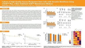 科学海报Culture of High-Quality Human Pluripotent Stem Cells with Versatile Workflows Using mTeSR™ Plus, a New Stabilized TeSR™ Maintenance Medium
 沪公网安备31010102008431号