Drury-Stewart D et al. (AUG 2013)
Stem cell research & therapy 4 4 93
Highly efficient differentiation of neural precursors from human embryonic stem cells and benefits of transplantation after ischemic stroke in mice.
INTRODUCTION: Ischemic stroke is a leading cause of death and disability,but treatment options are severely limited. Cell therapy offers an attractive strategy for regenerating lost tissues and enhancing the endogenous healing process. In this study,we investigated the use of human embryonic stem cell-derived neural precursors as a cell therapy in a murine stroke model.backslashnbackslashnMETHODS: Neural precursors were derived from human embryonic stem cells by using a fully adherent SMAD inhibition protocol employing small molecules. The efficiency of neural induction and the ability of these cells to further differentiate into neurons were assessed by using immunocytochemistry. Whole-cell patch-clamp recording was used to demonstrate the electrophysiological activity of human embryonic stem cell-derived neurons. Neural precursors were transplanted into the core and penumbra regions of a focal ischemic stroke in the barrel cortex of mice. Animals received injections of bromodeoxyuridine to track regeneration. Neural differentiation of the transplanted cells and regenerative markers were measured by using immunohistochemistry. The adhesive removal test was used to determine functional improvement after stroke and intervention.backslashnbackslashnRESULTS: After 11 days of neural induction by using the small-molecule protocol,over 95% of human embryonic stem-derived cells expressed at least one neural marker. Further in vitro differentiation yielded cells that stained for mature neuronal markers and exhibited high-amplitude,repetitive action potentials in response to depolarization. Neuronal differentiation also occurred after transplantation into the ischemic cortex. A greater level of bromodeoxyuridine co-localization with neurons was observed in the penumbra region of animals receiving cell transplantation. Transplantation also improved sensory recovery in transplant animals over that in control animals.backslashnbackslashnCONCLUSIONS: Human embryonic stem cell-derived neural precursors derived by using a highly efficient small-molecule SMAD inhibition protocol can differentiate into electrophysiologically functional neurons in vitro. These cells also differentiate into neurons in vivo,enhance regenerative activities,and improve sensory recovery after ischemic stroke.
View Publication
产品类型:
产品号#:
85850
85857
产品名:
mTeSR™1
mTeSR™1
Sun N and Zhao H (MAY 2014)
Biotechnology and Bioengineering 111 5 1048--53
Seamless correction of the sickle cell disease mutation of the HBB gene in human induced pluripotent stem cells using TALENs.
Sickle cell disease (SCD) is the most common human genetic disease which is caused by a single mutation of human β-globin (HBB) gene. The lack of long-term treatment makes the development of reliable cell and gene therapies highly desirable. Disease-specific patient-derived human induced pluripotent stem cells (hiPSCs) have great potential for developing novel cell and gene therapies. With the disease-causing mutations corrected in situ,patient-derived hiPSCs can restore normal cell functions and serve as a renewable autologous cell source for the treatment of genetic disorders. Here we successfully utilized transcription activator-like effector nucleases (TALENs),a recently emerged novel genome editing tool,to correct the SCD mutation in patient-derived hiPSCs. The TALENs we have engineered are highly specific and generate minimal off-target effects. In combination with piggyBac transposon,TALEN-mediated gene targeting leaves no residual ectopic sequences at the site of correction and the corrected hiPSCs retain full pluripotency and a normal karyotype. Our study demonstrates an important first step of using TALENs for the treatment of genetic diseases such as SCD,which represents a significant advance toward hiPSC-based cell and gene therapies.
View Publication
产品类型:
产品号#:
07920
07922
07923
100-0247
72252
72254
85850
85857
产品名:
ACCUTASE™
ACCUTASE™
Dispase (1 U/mL)
Thiazovivin
Thiazovivin
Thiazovivin
mTeSR™1
mTeSR™1
Nguyen TY et al. (OCT 2013)
PLoS ONE 8 10 e76547
An In Vitro Mechanism Study on the Proliferation and Pluripotency of Human Embryonic Stems Cells in Response to Magnesium Degradation
Magnesium (Mg) is a promising biodegradable metallic material for applications in cellular/tissue engineering and biomedical implants/devices. To advance clinical translation of Mg-based biomaterials,we investigated the effects and mechanisms of Mg degradation on the proliferation and pluripotency of human embryonic stem cells (hESCs). We used hESCs as the in vitro model system to study cellular responses to Mg degradation because they are sensitive to toxicants and capable of differentiating into any cell types of interest for regenerative medicine. In a previous study when hESCs were cultured in vitro with either polished metallic Mg (99.9% purity) or pre-degraded Mg,cell death was observed within the first 30 hours of culture. Excess Mg ions and hydroxide ions induced by Mg degradation may have been the causes for the observed cell death; hence,their respective effects on hESCs were investigated for the first time to reveal the potential mechanisms. For this purpose,the mTeSR®1 hESC culture media was either modified to an alkaline pH of 8.1 or supplemented with 0.4-40 mM of Mg ions. We showed that the initial increase of media pH to 8.1 had no adverse effect on hESC proliferation. At all tested Mg ion dosages,the hESCs grew to confluency and retained pluripotency as indicated by the expression of OCT4,SSEA3,and SOX2. When the supplemental Mg ion dosages increased to greater than 10 mM,however,hESC colony morphology changed and cell counts decreased. These results suggest that Mg-based implants or scaffolds are promising in combination with hESCs for regenerative medicine applications,providing their degradation rate is moderate. Additionally,the hESC culture system could serve as a standard model for cytocompatibility studies of Mg in vitro,and an identified 10 mM critical dosage of Mg ions could serve as a design guideline for safe degradation of Mg-based implants/scaffolds.
View Publication
产品类型:
产品号#:
85850
85857
产品名:
mTeSR™1
mTeSR™1
Liu B et al. (MAR 2014)
PLoS ONE 9 3 e90615
Nanog1 in NTERA-2 and recombinant NanogP8 from somatic cancer cells adopt multiple protein conformations and migrate at multiple M.W species
Human Nanog1 is a 305-amino acid (aa) homeodomain-containing transcription factor critical for the pluripotency of embryonic stem (ES) and embryonal carcinoma (EC) cells. Somatic cancer cells predominantly express a retrogene homolog of Nanog1 called NanogP8,which is ˜99% similar to Nanog at the aa level. Although the predicted M.W of Nanog1/NanogP8 is ∼35 kD,both have been reported to migrate,on Western blotting (WB),at apparent molecular masses of 29-80 kD. Whether all these reported protein bands represent authentic Nanog proteins is unclear. Furthermore,detailed biochemical studies on Nanog1/NanogpP8 have been lacking. By combining WB using 8 anti-Nanog1 antibodies,immunoprecipitation,mass spectrometry,and studies using recombinant proteins,here we provide direct evidence that the Nanog1 protein in NTERA-2 EC cells exists as multiple M.W species from ˜22 kD to 100 kD with a major 42 kD band detectable on WB. We then demonstrate that recombinant NanogP8 (rNanogP8) proteins made in bacteria using cDNAs from multiple cancer cells also migrate,on denaturing SDS-PAGE,at ˜28 kD to 180 kD. Interestingly,different anti-Nanog1 antibodies exhibit differential reactivity towards rNanogP8 proteins,which can spontaneously form high M.W protein species. Finally,we show that most long-term cultured cancer cell lines seem to express very low levels of or different endogenous NanogP8 protein that cannot be readily detected by immunoprecipitation. Altogether,the current study reveals unique biochemical properties of Nanog1 in EC cells and NanogP8 in somatic cancer cells.
View Publication
产品类型:
产品号#:
85850
85857
产品名:
mTeSR™1
mTeSR™1
Turner J et al. (NOV 2014)
PLoS ONE 9 11 e112757
Metabolic Profiling and Flux Analysis of MEL-2 Human Embryonic Stem Cells during Exponential Growth at Physiological and Atmospheric Oxygen Concentrations
As human embryonic stem cells (hESCs) steadily progress towards regenerative medicine applications there is an increasing emphasis on the development of bioreactor platforms that enable expansion of these cells to clinically relevant numbers. Surprisingly little is known about the metabolic requirements of hESCs,precluding the rational design and optimisation of such platforms. In this study,we undertook an in-depth characterisation of MEL-2 hESC metabolic behaviour during the exponential growth phase,combining metabolic profiling and flux analysis tools at physiological (hypoxic) and atmospheric (normoxic) oxygen concentrations. To overcome variability in growth profiles and the problem of closing mass balances in a complex environment,we developed protocols to accurately measure uptake and production rates of metabolites,cell density,growth rate and biomass composition,and designed a metabolic flux analysis model for estimating internal rates. hESCs are commonly considered to be highly glycolytic with inactive or immature mitochondria,however,whilst the results of this study confirmed that glycolysis is indeed highly active,we show that at least in MEL-2 hESC,it is supported by the use of oxidative phosphorylation within the mitochondria utilising carbon sources,such as glutamine to maximise ATP production. Under both conditions,glycolysis was disconnected from the mitochondria with all of the glucose being converted to lactate. No difference in the growth rates of cells cultured under physiological or atmospheric oxygen concentrations was observed nor did this cause differences in fluxes through the majority of the internal metabolic pathways associated with biogenesis. These results suggest that hESCs display the conventional Warburg effect,with high aerobic activity despite high lactate production,challenging the idea of an anaerobic metabolism with low mitochondrial activity. The results of this study provide new insight that can be used in rational bioreactor design and in the development of
View Publication
产品类型:
产品号#:
85850
85857
产品名:
mTeSR™1
mTeSR™1
van den Akker E et al. (AUG 2010)
Haematologica 95 8 1278--86
Investigating the key membrane protein changes during in vitro erythropoiesis of protein 4.2 (-) cells (mutations Chartres 1 and 2).
BACKGROUND: Protein 4.2 deficiency caused by mutations in the EPB42 gene results in hereditary spherocytosis with characteristic alterations of CD47,CD44 and RhAG. We decided to investigate at which stage of erythropoiesis these hallmarks of protein 4.2 deficiency arise in a novel protein 4.2 patient and whether they cause disruption to the band 3 macrocomplex. DESIGN AND METHODS: We used immunoprecipitations and detergent extractability to assess the strength of protein associations within the band 3 macrocomplex and with the cytoskeleton in erythrocytes. Patient erythroblasts were cultured from peripheral blood mononuclear cells to study the effects of protein 4.2 deficiency during erythropoiesis. RESULTS: We report a patient with two novel mutations in EPB42 resulting in complete protein 4.2 deficiency. Immunoprecipitations revealed a weakened ankyrin-1-band 3 interaction in erythrocytes resulting in increased band 3 detergent extractability. CD44 abundance and its association with the cytoskeleton were increased. Erythroblast differentiation revealed that protein 4.2 and band 3 appear simultaneously and associate early in differentiation. Protein 4.2 deficiency results in lower CD47,higher CD44 expression and increased RhAG glycosylation starting from the basophilic stage. The normal downregulation of CD44 expression was not seen during protein 4.2(-) erythroblast differentiation. Knockdown of CD47 did not increase CD44 expression,arguing against a direct reciprocal relationship. CONCLUSIONS: We have established that the characteristic changes caused by protein 4.2 deficiency occur early during erythropoiesis. We postulate that weakening of the ankyrin-1-band 3 association during protein 4.2 deficiency is compensated,in part,by increased CD44-cytoskeleton binding.
View Publication
产品类型:
产品号#:
产品名:
Sun Y et al. (MAR )
PLOS ONE 3 e0118771
Properties of Neurons Derived from Induced Pluripotent Stem Cells of Gaucher Disease Type 2 Patient Fibroblasts: Potential Role in Neuropathology
Gaucher disease (GD) is caused by insufficient activity of acid $\$-glucosidase (GCase) resulting from mutations in GBA1. To understand the pathogenesis of the neuronopathic GD,induced pluripotent stem cells (iPSCs) were generated from fibroblasts isolated from three GD type 2 (GD2) and 2 unaffected (normal and GD carrier) individuals. The iPSCs were converted to neural precursor cells (NPCs) which were further differentiated into neurons. Parental GD2 fibroblasts as well as iPSCs,NPCs,and neurons had similar degrees of GCase deficiency. Lipid analyses showed increases of glucosylsphingosine and glucosylceramide in the GD2 cells. In addition,GD2 neurons showed increased $\$-synuclein protein compared to control neurons. Whole cell patch-clamping of the GD2 and control iPSCs-derived neurons demonstrated excitation characteristics of neurons,but intriguingly,those from GD2 exhibited consistently less negative resting membrane potentials with various degree of reduction in action potential amplitudes,sodium and potassium currents. Culture of control neurons in the presence of the GCase inhibitor (conduritol B epoxide) recapitulated these findings,providing a functional link between decreased GCase activity in GD and abnormal neuronal electrophysiological properties. To our knowledge,this study is first to report abnormal electrophysiological properties in GD2 iPSC-derived neurons that may underlie the neuropathic phenotype in Gaucher disease.
View Publication
产品类型:
产品号#:
05835
05839
05854
05855
34811
34815
34821
34825
34850
34860
85850
85857
产品名:
STEMdiff™ 神经诱导培养基
STEMdiff™ 神经诱导培养基
mFreSR™
mFreSR™
AggreWell™ 800 24孔板,1个
AggreWell™ 800 24孔板,5个
AggreWell™ 800 6孔板,1个
AggreWell™ 800 6孔板,5个
AggreWell™ 800 24孔板启动套装
AggreWell™ 800 6孔板启动套装
mTeSR™1
mTeSR™1
Fenouille N et al. (DEC 2010)
Cancer research 70 23 9659--70
Persistent activation of the Fyn/ERK kinase signaling axis mediates imatinib resistance in chronic myelogenous leukemia cells through upregulation of intracellular SPARC.
SPARC is an extracellular matrix protein that exerts pleiotropic effects on extracellular matrix organization,growth factor availability,cell adhesion,differentiation,and immunity in cancer. Chronic myelogenous leukemia (CML) cells resistant to the BCR-ABL inhibitor imatinib (IM-R cells) were found to overexpress SPARC mRNA. In this study,we show that imatinib triggers SPARC accumulation in a variety of tyrosine kinase inhibitor (TKI)-resistant CML cell lines. SPARC silencing in IM-R cells restored imatinib sensitivity,whereas enforced SPARC expression in imatinib-sensitive cells promoted viability as well as protection against imatinib-mediated apoptosis. Notably,we found that the protective effect of SPARC required intracellular retention inside cells. Accordingly,SPARC was not secreted into the culture medium of IM-R cells. Increased SPARC expression was intimately linked to persistent activation of the Fyn/ERK kinase signaling axis. Pharmacologic inhibition of this pathway or siRNA-mediated knockdown of Fyn kinase resensitized IM-R cells to imatinib. In support of our findings,increased levels of SPARC mRNA were documented in blood cells from CML patients after 1 year of imatinib therapy compared with initial diagnosis. Taken together,our results highlight an important role for the Fyn/ERK signaling pathway in imatinib-resistant cells that is driven by accumulation of intracellular SPARC.
View Publication
产品类型:
产品号#:
04100
产品名:
MethoCult™ H4100
Sebastiano V et al. (NOV 2011)
Stem Cells 29 11 1717--1726
In situ genetic correction of the sickle cell anemia mutation in human induced pluripotent stem cells using engineered zinc finger nucleases.
The combination of induced pluripotent stem cell (iPSC) technology and targeted gene modification by homologous recombination (HR) represents a promising new approach to generate genetically corrected,patient-derived cells that could be used for autologous transplantation therapies. This strategy has several potential advantages over conventional gene therapy including eliminating the need for immunosuppression,avoiding the risk of insertional mutagenesis by therapeutic vectors,and maintaining expression of the corrected gene by endogenous control elements rather than a constitutive promoter. However,gene targeting in human pluripotent cells has remained challenging and inefficient. Recently,engineered zinc finger nucleases (ZFNs) have been shown to substantially increase HR frequencies in human iPSCs,raising the prospect of using this technology to correct disease causing mutations. Here,we describe the generation of iPSC lines from sickle cell anemia patients and in situ correction of the disease causing mutation using three ZFN pairs made by the publicly available oligomerized pool engineering method (OPEN). Gene-corrected cells retained full pluripotency and a normal karyotype following removal of reprogramming factor and drug-resistance genes. By testing various conditions,we also demonstrated that HR events in human iPSCs can occur as far as 82 bps from a ZFN-induced break. Our approach delineates a roadmap for using ZFNs made by an open-source method to achieve efficient,transgene-free correction of monogenic disease mutations in patient-derived iPSCs. Our results provide an important proof of principle that ZFNs can be used to produce gene-corrected human iPSCs that could be used for therapeutic applications.
View Publication
产品类型:
产品号#:
85850
85857
产品名:
mTeSR™1
mTeSR™1
Wang P et al. ( 2017)
Molecular autism 8 11
CRISPR/Cas9-mediated heterozygous knockout of the autism gene CHD8 and characterization of its transcriptional networks in cerebral organoids derived from iPS cells.
BACKGROUND CHD8 (chromodomain helicase DNA-binding protein 8),which codes for a member of the CHD family of ATP-dependent chromatin-remodeling factors,is one of the most commonly mutated genes in autism spectrum disorders (ASD) identified in exome-sequencing studies. Loss of function mutations in the gene have also been found in schizophrenia (SZ) and intellectual disabilities and influence cancer cell proliferation. We previously reported an RNA-seq analysis carried out on neural progenitor cells (NPCs) and monolayer neurons derived from induced pluripotent stem (iPS) cells that were heterozygous for CHD8 knockout (KO) alleles generated using CRISPR-Cas9 gene editing. A significant number of ASD and SZ candidate genes were among those that were differentially expressed in a comparison of heterozygous KO lines (CHD8(+/-)) vs isogenic controls (CHD8(+/-)),including the SZ and bipolar disorder (BD) candidate gene TCF4,which was markedly upregulated in CHD8(+/-) neuronal cells. METHODS In the current study,RNA-seq was carried out on CHD8(+/-) and isogenic control (CHD8(+/+)) cerebral organoids,which are 3-dimensional structures derived from iPS cells that model the developing human telencephalon. RESULTS TCF4 expression was,again,significantly upregulated. Pathway analysis carried out on differentially expressed genes (DEGs) revealed an enrichment of genes involved in neurogenesis,neuronal differentiation,forebrain development,Wnt/β-catenin signaling,and axonal guidance,similar to our previous study on NPCs and monolayer neurons. There was also significant overlap in our CHD8(+/-) DEGs with those found in a transcriptome analysis carried out by another group using cerebral organoids derived from a family with idiopathic ASD. Remarkably,the top DEG in our respective studies was the non-coding RNA DLX6-AS1,which was markedly upregulated in both studies; DLX6-AS1 regulates the expression of members of the DLX (distal-less homeobox) gene family. DLX1 was also upregulated in both studies. DLX genes code for transcription factors that play a key role in GABAergic interneuron differentiation. Significant overlap was also found in a transcriptome study carried out by another group using iPS cell-derived neurons from patients with BD,a condition characterized by dysregulated WNT/β-catenin signaling in a subgroup of affected individuals. CONCLUSIONS Overall,the findings show that distinct ASD,SZ,and BD candidate genes converge on common molecular targets-an important consideration for developing novel therapeutics in genetically heterogeneous complex traits.
View Publication
产品类型:
产品号#:
85850
85857
产品名:
mTeSR™1
mTeSR™1
Penicka M et al. (JUL 2007)
Heart (British Cardiac Society) 93 7 837--41
One-day kinetics of myocardial engraftment after intracoronary injection of bone marrow mononuclear cells in patients with acute and chronic myocardial infarction.
OBJECTIVE: To investigate the kinetics of myocardial engraftment of bone marrow-derived mononuclear cells (BMNCs) after intracoronary injection using 99mTc-d,l-hexamethylpropylene amine oxime (99mTc-HMPAO) nuclear imaging in patients with acute and chronic anterior myocardial infarction. DESIGN: Nuclear imaging-derived tracking of BMNCs at 2 and 20 h after injection in the left anterior descending (LAD) coronary artery. SETTING: Academical cardiocentre. PATIENTS: Five patients with acute (mean (SD) age 58 (11) years; ejection fraction range 33-45%) and five patients with chronic (mean (SD) age 50 (6) years; ejection fraction range 28-34%) anterior myocardial infarction. INTERVENTIONS: A total of 24.2 x 10(8)-57.0 x 10(8) BMNCs (20% labelled with 700-1000 MBq 99mTc-HMPAO) were injected in the LAD coronary artery. RESULTS: At 2 h after BMNC injection,myocardial activity was observed in all patients with acute (range 1.31-5.10%) and in all but one patient with chronic infarction (range 1.10-3.0%). At 20 h,myocardial engraftment was noted only in three patients with acute myocardial infarction,whereas no myocardial activity was noted in any patient with chronic infarction. CONCLUSIONS: Engraftment of BMNCs shows dynamic changes within the first 20 h after intracoronary injection. Persistent myocardial engraftment was noted only in a subset of patients with acute myocardial infarction.
View Publication

 EasySep™小鼠TIL(CD45)正选试剂盒


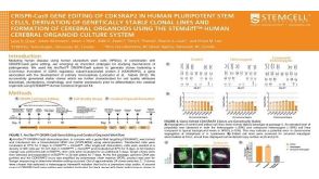 科学海报CRISPR-Cas9 Gene Editing Of CDK5RAP2 In Human Pluripotent Stem Cells, Derivation Of Genetically Stable Clonal Lines And Formation Of Cerebral Organoids
 沪公网安备31010102008431号