Genetic reprogramming of human amniotic cells with episomal vectors: neural rosettes as sentinels in candidate selection for validation assays.
The promise of genetic reprogramming has prompted initiatives to develop banks of induced pluripotent stem cells (iPSCs) from diverse sources. Sentinel assays for pluripotency could maximize available resources for generating iPSCs. Neural rosettes represent a primitive neural tissue that is unique to differentiating PSCs and commonly used to identify derivative neural/stem progenitors. Here,neural rosettes were used as a sentinel assay for pluripotency in selection of candidates to advance to validation assays. Candidate iPSCs were generated from independent populations of amniotic cells with episomal vectors. Phase imaging of living back up cultures showed neural rosettes in 2 of the 5 candidate populations. Rosettes were immunopositive for the Sox1,Sox2,Pax6 and Pax7 transcription factors that govern neural development in the earliest stage of development and for the Isl1/2 and Otx2 transcription factors that are expressed in the dorsal and ventral domains,respectively,of the neural tube in vivo. Dissociation of rosettes produced cultures of differentiation competent neural/stem progenitors that generated immature neurons that were immunopositive for βIII-tubulin and glia that were immunopositive for GFAP. Subsequent validation assays of selected candidates showed induced expression of endogenous pluripotency genes,epigenetic modification of chromatin and formation of teratomas in immunodeficient mice that contained derivatives of the 3 embryonic germ layers. Validated lines were vector-free and maintained a normal karyotype for more than 60 passages. The credibility of rosette assembly as a sentinel assay for PSCs is supported by coordinate loss of nuclear-localized pluripotency factors Oct4 and Nanog in neural rosettes that emerge spontaneously in cultures of self-renewing validated lines. Taken together,these findings demonstrate value in neural rosettes as sentinels for pluripotency and selection of promising candidates for advance to validation assays.
View Publication
Pipino C et al. (OCT 2014)
Cellular reprogramming 16 5 331--344
Trisomy 21 mid-trimester amniotic fluid induced pluripotent stem cells maintain genetic signatures during reprogramming: implications for disease modeling and cryobanking.
Trisomy 21 is the most common chromosomal abnormality and is associated primarily with cardiovascular,hematological,and neurological complications. A robust patient-derived cellular model is necessary to investigate the pathophysiology of the syndrome because current animal models are limited and access to tissues from affected individuals is ethically challenging. We aimed to derive induced pluripotent stem cells (iPSCs) from trisomy 21 human mid-trimester amniotic fluid stem cells (AFSCs) and describe their hematopoietic and neurological characteristics. Human AFSCs collected from women undergoing prenatal diagnosis were selected for c-KIT(+) and transduced with a Cre-lox-inducible polycistronic lentiviral vector encoding SOX2,OCT4,KLF-4,and c-MYC (50,000 cells at a multiplicity of infection (MOI) 1-5 for 72 h). The embryonic stem cell (ESC)-like properties of the AFSC-derived iPSCs were established in vitro by embryoid body formation and in vivo by teratoma formation in RAG2(-/-),$\$-chain(-/-),C2(-/-) immunodeficient mice. Reprogrammed cells retained their cytogenetic signatures and differentiated into specialized hematopoietic and neural precursors detected by morphological assessment,immunostaining,and RT-PCR. Additionally,the iPSCs expressed all pluripotency markers upon multiple rounds of freeze-thawing. These findings are important in establishing a patient-specific cellular platform of trisomy 21 to study the pathophysiology of the aneuploidy and for future drug discovery.
View Publication
产品类型:
产品号#:
04434
04444
05850
05857
05870
05875
07909
85850
85857
85870
85875
产品名:
MethoCult™ H4434 Classic
MethoCult™ H4434 Classic
IV型胶原酶(1mg /mL)
mTeSR™1
mTeSR™1
Taubert I et al. (APR 2011)
Cytotherapy 13 4 459--66
Characterization of hematopoietic stem cell subsets from patients with multiple myeloma after mobilization with plerixafor.
BACKGROUND AIMS: Previous studies have demonstrated that the combination of granulocyte-colony-stimulating factor (G-CSF) + plerixafor is more efficient in mobilizing CD34(+) hematopoietic stem cells (HSC) into the peripheral blood than G-CSF alone. In this study we analyzed the impact of adding plerixafor to G-CSF upon the mobilization of different HSC subsets. METHODS: We characterized the immunophenotype of HSC subsets isolated from the peripheral blood of eight patients with multiple myeloma (MM) before and after treatment with plerixafor. All patients were supposed to collect stem cells prior to high-dose chemotherapy and consecutive autologous stem cell transplantation,and therefore received front-line mobilization with 4 days of G-CSF followed by a single dose of plerixafor. Samples of peripheral blood were analyzed comparatively by flow cytometry directly before and 12 h after administration of plerixafor. RESULTS: The number of aldehyde dehydrogenase (ALDH)(bright) and CD34(+) cells was significantly higher after plerixafor treatment (1.2-5.0 and 1.5-6.0 times; both P textless 0.01) and an enrichment of the very primitive CD34(+) CD38(-) and ALDH(bright) CD34(+) CD38(-) HSC subsets was detectable. Additionally,two distinct ALDH(+) subsets could be clearly distinguished. The small ALDH(high) subset showed a higher number of CD34(+) CD38(-) cells in contrast to the total ALDH(bright) subpopulation and probably represented a very primitive subpopulation of HSC. CONCLUSIONS: A combined staining of ALDH,CD34 and CD38 might represent a powerful tool for the identification of a very rare and primitive hematopoietic stem cell subset. The addition of plerixafor mobilized not only more CD34(+) cells but was also able to increase the proportion of more primitive stem cell subsets.
View Publication
产品类型:
产品号#:
01700
01705
01702
产品名:
ALDEFLUOR™ 试剂盒
ALDEFLUOR™ DEAB试剂, 1.5 mM, 1 mL
ALDEFLUOR™检测缓冲液
Jiang S et al. (JAN 2011)
Blood 117 3 827--38
Cannabinoid receptor 2 and its agonists mediate hematopoiesis and hematopoietic stem and progenitor cell mobilization.
Endocannabinoids are arachidonic acid derivatives and part of a novel bioactive lipid signaling system,along with their G-coupled cannabinoid receptors (CB�? and CB₂) and the enzymes involved in their biosynthesis and degradation. However,their roles in hematopoiesis and hematopoietic stem and progenitor cell (HSPC) functions are not well characterized. Here,we show that bone marrow stromal cells express endocannabinoids (anandamide and 2-arachidonylglycerol),whereas CB₂ receptors are expressed in human and murine HSPCs. On ligand stimulation with CB₂ agonists,CB₂ receptors induced chemotaxis,migration,and enhanced colony formation of bone marrow cells,which were mediated via ERK,PI3-kinase,and Gαi-Rac1 pathways. In vivo,the CB₂ agonist AM1241 induced mobilization of murine HSPCs with short- and long-term repopulating abilities. In addition,granulocyte colony-stimulating factor -induced mobilization of HSPCs was significantly decreased by specific CB₂ antagonists and was impaired in Cnr2(-/-) cannabinoid type 2 receptor knockout mice. Taken together,these results demonstrate that the endocannabinoid system is involved in hematopoiesis and that CB₂/CB₂ agonist axis mediates repopulation of hematopoiesis and mobilization of HSPCs. Thus,CB₂ agonists may be therapeutically applied in clinical conditions,such as bone marrow transplantation.
View Publication
Ting S et al. (SEP 2014)
Stem Cell Research 13 2 202--213
An intermittent rocking platform for integrated expansion and differentiation of human pluripotent stem cells to cardiomyocytes in suspended microcarrier cultures
The development of novel platforms for large scale production of human embryonic stem cells (hESC) derived cardiomyocytes (CM) becomes more crucial as the demand for CMs in preclinical trials,high throughput cardio toxicity assays and future regenerative therapeutics rises. To this end,we have designed a microcarrier (MC) suspension agitated platform that integrates pluripotent hESC expansion followed by CM differentiation in a continuous,homogenous process.Hydrodynamic shear stresses applied during the hESC expansion and CM differentiation steps drastically reduced the capability of the cells to differentiate into CMs. Applying vigorous stirring during pluripotent hESC expansion on Cytodex 1 MC in spinner cultures resulted in low CM yields in the following differentiation step (cardiac troponin-T (cTnT): 22.83. ??. 2.56%; myosin heavy chain (MHC): 19.30. ??. 5.31%). Whereas the lower shear experienced in side to side rocker (wave type) platform resulted in higher CM yields (cTNT: 47.50. ??. 7.35%; MHC: 42.85. ??. 2.64%). The efficiency of CM differentiation is also affected by the hydrodynamic shear stress applied during the first 3. days of the differentiation stage. Even low shear applied continuously by side to side rocker agitation resulted in very low CM differentiation efficiency (cTnT. textless. 5%; MHC. textless. 2%). Simply by applying intermittent agitation during these 3. days followed by continuous agitation for the subsequent 9. days,CM differentiation efficiency can be substantially increased (cTNT: 65.73. ??. 10.73%; MHC: 59.73. ??. 9.17%). These yields are 38.3% and 39.3% higher (for cTnT and MHC respectively) than static culture control.During the hESC expansion phase,cells grew on continuously agitated rocker platform as pluripotent cell/MC aggregates (166??88??105??m2) achieving a cell concentration of 3.74??0.55??106cells/mL (18.89??2.82 fold expansion) in 7days. These aggregates were further differentiated into CMs using a WNT modulation differentiation protocol for the subsequent 12days on a rocking platform with an intermittent agitation regime during the first 3days. Collectively,the integrated MC rocker platform produced 190.5??58.8??106 CMs per run (31.75??9.74 CM/hESC seeded). The robustness of the system was demonstrated by using 2 cells lines,hESC (HES-3) and human induced pluripotent stem cell (hiPSC) IMR-90. The CM/MC aggregates formed extensive sarcomeres that exhibited cross-striations confirming cardiac ontogeny. Functionality of the CMs was demonstrated by monitoring the effect of inotropic drug,Isoproterenol on beating frequency.In conclusion,we have developed a simple robust and scalable platform that integrates both hESC expansion and CM differentiation in one unit process which is capable of meeting the need for large amounts of CMs. ?? 2014.
View Publication
产品类型:
产品号#:
05850
05857
05870
05875
85850
85857
85870
85875
产品名:
mTeSR™1
mTeSR™1
Radrizzani M et al. ( 2014)
Journal of translational medicine 12 276
Bone marrow-derived cells for cardiovascular cell therapy: an optimized GMP method based on low-density gradient improves cell purity and function.
BACKGROUND Cardiovascular cell therapy represents a promising field,with several approaches currently being tested. The advanced therapy medicinal product (ATMP) for the ongoing METHOD clinical study (Bone marrow derived cell therapy in the stable phase of chronic ischemic heart disease") consists of fresh mononuclear cells (MNC) isolated from autologous bone marrow (BM) through density gradient centrifugation on standard Ficoll-Paque. Cells are tested for safety (sterility�
View Publication
产品类型:
产品号#:
05420
05429
05424
05900
05950
产品名:
Orellana MD et al. (AUG 2015)
Cryobiology 71 1 151--160
Efficient recovery of undifferentiated human embryonic stem cell cryopreserved with hydroxyethyl starch, dimethyl sulphoxide and serum replacement
BACKGROUND The therapeutic use of human embryonic stem cells (hESCs) is dependent on an efficient cryopreservation protocol for long-term storage. The aim of this study was to determine whether the combination of three cryoprotecting reagents using two freezing systems might improve hESC recovery rates with maintenance of hESC pluripotency properties for potential cell therapy application. METHODS Recovery rates of hESC colonies which were frozen in three cryoprotective solutions: Me2SO/HES/SR medium,Defined-medium® and Me2SO/SFB in medium solution were evaluated in ultra-slow programmable freezing system (USPF) and a slow-rate freezing system (SRF). The hESC pluripotency properties after freezing-thawing were evaluated. RESULTS We estimated the distribution frequency of survival colonies and observed that independent of the freezing system used (USPF or SRF) the best results were obtained with Me2SO/HES/SR as cryopreservation medium. We showed a significant hESC recovery colonies rate after thawing in Me2SO/HES/SR medium were 3.88 and 2.9 in USPF and SRF,respectively. The recovery colonies rate with Defined-medium® were 1.05 and 1.07 however in classical Me2SO medium were 0.5 and 0.86 in USPF and SRF,respectively. We showed significant difference between Me2SO/HES/SR medium×Defined-medium® and between Me2SO/HES/SR medium×Me2SO medium,for two cryopreservation systems (Ptextless0.05). CONCLUSION We developed an in house protocol using the combination of Me2SO/HES/SR medium and ultra-slow programmable freezing system which resulted in hESC colonies that remain undifferentiated,maintain their in vitro and in vivo pluripotency properties and genetic stability. This approach may be suitable for cell therapy studies.
View Publication
产品类型:
产品号#:
05854
05855
05850
05857
05870
05875
85850
85857
85870
85875
产品名:
mFreSR™
mFreSR™
mTeSR™1
mTeSR™1
Baksh D et al. (NOV 2005)
Blood 106 9 3012--9
Soluble factor cross-talk between human bone marrow-derived hematopoietic and mesenchymal cells enhances in vitro CFU-F and CFU-O growth and reveals heterogeneity in the mesenchymal progenitor cell compartment.
The homeostatic adult bone marrow (BM) is a complex tissue wherein physical and biochemical interactions serve to maintain a balance between the hematopoietic and nonhematopoietic compartments. To focus on soluble factor interactions occurring between mesenchymal and hematopoietic cells,a serum-free adhesion-independent culture system was developed that allows manipulation of the growth of both mesenchymal and hematopoietic human BM-derived progenitors and the balance between these compartments. Factorial experiments demonstrated a role for stem cell factor (SCF) and interleukin 3 (IL-3) in the concomitant growth of hematopoietic (CD45+) and nonhematopoietic (CD45-) cells,as well as their derivatives. Kinetic tracking of IL-3alpha receptor (CD123) and SCF receptor (CD117) expression on a sorted CD45- cell population revealed the emergence of CD45-CD123+ cells capable of osteogenesis. Of the total fibroblast colony-forming units (CFU-Fs) and osteoblast colony-forming units (CFU-O),approximately 24% of CFU-Fs and about 22% of CFU-Os were recovered from this population. Cell-sorting experiments demonstrated that the CD45+ cell population secreted soluble factors that positively affect the survival and proliferation of CFU-Fs and CFU-Os generated from the CD45- cells. Together,our results provide insight into the intercellular cytokine network between hematopoietic and mesenchymal cells and provide a strategy to mutually culture both mesenchymal and hematopoietic cells in a defined scalable bioprocess.
View Publication
产品类型:
产品号#:
09850
产品名:
Wang L et al. (DEC 2016)
Materials science & engineering. C,Materials for biological applications 69 1125--1136
Injectable calcium phosphate with hydrogel fibers encapsulating induced pluripotent, dental pulp and bone marrow stem cells for bone repair.
Human induced pluripotent stem cell-derived mesenchymal stem cells (hiPSC-MSCs),dental pulp stem cells (hDPSCs) and bone marrow MSCs (hBMSCs) are exciting cell sources in regenerative medicine. However,there has been no report comparing hDPSCs,hBMSCs and hiPSC-MSCs for bone engineering in an injectable calcium phosphate cement (CPC) scaffold. The objectives of this study were to: (1) develop a novel injectable CPC containing hydrogel fibers encapsulating stem cells for bone engineering,and (2) compare cell viability,proliferation and osteogenic differentiation of hDPSCs,hiPSC-MSCs from bone marrow (BM-hiPSC-MSCs) and from foreskin (FS-hiPSC-MSCs),and hBMSCs in CPC for the first time. The results showed that the injection did not harm cell viability. The porosity of injectable CPC was 62%. All four types of cells proliferated and differentiated down the osteogenic lineage inside hydrogel fibers in CPC. hDPSCs,BM-hiPSC-MSCs,and hBMSCs exhibited high alkaline phosphatase,runt-related transcription factor,collagen I,and osteocalcin gene expressions. Cell-synthesized minerals increased with time (ptextless0.05),with no significant difference among hDPSCs,BM-hiPSC-MSCs and hBMSCs (ptextgreater0.1). Mineralization by hDPSCs,BM-hiPSC-MSCs,and hBMSCs inside CPC at 14d was 14-fold that at 1d. FS-hiPSC-MSCs were inferior in osteogenic differentiation compared to the other cells. In conclusion,hDPSCs,BM-hiPSC-MSCs and hBMSCs are similarly and highly promising for bone tissue engineering; however,FS-hiPSC-MSCs were relatively inferior in osteogenesis. The novel injectable CPC with cell-encapsulating hydrogel fibers may enhance bone regeneration in dental,craniofacial and orthopedic applications.
View Publication
产品类型:
产品号#:
05850
05857
05870
05875
85850
85857
85870
85875
产品名:
mTeSR™1
mTeSR™1
Mousa SA et al. (MAR 2010)
Cancer Letters 289 2 208--216
Stress resistant human embryonic stem cells as a potential source for the identification of novel cancer stem cell markers
Cancer stem cells are known for their inherent resistance to therapy. Here we investigated whether normal stem cells with acquired resistance to stress can be used to identify novel markers of cancer stem cells. For this,we generated a human embryonic stem cell line resistant to Trichostatin A and analyzed changes in its gene expression. The resistant cells over-expressed various genes associated with tumor aggressiveness,many of which are also expressed in the CD133+ glioma cancer stem cells. These findings suggest that stress-resistant stem cells generated in vitro may be useful for the discovery of novel markers of cancer stem cells.
View Publication

 EasySep™小鼠TIL(CD45)正选试剂盒


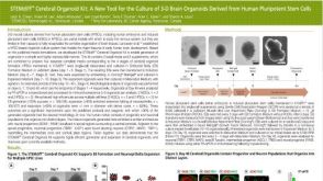 科学海报STEMdiff Cerebral Organoid Kit: A New Tool for the Culture of 3D Brain Organoids Derived from Human Pluripotent Stem Cells

 沪公网安备31010102008431号