Directed evolution of a recombinase that excises the provirus of most HIV-1 primary isolates with high specificity.
Current combination antiretroviral therapies (cART) efficiently suppress HIV-1 reproduction in humans,but the virus persists as integrated proviral reservoirs in small numbers of cells. To generate an antiviral agent capable of eradicating the provirus from infected cells,we employed 145 cycles of substrate-linked directed evolution to evolve a recombinase (Brec1) that site-specifically recognizes a 34-bp sequence present in the long terminal repeats (LTRs) of the majority of the clinically relevant HIV-1 strains and subtypes. Brec1 efficiently,precisely and safely removes the integrated provirus from infected cells and is efficacious on clinical HIV-1 isolates in vitro and in vivo,including in mice humanized with patient-derived cells. Our data suggest that Brec1 has potential for clinical application as a curative HIV-1 therapy.
View Publication
产品类型:
产品号#:
02697
17896
17896RF
17952
17952RF
21000
20119
20155
04435
04445
100-0696
产品名:
StemSpan™ CC110
EasySep™人脐带血CD34正选试剂盒II
RoboSep™ 人脐带血CD34正选试剂盒II
EasySep™人CD4+ T细胞分选试剂盒
RoboSep™ 人CD4+ T细胞分选试剂盒
RoboSep™- S
RoboSep™ 吸头组件抛光剂
RoboSep™分选管套装(9个塑料管)
MethoCult™ H4435 Enriched
MethoCult™ H4435 Enriched
EasySep™人CD4+ T细胞分离试剂盒
Beer PA et al. (JAN 2015)
Blood 125 3 504--15
Disruption of IKAROS activity in primitive chronic-phase CML cells mimics myeloid disease progression.
Without effective therapy,chronic-phase chronic myeloid leukemia (CP-CML) evolves into an acute leukemia (blast crisis [BC]) that displays either myeloid or B-lymphoid characteristics. This transition is often preceded by a clinically recognized,but biologically poorly characterized,accelerated phase (AP). Here,we report that IKAROS protein is absent or reduced in bone marrow blasts from most CML patients with advanced myeloid disease (AP or BC). This contrasts with primitive CP-CML cells and BCR-ABL1-negative acute myeloid leukemia blasts,which express readily detectable IKAROS. To investigate whether loss of IKAROS contributes to myeloid disease progression in CP-CML,we examined the effects of forced expression of a dominant-negative isoform of IKAROS (IK6) in CP-CML patients' CD34(+) cells. We confirmed that IK6 disrupts IKAROS activity in transduced CP-CML cells and showed that it confers on them features of AP-CML,including a prolonged increased output in vitro and in xenografted mice of primitive cells with an enhanced ability to differentiate into basophils. Expression of IK6 in CD34(+) CP-CML cells also led to activation of signal transducer and activator of transcription 5 and transcriptional repression of its negative regulators. These findings implicate loss of IKAROS as a frequent step and potential diagnostic harbinger of progressive myeloid disease in CML patients.
View Publication
产品类型:
产品号#:
18056
18056RF
产品名:
Wunderlich M et al. (SEP 2006)
Blood 108 5 1690--7
Human CD34+ cells expressing the inv(16) fusion protein exhibit a myelomonocytic phenotype with greatly enhanced proliferative ability.
The t(16:16) and inv(16) are associated with FAB M4Eo myeloid leukemias and result in fusion of the CBFB gene to the MYH11 gene (encoding smooth muscle myosin heavy chain [SMMHC]). Knockout of CBFbeta causes embryonic lethality due to lack of definitive hematopoiesis. Although knock-in of CBFB-MYH11 is not sufficient to cause disease,expression increases the incidence of leukemia when combined with cooperating events. Although mouse models are valuable tools in the study of leukemogenesis,little is known about the contribution of CBFbeta-SMMHC to human hematopoietic stem and progenitor cell self-renewal. We introduced the CBFbeta-MYH11 cDNA into human CD34+ cells via retroviral transduction. Transduced cells displayed an initial repression of progenitor activity but eventually dominated the culture,resulting in the proliferation of clonal populations for up to 7 months. Long-term cultures displayed a myelomonocytic morphology while retaining multilineage progenitor activity and engraftment in NOD/SCID-B2M-/- mice. Progenitor cells from long-term cultures showed altered expression of genes defining inv(16) identified in microarray studies of human patient samples. This system will be useful in examining the effects of CBFbeta-SMMHC on gene expression in the human preleukemic cell,in characterizing the effect of this oncogene on human stem cell biology,and in defining its contribution to the development of leukemia.
View Publication
产品类型:
产品号#:
04100
产品名:
MethoCult™ H4100
Maes C et al. (MAY 2006)
The Journal of clinical investigation 116 5 1230--42
Placental growth factor mediates mesenchymal cell development, cartilage turnover, and bone remodeling during fracture repair.
Current therapies for delayed- or nonunion bone fractures are still largely ineffective. Previous studies indicated that the VEGF homolog placental growth factor (PlGF) has a more significant role in disease than in health. Therefore we investigated the role of PlGF in a model of semi-stabilized bone fracture healing. Fracture repair in mice lacking PlGF was impaired and characterized by a massive accumulation of cartilage in the callus,reminiscent of delayed- or nonunion fractures. PlGF was required for the early recruitment of inflammatory cells and the vascularization of the fracture wound. Interestingly,however,PlGF also played a role in the subsequent stages of the repair process. Indeed in vivo and in vitro findings indicated that PlGF induced the proliferation and osteogenic differentiation of mesenchymal progenitors and stimulated cartilage turnover by particular MMPs. Later in the process,PlGF was required for the remodeling of the newly formed bone by stimulating osteoclast differentiation. As PlGF expression was increased throughout the process of bone repair and all the important cell types involved expressed its receptor VEGFR-1,the present data suggest that PlGF is required for mediating and coordinating the key aspects of fracture repair. Therefore PlGF may potentially offer therapeutic advantages for fracture repair.
View Publication
产品类型:
产品号#:
03334
03434
03444
03534
产品名:
MethoCult™ M3334
MethoCult™ GF M3434
MethoCult™ GF M3434
MethoCult™ GF M3534
Imren S et al. (OCT 2004)
The Journal of clinical investigation 114 7 953--62
High-level beta-globin expression and preferred intragenic integration after lentiviral transduction of human cord blood stem cells.
Transplantation of genetically corrected autologous hematopoietic stem cells is an attractive approach for the cure of sickle-cell disease and beta-thalassemia. Here,we infected human cord blood cells with a self-inactivating lentiviral vector encoding an anti-sickling betaA-T87Q-globin transgene and analyzed the transduced progeny produced over a 6-month period after transplantation of the infected cells directly into sublethally irradiated NOD/LtSz-scid/scid mice. Approximately half of the human erythroid and myeloid progenitors regenerated in the mice containing the transgene,and erythroid cells derived in vitro from these in vivo-regenerated cells produced high levels of betaA-T87Q-globin protein. Linker-mediated PCR analysis identified multiple transgene-positive clones in all mice analyzed with 2.1 +/- 0.1 integrated proviral copies per cell. Genomic sequencing of vector-containing fragments showed that 86% of the proviral inserts had occurred within genes,including several genes implicated in human leukemia. These findings indicate effective transduction of very primitive human cord blood cells with a candidate therapeutic lentiviral vector resulting in the long-term and robust,erythroid-specific production of therapeutically relevant levels of beta-globin protein. However,the frequency of proviral integration within genes that regulate hematopoiesis points to a need for additional safety modifications.
View Publication
产品类型:
产品号#:
18056
18056RF
产品名:
Uchida N et al. (JUN 2004)
Blood 103 12 4487--95
ABC transporter activities of murine hematopoietic stem cells vary according to their developmental and activation status.
Primitive hematopoietic cells from several species are known to efflux both Hoechst 33342 and Rhodamine-123. We now show that murine hematopoietic stem cells (HSCs) defined by long-term multilineage repopulation assays efflux both dyes variably according to their developmental or activation status. In day 14.5 murine fetal liver,very few HSCs efflux Hoechst 33342 efficiently,and they are thus not detected as side population" (SP) cells. HSCs in mouse fetal liver also fail to efflux Rhodamine-123. Both of these features are retained by most of the HSCs present until 4 weeks after birth but are reversed by 8 weeks of age or after a new HSC population is regenerated in adult mice that receive transplants with murine fetal liver cells. Activation of adult HSCs in vivo following 5-fluorouracil treatment�
View Publication
产品类型:
产品号#:
18756
18756RF
产品名:
EasySep™小鼠SCA1正选试剂盒
RoboSep™ 小鼠SCA1正选试剂盒含滤芯吸头
Stoklosa T et al. (APR 2008)
Cancer research 68 8 2576--80
BCR/ABL inhibits mismatch repair to protect from apoptosis and induce point mutations.
BCR/ABL kinase-positive chronic myelogenous leukemia (CML) cells display genomic instability leading to point mutations in various genes including bcr/abl and p53,eventually causing resistance to imatinib and malignant progression of the disease. Mismatch repair (MMR) is responsible for detecting misincorporated nucleotides,resulting in excision repair before point mutations occur and/or induction of apoptosis to avoid propagation of cells carrying excessive DNA lesions. To assess MMR activity in CML,we used an in vivo assay using the plasmid substrate containing enhanced green fluorescent protein (EGFP) gene corrupted by T:G mismatch in the start codon; therefore,MMR restores EGFP expression. The efficacy of MMR was reduced approximately 2-fold in BCR/ABL-positive cell lines and CD34(+) CML cells compared with normal counterparts. MMR was also challenged by N-methyl-N'-nitro-N-nitrosoguanidine (MNNG),which generates O(6)-methylguanine and O(4)-methylthymine recognized by MMR system. Impaired MMR activity in leukemia cells was associated with better survival,accumulation of p53 but not of p73,and lack of activation of caspase 3 after MNNG treatment. In contrast,parental cells displayed accumulation of p53,p73,and activation of caspase 3,resulting in cell death. Ouabain-resistance test detecting mutations in the Na(+)/K(+) ATPase was used to investigate the effect of BCR/ABL kinase-mediated inhibition of MMR on mutagenesis. BCR/ABL-positive cells surviving the treatment with MNNG displayed approximately 15-fold higher mutation frequency than parental counterparts and predominantly G:C--textgreaterA:T and A:T--textgreaterG:C mutator phenotype typical for MNNG-induced unrepaired lesions. In conclusion,these results suggest that BCR/ABL kinase abrogates MMR activity to inhibit apoptosis and induce mutator phenotype.
View Publication
产品类型:
产品号#:
18056
18056RF
产品名:
Popovic R et al. (APR 2009)
Blood 113 14 3314--22
Regulation of mir-196b by MLL and its overexpression by MLL fusions contributes to immortalization.
Chromosomal translocations involving the Mixed Lineage Leukemia (MLL) gene produce chimeric proteins that cause abnormal expression of a subset of HOX genes and leukemia development. Here,we show that MLL normally regulates expression of mir-196b,a hematopoietic microRNA located within the HoxA cluster,in a pattern similar to that of the surrounding 5' Hox genes,Hoxa9 and Hoxa10,during embryonic stem (ES) cell differentiation. Within the hematopoietic lineage,mir-196b is most abundant in short-term hematopoietic stem cells and is down-regulated in more differentiated hematopoietic cells. Leukemogenic MLL fusion proteins cause overexpression of mir-196b,while treatment of MLL-AF9 transformed bone marrow cells with mir-196-specific antagomir abrogates their replating potential in methylcellulose. This demonstrates that mir-196b function is necessary for MLL fusion-mediated immortalization. Furthermore,overexpression of mir-196b was found specifically in patients with MLL associated leukemias as determined from analysis of 55 primary leukemia samples. Overexpression of mir-196b in bone marrow progenitor cells leads to increased proliferative capacity and survival,as well as a partial block in differentiation. Our results suggest a mechanism whereby increased expression of mir-196b by MLL fusion proteins significantly contributes to leukemia development.
View Publication
c-myb supports erythropoiesis through the transactivation of KLF1 and LMO2 expression.
The c-myb transcription factor is highly expressed in immature hematopoietic cells and down-regulated during differentiation. To define its role during the hematopoietic lineage commitment,we silenced c-myb in human CD34(+) hematopoietic stem/progenitor cells. Noteworthy,c-myb silencing increased the commitment capacity toward the macrophage and megakaryocyte lineages,whereas erythroid differentiation was impaired,as demonstrated by clonogenic assay,morphologic and immunophenotypic data. Gene expression profiling and computational analysis of promoter regions of genes modulated in c-myb-silenced CD34(+) cells identified the transcription factors Kruppel-Like Factor 1 (KLF1) and LIM Domain Only 2 (LMO2) as putative targets,which can account for c-myb knockdown effects. Indeed,chromatin immunoprecipitation and luciferase reporter assay demonstrated that c-myb binds to KLF1 and LMO2 promoters and transactivates their expression. Consistently,the retroviral vector-mediated overexpression of either KLF1 or LMO2 partially rescued the defect in erythropoiesis caused by c-myb silencing,whereas only KLF1 was also able to repress the megakaryocyte differentiation enhanced in Myb-silenced CD34(+) cells. Our data collectively demonstrate that c-myb plays a pivotal role in human primary hematopoietic stem/progenitor cells lineage commitment,by enhancing erythropoiesis at the expense of megakaryocyte diffentiation. Indeed,we identified KLF1 and LMO2 transactivation as the molecular mechanism underlying Myb-driven erythroid versus megakaryocyte cell fate decision.
View Publication

 EasySep™小鼠TIL(CD45)正选试剂盒


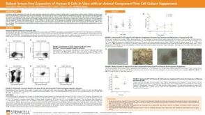 科学海报Robust Serum-Free Expansion of Human B Cells In Vitro with an Animal Component-Free Cell Culture Supplement

 沪公网安备31010102008431号