Polyglutamine Disease Modeling: Epitope Based Screen for Homologous Recombination using CRISPR/Cas9 System.
We have previously reported the genetic correction of Huntington's disease (HD) patient-derived induced pluripotent stem cells using traditional homologous recombination (HR) approaches. To extend this work,we have adopted a CRISPR-based genome editing approach to improve the efficiency of recombination in order to generate allelic isogenic HD models in human cells. Incorporation of a rapid antibody-based screening approach to measure recombination provides a powerful method to determine relative efficiency of genome editing for modeling polyglutamine diseases or understanding factors that modulate CRISPR/Cas9 HR.
View Publication
产品类型:
产品号#:
85850
85857
产品名:
mTeSR™1
mTeSR™1
Mormone E et al. (NOV 2014)
Stem cells and development 23 21 2626--36
Footprint-free" human induced pluripotent stem cell-derived astrocytes for in vivo cell-based therapy."
The generation of human induced pluripotent stem cells (hiPSC) from somatic cells has enabled the possibility to provide patient-specific hiPSC for cell-based therapy,drug discovery,and other translational applications. Two major obstacles in using hiPSC for clinical application reside in the risk of genomic modification when they are derived with viral transgenes and risk of teratoma formation if undifferentiated cells are engrafted. In this study,we report the generation of footprint-free" hiPSC-derived astrocytes. These are efficiently generated�
View Publication
产品类型:
产品号#:
85850
85857
产品名:
mTeSR™1
mTeSR™1
Baatz JE et al. (JUL 2014)
In vivo (Athens,Greece) 28 4 411--423
Cryopreservation of viable human lung tissue for versatile post-thaw analyses and culture.
Clinical trials are currently used to test therapeutic efficacies for lung cancer,infections and diseases. Animal models are also used as surrogates for human disease. Both approaches are expensive and time-consuming. The utility of human biospecimens as models is limited by specialized tissue processing methods that preserve subclasses of analytes (e.g. RNA,protein,morphology) at the expense of others. We present a rapid and reproducible method for the cryopreservation of viable lung tissue from patients undergoing lobectomy or transplant. This method involves the pseudo-diaphragmatic expansion of pieces of fresh lung tissue with cryoprotectant formulation (pseudo-diaphragmatic expansion-cryoprotectant perfusion or PDX-CP) followed by controlled-rate freezing in cryovials. Expansion-perfusion rates,volumes and cryoprotectant formulation were optimized to maintain tissue architecture,decrease crystal formation and increase long-term cell viability. Rates of expansion of 4 cc/min or less and volumes ranging from 0.8-1.2 × tissue volume were well-tolerated by lung tissue obtained from patients with chronic obstructive pulmonary disease or idiopathic pulmonary fibrosis,showing minimal differences compared to standard histopathology. Morphology was greatly improved by the PDX-CP procedure compared to simple fixation. Fresh versus post-thawed lung tissue showed minimal differences in histology,RNA integrity numbers and post-translational modified protein integrity (2-dimensional differential gel electrophoresis). It was possible to derive numerous cell types,including alveolar epithelial cells,fibroblasts and stem cells,from the tissue for at least three months after cryopreservation. This new method should provide a uniform,cost-effective approach to the banking of biospecimens,with versatility to be amenable to any post-acquisition process applicable to fresh tissue samples.
View Publication
产品类型:
产品号#:
85850
85857
产品名:
mTeSR™1
mTeSR™1
Gorman BR et al. (DEC 2014)
PLoS ONE 9 12 e116037
Multi-scale imaging and informatics pipeline for in situ pluripotent stem cell analysis
Human pluripotent stem (hPS) cells are a potential source of cells for medical therapy and an ideal system to study fate decisions in early development. However,hPS cells cultured in vitro exhibit a high degree of heterogeneity,presenting an obstacle to clinical translation. hPS cells grow in spatially patterned colony structures,necessitating quantitative single-cell image analysis. We offer a tool for analyzing the spatial population context of hPS cells that integrates automated fluorescent microscopy with an analysis pipeline. It enables high-throughput detection of colonies at low resolution,with single-cellular and sub-cellular analysis at high resolutions,generating seamless in situ maps of single-cellular data organized by colony. We demonstrate the tool's utility by analyzing inter- and intra-colony heterogeneity of hPS cell cycle regulation and pluripotency marker expression. We measured the heterogeneity within individual colonies by analyzing cell cycle as a function of distance. Cells loosely associated with the outside of the colony are more likely to be in G1,reflecting a less pluripotent state,while cells within the first pluripotent layer are more likely to be in G2,possibly reflecting a G2/M block. Our multi-scale analysis tool groups colony regions into density classes,and cells belonging to those classes have distinct distributions of pluripotency markers and respond differently to DNA damage induction. Lastly,we demonstrate that our pipeline can robustly handle high-content,high-resolution single molecular mRNA FISH data by using novel image processing techniques. Overall,the imaging informatics pipeline presented offers a novel approach to the analysis of hPS cells that includes not only single cell features but also colony wide,and more generally,multi-scale spatial configuration.
View Publication
Fischbach NA et al. (FEB 2005)
Blood 105 4 1456--66
HOXB6 overexpression in murine bone marrow immortalizes a myelomonocytic precursor in vitro and causes hematopoietic stem cell expansion and acute myeloid leukemia in vivo.
The HOX family of homeobox genes plays an important role in normal and malignant hematopoiesis. Dysregulated HOX gene expression profoundly effects the proliferation and differentiation of hematopoietic stem cells (HSCs) and committed progenitors,and aberrant activation of HOX genes is a common event in human myeloid leukemia. HOXB6 is frequently overexpressed in human acute myeloid leukemia (AML). To gain further insight into the role of HOXB6 in hematopoiesis,we overexpressed HOXB6 in murine bone marrow using retrovirus-mediated gene transfer. We also explored structure-function relationships using mutant HOXB6 proteins unable to bind to DNA or a key HOX-binding partner,pre-B-cell leukemia transcription factor-1 (PBX1). Additionally,we investigated the potential cooperative interaction with myeloid ecotropic viral integration site 1 homolog (MEIS1). In vivo,HOXB6 expanded HSCs and myeloid precursors while inhibiting erythropoiesis and lymphopoiesis. Overexpression of HOXB6 resulted in AML with a median latency of 223 days. Coexpression of MEIS1 dramatically shortened the onset of AML. Cytogenetic analysis of a subset of HOXB6-induced AMLs revealed recurrent deletions of chromosome bands 2D-E4,a region frequently deleted in HOXA9-induced AMLs. In vitro,HOXB6 immortalized a factor-dependent myelomonocytic precursor capable of granulocytic and monocytic differentiation. These biologic effects of HOXB6 were largely dependent on DNA binding but independent of direct interaction with PBX1.
View Publication
Critical role for PI 3-kinase in the control of erythropoietin-induced erythroid progenitor proliferation.
The production of red blood cells is tightly regulated by erythropoietin (Epo). The phosphoinositide 3-kinase (PI 3-kinase) pathway was previously shown to be activated in response to Epo. We studied the role of this pathway in the control of Epo-induced survival and proliferation of primary human erythroid progenitors. We show that phosphoinositide 3 (PI 3)-kinase associates with 4 tyrosine-phosphorylated proteins in primary human erythroid progenitors,namely insulin receptor substrate-2 (IRS2),Src homology 2 domain-containing inositol 5'-phosphatase (SHIP),Grb2-associated binder-1 (Gab1),and the Epo receptor (EpoR). Using different in vitro systems,we demonstrate that 3 alternative pathways independently lead to Epo-induced activation of PI 3-kinase and phosphorylation of its downstream effectors,Akt,FKHRL1,and P70S6 kinase: through direct association of PI 3-kinase with the last tyrosine residue (Tyr479) of the Epo receptor (EpoR),through recruitment and phosphorylation of Gab proteins via either Tyr343 or Tyr401 of the EpoR,or through phosphorylation of IRS2 adaptor protein. The mitogen-activated protein (MAP) kinase pathway was also activated by Epo in erythroid progenitors,but we found that this process is independent of PI 3-kinase activation. In erythroid progenitors,the functional role of PI 3-kinase was both to prevent apoptosis and to stimulate cell proliferation in response to Epo stimulation. Finally,our results show that PI 3-kinase-mediated proliferation of erythroid progenitors in response to Epo occurs mainly through modulation of the E3 ligase SCF(SKP2),which,in turn,down-regulates p27(Kip1) cyclin-dependent kinase (CDK) inhibitor via proteasome degradation.
View Publication
产品类型:
产品号#:
09500
09600
09650
产品名:
BIT 9500血清替代物
StemSpan™ SFEM
StemSpan™ SFEM
Christoffersson J et al. (APR 2016)
Methods in molecular biology (Clifton,N.J.)
A Microfluidic Bioreactor for Toxicity Testing of Stem Cell Derived 3D Cardiac Bodies.
Modeling tissues and organs using conventional 2D cell cultures is problematic as the cells rapidly lose their in vivo phenotype. In microfluidic bioreactors the cells reside in microstructures that are continuously perfused with cell culture medium to provide a dynamic environment mimicking the cells natural habitat. These micro scale bioreactors are sometimes referred to as organs-on-chips and are developed in order to improve and extend cell culture experiments. Here,we describe the two manufacturing techniques photolithography and soft lithography that are used in order to easily produce microfluidic bioreactors. The use of these bioreactors is exemplified by a toxicity assessment on 3D clustered human pluripotent stem cells (hPSC)-derived cardiomyocytes by beating frequency imaging.
View Publication
产品类型:
产品号#:
85850
85857
产品名:
mTeSR™1
mTeSR™1
Wang YI et al. (JUL 2016)
Biotechnology and Bioengineering
Microfluidic blood-brain barrier model provides in vivo-like barrier properties for drug permeability screening
Efficient delivery of therapeutics across the neuroprotective blood-brain barrier (BBB) remains a formidable challenge for central nervous system drug development. High-fidelity in vitro models of the BBB could facilitate effective early screening of drug candidates targeting the brain. In this study,we developed a microfluidic BBB model that is capable of mimicking in vivo BBB characteristics for a prolonged period and allows for reliable in vitro drug permeability studies under recirculating perfusion. We derived brain microvascular endothelial cells (BMECs) from human induced pluripotent stem cells (hiPSCs) and cocultured them with rat primary astrocytes on the two sides of a porous membrane on a pumpless microfluidic platform for up to 10 days. The microfluidic system was designed based on the blood residence time in human brain tissues,allowing for medium recirculation at physiologically relevant perfusion rates with no pumps or external tubing,meanwhile minimizing wall shear stress to test whether shear stress is required for in vivo-like barrier properties in a microfluidic BBB model. This BBB-on-a-chip model achieved significant barrier integrity as evident by continuous tight junction formation and in vivo-like values of trans-endothelial electrical resistance (TEER). The TEER levels peaked above 4000 $$ textperiodcentered cm(2) on day 3 on chip and were sustained above 2000 $$ textperiodcentered cm(2) up to 10 days,which are the highest sustained TEER values reported in a microfluidic model. We evaluated the capacity of our microfluidic BBB model to be used for drug permeability studies using large molecules (FITC-dextrans) and model drugs (caffeine,cimetidine,and doxorubicin). Our analyses demonstrated that the permeability coefficients measured using our model were comparable to in vivo values. Our BBB-on-a-chip model closely mimics physiological BBB barrier functions and will be a valuable tool for screening of drug candidates. The residence time-based design of a microfluidic platform will enable integration with other organ modules to simulate multi-organ interactions on drug response. Biotechnol. Bioeng. 2016;9999: 1-11. textcopyright 2016 Wiley Periodicals,Inc.
View Publication
产品类型:
产品号#:
85850
85857
产品名:
mTeSR™1
mTeSR™1
Modlich U et al. (OCT 2006)
Blood 108 8 2545--53
Cell-culture assays reveal the importance of retroviral vector design for insertional genotoxicity.
Retroviral vectors with long terminal repeats (LTRs),which contain strong enhancer/promoter sequences at both ends of their genome,are widely used for stable gene transfer into hematopoietic cells. However,recent clinical data and mouse models point to insertional activation of cellular proto-oncogenes as a dose-limiting side effect of retroviral gene delivery that potentially induces leukemia. Self-inactivating (SIN) retroviral vectors do not contain the terminal repetition of the enhancer/promoter,theoretically attenuating the interaction with neighboring cellular genes. With a new assay based on in vitro expansion of primary murine hematopoietic cells and selection in limiting dilution,we showed that SIN vectors using a strong internal retroviral enhancer/promoter may also transform cells by insertional mutagenesis. Most transformed clones,including those obtained after dose escalation of SIN vectors,showed insertions upstream of the third exon of Evi1 and in reverse orientation to its transcriptional orientation. Normalizing for the vector copy number,we found the transforming capacity of SIN vectors to be significantly reduced when compared with corresponding LTR vectors. Additional modifications of SIN vectors may further increase safety. Improved cell-culture assays will likely play an important role in the evaluation of insertional mutagenesis.
View Publication
产品类型:
产品号#:
28600
产品名:
L-Calc™有限稀释软件
Li H et al. (MAY 2007)
The Journal of clinical investigation 117 5 1314--23
Ewing sarcoma gene EWS is essential for meiosis and B lymphocyte development.
Ewing sarcoma gene EWS encodes a putative RNA-binding protein with proposed roles in transcription and splicing,but its physiological role in vivo remains undefined. Here,we have generated Ews-deficient mice and demonstrated that EWS is required for the completion of B cell development and meiosis. Analysis of Ews(-/-) lymphocytes revealed a cell-autonomous defect in precursor B lymphocyte (pre-B lymphocyte) development. During meiosis,Ews-null spermatocytes were deficient in XY bivalent formation and showed reduced meiotic recombination,resulting in massive apoptosis and complete arrest in gamete maturation. Inactivation of Ews in mouse embryonic fibroblasts resulted in premature cellular senescence,and the mutant animals showed hypersensitivity to ionizing radiation. Finally,we showed that EWS interacts with lamin A/C and that loss of EWS results in a reduced lamin A/C expression. Our findings reveal essential functions for EWS in pre-B cell development and meiosis,with proposed roles in DNA pairing and recombination/repair mechanisms. Furthermore,we demonstrate a novel role of EWS in cellular senescence,possibly through its interaction and modulation of lamin A/C.
View Publication

 EasySep™小鼠TIL(CD45)正选试剂盒


 20:24
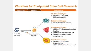
线上讲座STEMdiff™ Kits for Robust and Efficient Differentiation of hPSCs to Multiple Cell Types发布日期: 06/24/2016
20:24
线上讲座STEMdiff™ Kits for Robust and Efficient Differentiation of hPSCs to Multiple Cell Types发布日期: 06/24/2016

 沪公网安备31010102008431号