Garcí et al. (NOV 2010)
American journal of respiratory and critical care medicine 182 9 1144--52
Expression of matrix metalloproteases by fibrocytes: possible role in migration and homing.
RATIONALE: Fibrocytes are progenitor cells characterized by the simultaneous expression of mesenchymal,monocyte,and hematopoietic stem cell markers. We previously documented their presence in lungs of patients with idiopathic pulmonary fibrosis. However,the mechanisms involved in their migration,subsequent homing,and local role remain unclear. Matrix metalloproteinases (MMPs) facilitate cell migration and have been implicated in the pathogenesis of pulmonary fibrosis. OBJECTIVES: To evaluate the expression and role of matrix metalloproteinases in human fibrocytes. METHODS: Fibrocytes were purified from CD14(+) monocytes and cultured for 8 days; purity of fibrocyte cultures was 95% or greater as determined by flow cytometry. Conditioned media and total RNA were collected and the expression of MMP-1,MMP-2,MMP-7,MMP-8,and MMP-9 was evaluated by real-time polymerase chain reaction. Protein synthesis was examined using a Multiplex assay,Western blot,fluorescent immunocytochemistry,and confocal microscopy. MMP-2 and MMP-9 enzymatic activities were evaluated by gelatin zymography. Migration was assessed using collagen I-coated Boyden chambers. Stromal cell-derived factor-1α and platelet-derived growth factor-B were used as chemoattractant with or without a specific MMP-8 inhibitor. MEASUREMENTS AND MAIN RESULTS: Fibrocytes showed gene and protein expression of MMP-2,MMP-9,MMP-8,and MMP-7. MMP-2 and MMP-9 enzymatic activities were also demonstrated by gelatin zymography. Likewise,we found colocalization of MMP-8 and MMP-7 with type I collagen in fibrocytes. Fibrocyte migration toward platelet-derived growth factor-B or Stromal cell-derived factor-1α in collagen I-coated Boyden chambers was significantly reduced by a specific MMP-8 inhibitor. CONCLUSIONS: Our findings reveal that fibrocytes express a variety of MMPs and that MMP-8 actively participates in the process of fibrocyte migration.
View Publication
产品类型:
产品号#:
19058
19058RF
100-1525
产品名:
EasySep™人单核细胞富集试剂盒(不去除CD16)
RoboSep™ 人单核细胞富集试剂盒(不去除CD16)含滤芯吸头
EasySep™人单核细胞富集试剂盒(不去除CD16)
Wang X et al. (OCT 2009)
Cancer research 69 19 7612--8
Correction of the abnormal trafficking of primary myelofibrosis CD34+ cells by treatment with chromatin-modifying agents.
The abnormal trafficking of CD34+ cells is a unique characteristic of primary myelofibrosis (PMF). We have further studied the behavior of PMF CD34+ cells by examining their homing to the marrow and the spleens of nonobese diabetic/severe combined immunodeficient (NOD/SCID) mice. Following the infusion of PMF and normal granulocyte colony-stimulating factor-mobilized peripheral blood (mPB) CD34+ cells into NOD/SCID mice,reduced numbers of PMF CD34+ cells and granulocyte-macrophage colony-forming unit (CFU-GM) compared with mPB were detected in the marrow of these mice,whereas similar numbers of PMF and mPB CD34+ cells and CFU-GM homed to their spleens. The abnormal homing of PMF CD34+ cells was associated with reduced expression of CXCR4,but was not related to the presence of JAK2V617F. The sequential treatment of PMF CD34+ cells with the chromatin-modifying agents 5-aza-2'-deoxycytidine (5azaD) and trichostatin A (TSA),but not treatment with small molecule inhibitors of JAK2,resulted in the generation of increased numbers of CD34+CXCR4+ cells,which was accompanied by enhanced homing of PMF CD34+ cells to the marrow but not the spleens of NOD/SCID mice. Following 5azaD/TSA treatment,JAK2V617F-negative PMF hematopoietic progenitor cells preferentially homed to the marrow but not the spleens of recipient mice. Our data suggest that PMF CD34+ cells are characterized by a reduced ability to home to the marrow but not the spleens of NOD/SCID mice and that this homing defect can be corrected by sequential treatment with chromatin-modifying agents.
View Publication
产品类型:
产品号#:
18056
18056RF
产品名:
Grinshtein N et al. (MAY 2009)
Cancer research 69 9 3979--85
Neoadjuvant vaccination provides superior protection against tumor relapse following surgery compared with adjuvant vaccination.
Tumors that recur following surgical resection of melanoma are typically metastatic and associated with poor prognosis. Using the murine B16F10 melanoma and a robust antimelanoma vaccine,we evaluated immunization as a tool to improve tumor-free survival following surgery. We investigated the utility of vaccination in both neoadjuvant and adjuvant settings. Surprisingly,neoadjuvant vaccination was far superior and provided approximately 100% protection against tumor relapse. Neoadjuvant vaccination was associated with enhanced frequencies of tumor-specific T cells within the tumor and the tumor-draining lymph nodes following resection. We also observed increased infiltration of antigen-specific T cells into the area of surgery. This method should be amenable to any vaccine platform and can be readily extended to the clinic.
View Publication
产品类型:
产品号#:
18751
18751RF
产品名:
Luo M et al. (JAN 2009)
Cancer research 69 2 466--74
Mammary epithelial-specific ablation of the focal adhesion kinase suppresses mammary tumorigenesis by affecting mammary cancer stem/progenitor cells.
Focal adhesion kinase (FAK) has been implicated in the development of cancers,including those of the breast. Nevertheless,the molecular and cellular mechanisms by which FAK promotes mammary tumorigenesis in vivo are not well understood. Here,we show that targeted deletion of FAK in mouse mammary epithelium significantly suppresses mammary tumorigenesis in a well-characterized breast cancer model. Ablation of FAK leads to the depletion of a subset of bipotent cells in the tumor that express both luminal marker keratin 8/18 and basal marker keratin 5. Using mammary stem/progenitor markers,including aldehyde dehydrogenase,CD24,CD29,and CD61,we further revealed that ablation of FAK reduced the pool of cancer stem/progenitor cells in primary tumors of FAK-targeted mice and impaired their self-renewal and migration in vitro. Finally,through transplantation in NOD-SCID mice,we found that cancer stem/progenitor cells isolated from FAK-targeted mice have compromised tumorigenicity and impaired maintenance in vivo. Together,these results show a novel function of FAK in maintaining the mammary cancer stem/progenitor cell population and provide a novel mechanism by which FAK may promote breast cancer development and progression.
View Publication
产品类型:
产品号#:
18556
18556RF
产品名:
Cammenga J et al. (JAN 2007)
Cancer research 67 2 537--45
Mutations in the RUNX1 gene are found at high frequencies in minimally differentiated acute myelogenous leukemia. In addition to null mutations,many of the mutations generate Runx1 DNA-binding (RDB) mutants. To determine if these mutants antagonize wild-type protein activity,cDNAs were transduced into murine bone marrow or human cord blood cells using retroviral vectors. Significantly,the RDB mutants did not act in a transdominant fashion in vivo to disrupt Runx1 activity in either T-cell or platelet development,which are highly sensitive to Runx1 dosage. However,RDB mutant expression impaired expansion and differentiation of the erythroid compartment in which Runx1 expression is normally down-regulated,showing that a RDB-independent function is incompatible with erythroid differentiation. Significantly,both bone marrow progenitors expressing RDB mutants or deficient for Runx1 showed increased replating efficiencies in vitro,accompanied by the accumulation of myeloblasts and dysplastic progenitors,but the effect was more pronounced in RDB cultures. Disruption of the interface that binds CBFbeta,an important cofactor of Runx1,did not impair RDB mutant replating activity,arguing against inactivation of Runx1 function by CBFbeta sequestration. We propose that RDB mutants antagonize Runx1 function in early progenitors by disrupting a critical balance between DNA-binding-independent and DNA-binding-dependent signaling.
View Publication
产品类型:
产品号#:
03434
03444
09500
09600
09650
产品名:
MethoCult™ GF M3434
MethoCult™ GF M3434
BIT 9500血清替代物
StemSpan™ SFEM
StemSpan™ SFEM
Dykstra B et al. (MAY 2006)
Proceedings of the National Academy of Sciences of the United States of America 103 21 8185--90
High-resolution video monitoring of hematopoietic stem cells cultured in single-cell arrays identifies new features of self-renewal.
To search for new indicators of self-renewing hematopoietic stem cells (HSCs),highly purified populations were isolated from adult mouse marrow,micromanipulated into a specially designed microscopic array,and cultured for 4 days in 300 ng/ml Steel factor,20 ng/ml IL-11,and 1 ng/ml flt3-ligand. During this period,each cell and its progeny were imaged at 3-min intervals by using digital time-lapse photography. Individual clones were then harvested and assayed for HSCs in mice by using a 4-month multilineage repopulation endpoint (textgreater1% contribution to lymphoid and myeloid lineages). In a first experiment,6 of 14 initial cells (43%) and 17 of 61 clones (28%) had HSC activity,demonstrating that HSC self-renewal divisions had occurred in vitro. Characteristics associated with HSC activity included longer cell-cycle times and the absence of uropodia on a majority of cells within the clone during the final 12 h of culture. Combining these criteria maximized the distinction of clones with HSC activity from those without and identified a subset of 27 of the 61 clones. These 27 clones included all 17 clones that had HSC activity; a detection efficiency of 63% (2.26 times more frequently than in the original group). The utility of these characteristics for discriminating HSC-containing clones was confirmed in two independent experiments where all HSC-containing clones were identified at a similar 2- to 3-fold-greater efficiency. These studies illustrate the potential of this monitoring system to detect new features of proliferating HSCs that are predictive of self-renewal divisions.
View Publication
产品类型:
产品号#:
19756
19756RF
产品名:
Wu X et al. (JAN 2018)
Cell 172 3 423--438.e25
Intrinsic Immunity Shapes Viral Resistance of Stem Cells.
Stem cells are highly resistant to viral infection compared to their differentiated progeny; however,the mechanism is mysterious. Here,we analyzed gene expression in mammalian stem cells and cells at various stages of differentiation. We find that,conserved across species,stem cells express a subset of genes previously classified as interferon (IFN) stimulated genes (ISGs) but that expression is intrinsic,as stem cells are refractory to interferon. This intrinsic ISG expression varies in a cell-type-specific manner,and many ISGs decrease upon differentiation,at which time cells become IFN responsive,allowing induction of a broad spectrum of ISGs by IFN signaling. Importantly,we show that intrinsically expressed ISGs protect stem cells against viral infection. We demonstrate the in vivo importance of intrinsic ISG expression for protecting stem cells and their differentiation potential during viral infection. These findings have intriguing implications for understanding stem cell biology and the evolution of pathogen resistance.
View Publication

 EasySep™小鼠TIL(CD45)正选试剂盒


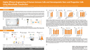 科学海报Highly Efficient Engineering of Human Immune Cells and Hematopoietic Stem and Progenitor Cells Using Microfluidic Transfection
 产品手册血细胞重编程的完整流程
 产品手册hPSC 培养 人多能干细胞的来源和维持培养
 沪公网安备31010102008431号