Chung J et al. (AUG 2009)
Current protocols in stem cell biology Chapter 5 August Unit 5A.3
Magnetic resonance imaging of human embryonic stem cells.
Magnetic resonance imaging (MRI) may emerge as an ideal non-invasive imaging modality to monitor stem cell therapy in the failing heart. This imaging modality generates any arbitrary tomographic view at high spatial and temporal resolution with exquisite intrinsic tissue contrast. This capability enables robust evaluation of both the cardiac anatomy and function. Traditionally,superparamagnetic iron oxide nanoparticle (SPIO) has been widely used for cellular MRI due to SPIO's ability to enhance sensitivity of MRI by inducing remarkable hypointense,negative signal,blooming effect" on T2*-weighted MRI acquisition. Recently�
View Publication
Reprogramming of T cells from human peripheral blood.
Vogt-Koyanagi-Harada (VKH) disease (and sympathetic ophthalmia) is an ocular inflammatory disease that is considered to be a cell-mediated autoimmune disease against melanocytes. The purpose of this study was to determine the Ags specific to VKH disease and to develop an animal model of VKH disease. We found that exposure of lymphocytes from patients with VKH disease to peptides (30-mer) derived from the tyrosinase family proteins led to significant proliferation of the lymphocytes. Immunization of these peptides into pigmented rats induced ocular and extraocular changes that highly resembled human VKH disease,and we suggest that an experimental VKH disease was induced in these rats. We conclude that VKH disease is an autoimmune disease against the tyrosinase family proteins.
View Publication
产品类型:
产品号#:
85850
85857
产品名:
mTeSR™1
mTeSR™1
Sokolov MV and Neumann RD (JAN 2010)
PLoS ONE 5 12 e14195
Radiation-induced bystander effects in cultured human stem cells.
BACKGROUND: The radiation-induced bystander effect" (RIBE) was shown to occur in a number of experimental systems both in vitro and in vivo as a result of exposure to ionizing radiation (IR). RIBE manifests itself by intercellular communication from irradiated cells to non-irradiated cells which may cause DNA damage and eventual death in these bystander cells. It is known that human stem cells (hSC) are ultimately involved in numerous crucial biological processes such as embryologic development; maintenance of normal homeostasis; aging; and aging-related pathologies such as cancerogenesis and other diseases. However�
View Publication
产品类型:
产品号#:
85850
85857
产品名:
mTeSR™1
mTeSR™1
Mangeot P-E et al. (SEP 2011)
Molecular therapy : the journal of the American Society of Gene Therapy 19 9 1656--66
Protein Transfer Into Human Cells by VSV-G-induced Nanovesicles.
Identification of new techniques to express proteins into mammal cells is of particular interest for both research and medical purposes. The present study describes the use of engineered vesicles to deliver exogenous proteins into human cells. We show that overexpression of the spike glycoprotein of the vesicular stomatitis virus (VSV-G) in human cells induces the release of fusogenic vesicles named gesicles. Biochemical and functional studies revealed that gesicles incorporated proteins from producer cells and could deliver them to recipient cells. This protein-transduction method allows the direct transport of cytoplasmic,nuclear or surface proteins in target cells. This was demonstrated by showing that the TetR transactivator and the receptor for the murine leukemia virus (MLV) envelope [murine cationic amino acid transporter-1 (mCAT-1)] were efficiently delivered by gesicles in various cell types. We further shows that gesicle-mediated transfer of mCAT-1 confers to human fibroblasts a robust permissiveness to ecotropic vectors,allowing the generation of human-induced pluripotent stem cells in level 2 biosafety facilities. This highlights the great potential of mCAT-1 gesicles to increase the safety of experiments using retro/lentivectors. Besides this,gesicles is a versatile tool highly valuable for the nongenetic delivery of functions such as transcription factors or genome engineering agents.
View Publication
产品类型:
产品号#:
85850
85857
产品名:
mTeSR™1
mTeSR™1
Chen C et al. (AUG 2013)
Journal of Neurochemistry 126 3 318--330
Inhibition of neuronal nitric oxide synthase activity promotes migration of human-induced pluripotent stem cell-derived neural stem cells toward cancer cells
The breakthrough in derivation of human-induced pluripotent stem cells (hiPSCs) provides an approach that may help overcome ethical and allergenic challenges posed in numerous medical applications involving human cells,including neural stem/progenitor cells (NSCs). Considering the great potential of NSCs in targeted cancer gene therapy,we investigated in this study the tumor tropism of hiPSC-derived NSCs and attempted to enhance the tropism by manipulation of biological activities of proteins that are involved in regulating the migration of NSCs toward cancer cells. We first demonstrated that hiPSC-NSCs displayed tropism for both glioblastoma cells and breast cancer cells in vitro and in vivo. We then compared gene expression profiles between migratory and non-migratory hiPSC-NSCs toward these cancer cells and observed that the gene encoding neuronal nitric oxide synthase (nNOS) was down-regulated in migratory hiPSC-NSCs. Using nNOS inhibitors and nNOS siRNAs,we demonstrated that this protein is a relevant regulator in controlling migration of hiPSC-NSCs toward cancer cells,and that inhibition of its activity or down-regulation of its expression can sensitize poorly migratory NSCs and be used to improve their tumor tropism. These findings suggest a novel application of nNOS inhibitors in neural stem cell-mediated cancer therapy.
View Publication
产品类型:
产品号#:
85850
85857
产品名:
mTeSR™1
mTeSR™1
Alla RK and Cairns BR (JAN 2014)
PloS one 9 1 e85648
RNA polymerase III transcriptomes in human embryonic stem cells and induced pluripotent stem cells, and relationships with pluripotency transcription factors
Recent genomic approaches have revealed that the repertoire of RNA Pol III-transcribed genes varies in different human cell types,and that this variation is likely determined by a combination of the chromatin landscape,cell-specific DNA-binding transcription factors,and collaboration with RNA Pol II. Although much is known about this regulation in differentiated human cells,there is presently little understanding of this aspect of the Pol III system in human ES cells. Here,we determine the occupancy profiles of Pol III components in human H1 ES cells,and also induced pluripotent cells,and compare to known profiles of chromatin,transcription factors,and RNA expression. We find a relatively large fraction of the Pol III repertoire occupied in human embryonic stem cells (hESCs) and induced pluripotent stem cells (iPSCs). In ES cells we find clear correlations between Pol III occupancy and active chromatin. Interestingly,we find a highly significant fraction of Pol III-occupied genes with adjacent binding events by pluripotency factors in ES cells,especially NANOG. Notably,in human ES cells we find H3K27me3 adjacent to but not overlapping many active Pol III loci. We observe in all such cases,a peak of H3K4me3 and/or RNA Pol II,between the H3K27me3 and Pol III binding peaks,suggesting that H3K4me3 and Pol II activity may “insulate�? Pol III from neighboring repressive H3K27me3. Further,we find iPSCs have a larger Pol III repertoire than their precursors. Finally,the active Pol III genome in iPSCs is not completely reprogrammed to a hESC like state and partially retains the transcriptional repertoire of the precursor. Together,our correlative results are consistent with Pol III binding and activity in human ES cells being enabled by active/permissive chromatin that is shaped in part by the pluripotency network of transcription factors and RNA Pol II activity.
View Publication
产品类型:
产品号#:
85850
85857
产品名:
mTeSR™1
mTeSR™1
Wilson HK et al. (DEC 2016)
Tissue engineering. Part C,Methods 22 12 1085--1094
Cryopreservation of Brain Endothelial Cells Derived from Human Induced Pluripotent Stem Cells Is Enhanced by Rho-Associated Coiled Coil-Containing Kinase Inhibition.
The blood-brain barrier (BBB) maintains brain homeostasis but also presents a major obstacle to brain drug delivery. Brain microvascular endothelial cells (BMECs) form the principal barrier and therefore represent the major cellular component of in vitro BBB models. Such models are often used for mechanistic studies of the BBB in health and disease and for drug screening. Recently,human induced pluripotent stem cells (iPSCs) have emerged as a new source for generating BMEC-like cells for use in in vitro human BBB studies. However,the inability to cryopreserve iPSC-BMECs has impeded implementation of this model by requiring a fresh differentiation to generate cells for each experiment. Cryopreservation of differentiated iPSC-BMECs would have a number of distinct advantages,including enabling production of larger scale lots,decreasing lead time to generate purified iPSC-BMEC cultures,and facilitating use of iPSC-BMECs in large-scale screening. In this study,we demonstrate that iPSC-BMECs can be successfully cryopreserved at multiple differentiation stages. Cryopreserved iPSC-BMECs retain high viability,express standard endothelial and BBB markers,and reach a high transendothelial electrical resistance (TEER) of ∼3000 Ωtextperiodcenteredcm(2),equivalent to nonfrozen controls. Rho-associated coiled coil-containing kinase (ROCK) inhibitor Y-27632 substantially increased survival and attachment of cryopreserved iPSC-BMECs,as well as stabilized TEER above 800 Ωtextperiodcenteredcm(2) out to 7 days post-thaw. Overall,cryopreservation will ease handling and storage of high-quality iPSC-BMECs,reducing a key barrier to greater implementation of these cells in modeling the human BBB.
View Publication
产品类型:
产品号#:
85850
85857
产品名:
mTeSR™1
mTeSR™1
Carpenter L et al. (APR 2012)
Stem cells and development 21 6 977--86
Efficient differentiation of human induced pluripotent stem cells generates cardiac cells that provide protection following myocardial infarction in the rat.
Induced pluripotent stem (iPS) cells are being used increasingly to complement their embryonic counterparts to understand and develop the therapeutic potential of pluripotent cells. Our objectives were to identify an efficient cardiac differentiation protocol for human iPS cells as monolayers,and demonstrate that the resulting cardiac progenitors could provide a therapeutic benefit in a rodent model of myocardial infarction. Herein,we describe a 14-day protocol for efficient cardiac differentiation of human iPS cells as a monolayer,which routinely yielded a mixed population in which over 50% were cardiomyocytes,endothelium,or smooth muscle cells. When differentiating,cardiac progenitors from day 6 of this protocol were injected into the peri-infarct region of the rat heart; after coronary artery ligation and reperfusion,we were able to show that human iPS cell-derived cardiac progenitor cells engrafted,differentiated into cardiomyocytes and smooth muscle,and persisted for at least 10 weeks postinfarct. Hearts injected with iPS-derived cells showed a nonsignificant trend toward protection from decline in function after myocardial infarction,as assessed by magnetic resonance imaging at 10 weeks,such that the ejection fraction at 10 weeks in iPS treated hearts was 62%±4%,compared to that of control infarcted hearts at 45%±9% (Ptextless0.2). In conclusion,we demonstrated efficient cardiac differentiation of human iPS cells that gave rise to progenitors that were retained within the infarcted rat heart,and reduced remodeling of the heart after ischemic damage.
View Publication
West FD et al. (AUG 2010)
Stem cells and development 19 8 1211--1220
Porcine induced pluripotent stem cells produce chimeric offspring.
Ethical and moral issues rule out the use of human induced pluripotent stem cells (iPSCs) in chimera studies that would determine the full extent of their reprogrammed state,instead relying on less rigorous assays such as teratoma formation and differentiated cell types. To date,only mouse iPSC lines are known to be truly pluripotent. However,initial mouse iPSC lines failed to form chimeric offspring,but did generate teratomas and differentiated embryoid bodies,and thus these specific iPSC lines were not completely reprogrammed or truly pluripotent. Therefore,there is a need to address whether the reprogramming factors and process used eventually to generate chimeric mice are universal and sufficient to generate reprogrammed iPSC that contribute to chimeric offspring in additional species. Here we show that porcine mesenchymal stem cells transduced with 6 human reprogramming factors (POU5F1,SOX2,NANOG,KLF4,LIN28,and C-MYC) injected into preimplantation-stage embryos contributed to multiple tissue types spanning all 3 germ layers in 8 of 10 fetuses. The chimerism rate was high,85.3% or 29 of 34 live offspring were chimeras based on skin and tail biopsies harvested from 2- to 5-day-old pigs. The creation of pluripotent porcine iPSCs capable of generating chimeric offspring introduces numerous opportunities to study the facets significantly affecting cell therapies,genetic engineering,and other aspects of stem cell and developmental biology.
View Publication
产品类型:
产品号#:
85850
85857
产品名:
mTeSR™1
mTeSR™1
Ishikawa T et al. (JAN 2012)
Methods in molecular biology (Clifton,N.J.) 826 103--114
Generation and hepatic differentiation of human iPS cells.
A method for the generation of human induced pluripotent stem (iPS) cells was established. This method employs adenovirus carrying the ecotropic retrovirus receptor mCAT1 and Moloney murine leukemia virus (MMLV)-based retroviral vectors carrying the four transcription factors POU5F1 (OCT3/4),KLF4,SOX2,and MYC (c-Myc) (Masaki H & Ishikawa T Stem Cell Res 1:105-15,2007). The differentiation of human iPS cells into hepatic cells was performed by a stepwise protocol (Song Z et al. Cell Res 19:1233-42,2009). These cells have potential as patient-specific in vitro models for studying disease etiology and could be used in drug discovery programs tailored to deal with genetic variations in drug efficacy and toxicity.
View Publication
产品类型:
产品号#:
85850
85857
产品名:
mTeSR™1
mTeSR™1
Deglincerti A et al. (NOV 2016)
Nature protocols 11 11 2223--2232
Self-organization of human embryonic stem cells on micropatterns.
Fate allocation in the gastrulating embryo is spatially organized as cells differentiate into specialized cell types depending on their positions with respect to the body axes. There is a need for in vitro protocols that allow the study of spatial organization associated with this developmental transition. Although embryoid bodies and organoids can exhibit some spatial organization of differentiated cells,methods that generate embryoid bodies or organoids do not yield consistent and fully reproducible results. Here,we describe a micropatterning approach in which human embryonic stem cells are confined to disk-shaped,submillimeter colonies. After 42 h of BMP4 stimulation,cells form self-organized differentiation patterns in concentric radial domains,which express specific markers associated with the embryonic germ layers,reminiscent of gastrulating embryos. Our protocol takes 3 d; it uses commercial microfabricated slides (from CYTOO),human laminin-521 (LN-521) as extracellular matrix coating,and either conditioned or chemically defined medium (mTeSR). Differentiation patterns within individual colonies can be determined by immunofluorescence and analyzed with cellular resolution. Both the size of the micropattern and the type of medium affect the patterning outcome. The protocol is appropriate for personnel with basic stem cell culture training. This protocol describes a robust platform for quantitative analysis of the mechanisms associated with pattern formation at the onset of gastrulation.
View Publication

 EasySep™小鼠TIL(CD45)正选试剂盒


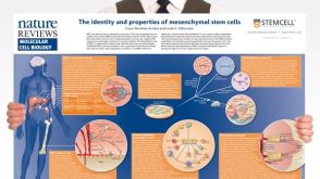 挂图The Identity and Properties of Mesenchymal Stem Cells Overview of MSC expansion, differentiation, immunoregulatory properties and therapeutic potential
 沪公网安备31010102008431号