Darabi R and Perlingeiro RCR ( 2016)
1357 423--439
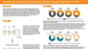
Derivation of Skeletal Myogenic Precursors from Human Pluripotent Stem Cells Using Conditional Expression of PAX7.
Cell-based therapies are considered as one of the most promising approaches for the treatment of degenerating pathologies including muscle disorders and dystrophies. Advances in the approach of reprogramming somatic cells into induced pluripotent stem (iPS) cells allow for the possibility of using the patient's own pluripotent cells to generate specific tissues for autologous transplantation. In addition,patient-specific tissue derivatives have been shown to represent valuable material for disease modeling and drug discovery. Nevertheless,directed differentiation of pluripotent stem cells into a specific lineage is not a trivial task especially in the case of skeletal myogenesis,which is generally poorly recapitulated during the in vitro differentiation of pluripotent stem cells.Here,we describe a practical and efficient method for the derivation of skeletal myogenic precursors from differentiating human pluripotent stem cells using controlled expression of PAX7. Flow cytometry (FACS) purified myogenic precursors can be expanded exponentially and differentiated in vitro into myotubes,enabling researchers to use these cells for disease modeling as well as therapeutic purposes.
View Publication
产品类型:
产品号#:
85850
85857
产品名:
mTeSR™1
mTeSR™1
Wrighton PJ et al. (DEC 2014)
Proceedings of the National Academy of Sciences of the United States of America 111 51 18126--18131
Signals from the surface modulate differentiation of human pluripotent stem cells through glycosaminoglycans and integrins.
The fate decisions of human pluripotent stem (hPS) cells are governed by soluble and insoluble signals from the microenvironment. Many hPS cell differentiation protocols use Matrigel,a complex and undefined substrate that engages multiple adhesion and signaling receptors. Using defined surfaces programmed to engage specific cell-surface ligands (i.e.,glycosaminoglycans and integrins),the contribution of specific matrix signals can be dissected. For ectoderm and motor neuron differentiation,peptide-modified surfaces that can engage both glycosaminoglycans and integrins are effective. In contrast,surfaces that interact selectively with glycosaminoglycans are superior to Matrigel in promoting hPS cell differentiation to definitive endoderm and mesoderm. The modular surfaces were used to elucidate the signaling pathways underlying these differences. Matrigel promotes integrin signaling,which in turn inhibits mesendoderm differentiation. The data indicate that integrin-activating surfaces stimulate Akt signaling via integrin-linked kinase (ILK),which is antagonistic to endoderm differentiation. The ability to attribute cellular responses to specific interactions between the cell and the substrate offers new opportunities for revealing and controlling the pathways governing cell fate.
View Publication
Meng A et al. (DEC 2003)
Experimental hematology 31 12 1348--56
Ionizing radiation and busulfan inhibit murine bone marrow cell hematopoietic function via apoptosis-dependent and -independent mechanisms.
OBJECTIVE: Ionizing radiation (IR) and busulfan (BU) are commonly used as preconditioning regimens for bone marrow transplantation (BMT). We examined whether induction of apoptosis in murine bone marrow (BM) hematopoietic cells contributes to IR- and BU-induced suppression of their hematopoietic function. METHODS: The hematopoietic functions of hematopoietic stem cells (HSCs) and progenitors were analyzed by the cobblestone area-forming cell (CAFC) assay. Apoptosis was determined by measuring 3,3'-dihexyloxacarbocyanine iodide (DiCO6) uptake,annexin V staining,and/or sub-G(0/1) cells. Four cell types were studied: murine BM mononuclear cells (BM-MNCs),linage-negative hematopoietic cells (Lin-) cells),Lin- Scal+ c-kit+ cells,and Lin- Scal- c-kit+ cells by flow cytometry. RESULTS: Exposure of BM-MNCs to IR (4 Gy) or incubation of the cells with BU (30 microM) resulted in a significant reduction in CAFC frequency (ptextless0.001). The survival fractions of various day-types of CAFC for the irradiated cells were less than 10%,while that for BU-treated cells was 71.3% on day 7 and progressively declined to 5.3% on day 35. Interestingly,IR significantly induced apoptosis in BM-MNCs,Lin- cells,HSCs,and progenitors,whereas BU failed to increase apoptosis in these cells. In addition,preincubation of BM-MNCs with z-Val-Ala-Asp (OCH3)-fluoromethylketone,methyl ester (z-VAD) attenuated IR-induced reduction in CAFC but not that induced by BU. CONCLUSION: IR and BU differentially suppress the hematopoietic function of HSCs and progenitors by fundamentally different mechanisms. IR inhibits the function primarily by the induction of HSC and progenitor apoptosis. In contrast,BU suppresses HSC and progenitor function via an apoptosis-independent mechanism.
View Publication
产品类型:
产品号#:
03534
产品名:
MethoCult™GF M3534
Lam BS et al. (JAN 2011)
Blood 117 4 1167--75
Pharmacologic modulation of the calcium-sensing receptor enhances hematopoietic stem cell lodgment in the adult bone marrow.
The ability of hematopoietic stem cells (HSCs) to undergo self-renewal is partly regulated by external signals originating from the stem cell niche. Our previous studies with HSCs obtained from fetal liver of mice deficient for the calcium-sensing receptor (CaR) have shown the crucial role of this receptor in HSC lodgment and engraftment in the bone marrow (BM) endosteal niche. Using a CaR agonist,Cinacalcet,we assessed the effects of stimulating the CaR on the function of murine HSCs. Our results show that CaR stimulation increases primitive hematopoietic cell activity in vitro,including growth in stromal cell cocultures,adhesion to extracellular matrix molecules such as collagen I and fibronectin,and migration toward the chemotactic stimulus,stromal cell-derived factor 1α. Receptor stimulation also led to augmented in vivo homing,CXCR4-mediated lodgment at the endosteal niche,and engraftment capabilities. These mechanisms by which stimulating the CaR dictates preferential localization of HSCs in the BM endosteal niche provide additional insights into the fundamental interrelationship between the stem cell and its niche. These studies also have implications in the area of clinical stem cell transplantation,where ex vivo modulation of the CaR may be envisioned as a strategy to enhance HSC engraftment in the BM.
View Publication
产品类型:
产品号#:
03434
03444
产品名:
MethoCult™GF M3434
MethoCult™GF M3434
Kumar A et al. (JAN 2012)
Breast cancer research : BCR 14 1 R4
Evidence that GTP-binding domain but not catalytic domain of transglutaminase 2 is essential for epithelial-to-mesenchymal transition in mammary epithelial cells.
INTRODUCTION: The expression of proinflammatory protein tissue transglutaminase 2 (TG2) is frequently upregulated in multiple cancer cell types. However,the exact role of TG2 in cancer cells is not well-understood. We recently initiated studies to determine the significance of TG2 in cancer cells and observed that sustained expression of TG2 resulted in epithelial-to-mesenchymal transition (EMT) and promoted cancer stem cell (CSC) traits in mammary epithelial cells. These results suggested that TG2 could serve as a promising therapeutic target for overcoming chemoresistance and inhibiting metastatic spread of cancer cells. METHODS: Using various mutant constructs,we analyzed the activity of TG2 that is essential for promoting the EMT-CSC phenotype. RESULTS: Our results suggest that catalytically inactive TG2 (TG2-C277S) is as effective as wild-type TG2 (TG2-WT) in inducing the EMT-CSC in mammary epithelial cells. In contrast,overexpression of a GTP-binding-deficient mutant (TG2-R580A) was completely incompetent in this regard. Moreover,TG2-dependent activation of the proinflammatory transcription factor NF-κB is deemed essential for promoting the EMT-CSC phenotype in mammary epithelial cells. CONCLUSIONS: Our results suggest that the transamidation activity of TG2 is not essential for promoting its oncogenic functions and provide a strong rationale for developing small-molecule inhibitors to block GTP-binding pockets of TG2. Such inhibitors may have great potential for inhibiting the TG2-regulated pathways,reversing drug resistance and inhibiting the metastasis of cancer cells.
View Publication
产品类型:
产品号#:
05620
产品名:
MammoCult™人培养基试剂盒
Francis KR et al. (APR 2016)
Nature medicine 22 4 388--396
Modeling Smith-Lemli-Opitz syndrome with induced pluripotent stem cells reveals a causal role for Wnt/$$-catenin defects in neuronal cholesterol synthesis phenotypes.
Smith-Lemli-Opitz syndrome (SLOS) is a malformation disorder caused by mutations in DHCR7,which impair the reduction of 7-dehydrocholesterol (7DHC) to cholesterol. SLOS results in cognitive impairment,behavioral abnormalities and nervous system defects,though neither affected cell types nor impaired signaling pathways are fully understood. Whether 7DHC accumulation or cholesterol loss is primarily responsible for disease pathogenesis is also unclear. Using induced pluripotent stem cells (iPSCs) from subjects with SLOS,we identified cellular defects that lead to precocious neuronal specification within SLOS derived neural progenitors. We also demonstrated that 7DHC accumulation,not cholesterol deficiency,is critical for SLOS-associated defects. We further identified downregulation of Wnt/$$-catenin signaling as a key initiator of aberrant SLOS iPSC differentiation through the direct inhibitory effects of 7DHC on the formation of an active Wnt receptor complex. Activation of canonical Wnt signaling prevented the neural phenotypes observed in SLOS iPSCs,suggesting that Wnt signaling may be a promising therapeutic target for SLOS.
View Publication
产品类型:
产品号#:
07923
85850
85857
产品名:
Dispase (1 U/mL)
mTeSR™1
mTeSR™1
Carvalho JL et al. (NOV 2012)
Journal of tissue science & engineering Suppl 11 002
Characterization of Decellularized Heart Matrices as Biomaterials for Regular and Whole Organ Tissue Engineering and Initial In-vitro Recellularization with Ips Cells.
Tissue engineering strategies,based on solid/porous scaffolds,suffer from several limitations,such as ineffective vascularization,poor cell distribution and organization within scaffold,in addition to low final cell density,among others. Therefore,the search for other tissue engineering approaches constitutes an active area of investigation. Decellularized matrices (DM) present major advantages compared to solid scaffolds,such as ideal chemical composition,the preservation of vascularization structure and perfect three-dimensional structure. In the present study,we aimed to characterize and investigate murine heart decellularized matrices as biomaterials for regular and whole organ tissue engineering. Heart decellularized matrices were characterized according to: 1. DNA content,through DNA quantificationo and PCR of isolated genomic DNA; 2. Histological structure,assessed after Hematoxylin and Eosin,as well as Masson's Trichrome stainings; 3. Surface nanostructure analysis,performed,using SEM. Those essays allowed us to conclude that DM was indeed decellularized,with preserved extracellular matrix structure. Following characterization,decellularized heart slices were seeded with induced Pluripotent Stem cells (iPS). As expected,but - to the best of our knowledge - never shown before,decellularization of murine heart matrices maintained matrix biocompatibility,as iPS cells rapidly attached to the surface of the material and proliferated. Strikingly though,heart DM presented a differentiation induction effect over those cells,which lost their pluripotency markers after 7 days of culture in the DM. Such loss of differentiation markers was observed,even though bFGF containing media mTSR was used during such period. Gene expression of iPS cells cultured on DM will be further analyzed,in order to assess the effects of culturing pluripotent stem cells in decellularized heart matrices.
View Publication
3D printing of soft lithography mold for rapid production of polydimethylsiloxane-based microfluidic devices for cell stimulation with concentration gradients
Three-dimensional (3D) printing is advantageous over conventional technologies for the fabrication of sophisticated structures such as 3D micro-channels for future applications in tissue engineering and drug screening. We aimed to apply this technology to cell-based assays using polydimethylsiloxane (PDMS),the most commonly used material for fabrication of micro-channels used for cell culture experiments. Useful properties of PDMS include biocompatibility,gas permeability and transparency. We developed a simple and robust protocol to generate PDMS-based devices using a soft lithography mold produced by 3D printing. 3D chemical gradients were then generated to stimulate cells confined to a micro-channel. We demonstrate that concentration gradients of growth factors,important regulators of cell/tissue functions in vivo,influence the survival and growth of human embryonic stem cells. Thus,this approach for generation of 3D concentration gradients could have strong implications for tissue engineering and drug screening.
View Publication
产品类型:
产品号#:
85850
85857
产品名:
mTeSR™1
mTeSR™1
Nakamura H et al. (OCT 2013)
Herpesviridae 4 1 2
Human cytomegalovirus induces apoptosis in neural stem/progenitor cells derived from induced pluripotent stem cells by generating mitochondrial dysfunction and endoplasmic reticulum stress
BACKGROUND Congenital human cytomegalovirus (HCMV) infection,a leading cause of birth defects,is most often manifested as neurological disorders. The pathogenesis of HCMV-induced neurological disorders is,however,largely unresolved,primarily because of limited availability of model systems to analyze the effects of HCMV infection on neural cells. METHODS An induced pluripotent stem cell (iPSC) line was established from the human fibroblast line MRC5 by introducing the Yamanaka's four factors and then induced to differentiate into neural stem/progenitor cells (NSPCs) by dual inhibition of the SMAD signaling pathway using Noggin and SB-431542. RESULTS iPSC-derived NSPCs (NSPC/iPSCs) were susceptible to HCMV infection and allowed the expression of both early and late viral gene products. HCMV-infected NSPC/iPSCs underwent apoptosis with the activation of caspase-3 and -9 as well as positive staining by the terminal deoxynucleotidyl transferase-mediated dUTP nick-end labeling (TUNEL). Cytochrome c release from mitochondria to cytosol was observed in these cells,indicating the involvement of mitochondrial dysfunction in their apoptosis. In addition,phosphorylation of proteins involved in the unfolded protein response (UPR),such as PKR-like eukaryotic initiation factor 2a kinase (PERK),c-Jun NH2-terminal kinase (JNK),inositol-requiring enzyme 1 (IRE1),and the alpha subunit of eukaryotic initiation factor 2 (eIF2$$) was observed in HCMV-infected NSPC/iPSCs. These results,coupled with the finding of increased expression of mRNA encoding the C/EBP-homologous protein (CHOP) and the detection of a spliced form of X-box binding protein 1 (XBP1) mRNA,suggest that endoplasmic reticulum (ER) stress is also involved in HCMV-induced apoptosis of these cells. CONCLUSIONS iPSC-derived NSPCs are thought to be a useful model to study HCMV neuropathogenesis and to analyze the mechanisms of HCMV-induced apoptosis in neural cells.
View Publication
产品类型:
产品号#:
85850
85857
产品名:
mTeSR™1
mTeSR™1
Giassi LJ et al. (AUG 2008)
Experimental biology and medicine (Maywood,N.J.) 233 8 997--1012
Expanded CD34+ human umbilical cord blood cells generate multiple lymphohematopoietic lineages in NOD-scid IL2rgamma(null) mice.
Umbilical cord blood (UCB) is increasingly being used for human hematopoietic stem cell (HSC) transplantation in children but often requires pooling multiple cords to obtain sufficient numbers for transplantation in adults. To overcome this limitation,we have used an ex vivo two-week culture system to expand the number of hematopoietic CD34(+) cells in cord blood. To assess the in vivo function of these expanded CD34(+) cells,cultured human UCB containing 1 x 10(6) CD34(+) cells were transplanted into conditioned NOD-scid IL2rgamma(null) mice. The expanded CD34(+) cells displayed short- and long-term repopulating cell activity. The cultured human cells differentiated into myeloid,B-lymphoid,and erythroid lineages,but not T lymphocytes. Administration of human recombinant TNFalpha to recipient mice immediately prior to transplantation promoted human thymocyte and T-cell development. These T cells proliferated vigorously in response to TCR cross-linking by anti-CD3 antibody. Engrafted TNFalpha-treated mice generated antibodies in response to T-dependent and T-independent immunization,which was enhanced when mice were co-treated with the B cell cytokine BLyS. Ex vivo expanded CD34(+) human UCB cells have the capacity to generate multiple hematopoietic lineages and a functional human immune system upon transplantation into TNFalpha-treated NOD-scid IL2rgamma(null) mice.
View Publication
产品类型:
产品号#:
09600
09650
产品名:
StemSpan™ SFEM
StemSpan™ SFEM
Vauchez K et al. (NOV 2009)
Molecular therapy : the journal of the American Society of Gene Therapy 17 11 1948--58
Aldehyde dehydrogenase activity identifies a population of human skeletal muscle cells with high myogenic capacities.
Aldehyde dehydrogenase 1A1 (ALDH) activity is one hallmark of human bone marrow (BM),umbilical cord blood (UCB),and peripheral blood (PB) primitive progenitors presenting high reconstitution capacities in vivo. In this study,we have identified ALDH(+) cells within human skeletal muscles,and have analyzed their phenotypical and functional characteristics. Immunohistofluorescence analysis of human muscle tissue sections revealed rare endomysial cells. Flow cytometry analysis using the fluorescent substrate of ALDH,Aldefluor,identified brightly stained (ALDH(br)) cells with low side scatter (SSC(lo)),in enzymatically dissociated muscle biopsies,thereafter abbreviated as SMALD(+) (for skeletal muscle ALDH(+)) cells. Phenotypical analysis discriminated two sub-populations according to CD34 expression: SMALD(+)/CD34(-) and SMALD(+)/CD34(+) cells. These sub-populations did not initially express endothelial (CD31),hematopoietic (CD45),and myogenic (CD56) markers. Upon sorting,however,whereas SMALD(+)/CD34(+) cells developed in vitro as a heterogeneous population of CD56(-) cells able to differentiate in adipoblasts,the SMALD(+)/CD34(-) fraction developed in vitro as a highly enriched population of CD56(+) myoblasts able to form myotubes. Moreover,only the SMALD(+)/CD34(-) population maintained a strong myogenic potential in vivo upon intramuscular transplantation. Our results suggest that ALDH activity is a novel marker for a population of new human skeletal muscle progenitors presenting a potential for cell biology and cell therapy.
View Publication

 EasySep™小鼠TIL(CD45)正选试剂盒


 沪公网安备31010102008431号