Garidou L et al. (SEP 2009)
Journal of virology 83 17 8905--15
Therapeutic memory T cells require costimulation for effective clearance of a persistent viral infection.
Persistent viral infections are a major health concern worldwide. During persistent infection,overwhelming viral replication and the rapid loss of antiviral T-cell function can prevent immune-mediated clearance of the infection,and therapies to reanimate the immune response and purge persistent viruses have been largely unsuccessful. Adoptive immunotherapy using memory T cells is a highly successful therapeutic approach to eradicate a persistent viral infection. Understanding precisely how therapeutically administered memory T cells achieve clearance should improve our ability to terminate states of viral persistence in humans. Mice persistently infected from birth with lymphocytic choriomeningitis virus are tolerant to the pathogen at the T-cell level and thus provide an excellent model to evaluate immunotherapeutic regimens. Previously,we demonstrated that adoptively transferred memory T cells require recipient dendritic cells to effectively purge an established persistent viral infection. However,the mechanisms that reactivate and sustain memory T-cell responses during clearance of such an infection remain unclear. Here we establish that therapeutic memory T cells require CD80 and CD86 costimulatory signals to efficiently clear an established persistent viral infection in vivo. Early blockade of costimulatory pathways with CTLA-4-Fc decreased the secondary expansion of virus-specific CD8(+) and CD4(+) memory T cells as well as their ability to produce antiviral cytokines and purge the persistent infection. Late costimulation blockade also reduced virus-specific T-cell numbers,illustrating that sustained interactions with costimulatory molecules is required for efficient T-cell expansion. These findings indicate that antiviral memory T cells require costimulation to efficiently clear a persistent viral infection and that costimulatory pathways can be targeted to modulate the magnitude of an adoptive immunotherapeutic regimen.
View Publication
产品类型:
产品号#:
18758
18758RF
18768
18768RF
产品名:
Safinia N et al. (FEB 2016)
Oncotarget 7 7 7563--77
Successful expansion of functional and stable regulatory T cells for immunotherapy in liver transplantation.
Strategies to prevent organ transplant rejection whilst minimizing long-term immunosuppression are currently under intense investigation with regulatory T cells (Tregs) nearing clinical application. The clinical trial,ThRIL,recently commenced at King's College London,proposes to use Treg cell therapy to induce tolerance in liver transplant recipients,the success of which has the potential to revolutionize the management of these patients and enable a future of drug-free transplants. This is the first report of the manufacture of clinical grade Tregs from prospective liver transplant recipients via a CliniMACS-based GMP isolation technique and expanded using anti-CD3/CD28 beads,IL-2 and rapamycin. We report the enrichment of a pure,stable population of Tregs (textgreater95% CD4(+)CD25(+)FOXP3(+)),reaching adequate numbers for their clinical application. Our protocol proved successful in,influencing the expansion of superior functional Tregs,as compared to freshly isolated cells,whilst also preventing their conversion to Th17 cells under pro-inflammatory conditions. We conclude with the manufacture of the final Treg product in the clinical research facility (CRF),a prerequisite for the clinical application of these cells. The data presented in this manuscript together with the much-anticipated clinical results from ThRIL,will undoubtedly inform the improved management of the liver transplant recipient.
View Publication
产品类型:
产品号#:
07930
07931
07940
07955
07956
07959
07954
100-1061
07952
产品名:
CryoStor® CS10
CryoStor® CS10
CryoStor® CS10
CryoStor® CS10
CryoStor® CS10
CryoStor® CS10
CryoStor® CS10
Anderson AE et al. (FEB 2009)
Journal of leukocyte biology 85 2 243--50
LPS activation is required for migratory activity and antigen presentation by tolerogenic dendritic cells.
Autoimmune pathologies are caused by a breakdown in self-tolerance. Tolerogenic dendritic cells (tolDC) are a promising immunotherapeutic tool for restoring self-tolerance in an antigen-specific manner. Studies about tolDC have focused largely on generating stable maturation-resistant DC,but few have fully addressed questions about the antigen-presenting and migratory capacities of these cells,prerequisites for successful immunotherapy. Here,we investigated whether human tolDC,generated with dexamethasone and the active form of vitamin D3,maintained their tolerogenic function upon activation with LPS (LPS-tolDC),while acquiring the ability to present exogenous autoantigen and to migrate in response to the CCR7 ligand CCL19. LPS activation led to important changes in the tolDC phenotype and function. LPS-tolDC,but not tolDC,expressed the chemokine receptor CCR7 and migrated in response to CCL19. Furthermore,LPS-tolDC were superior to tolDC in their ability to present type II collagen,a candidate autoantigen in rheumatoid arthritis. tolDC and LPS-tolDC had low stimulatory capacity for allogeneic,naïve T cells and skewed T cell polarization toward an anti-inflammatory phenotype,although LPS-tolDC induced significantly higher levels of IL-10 production by T cells. Our finding that LPS activation is essential for inducing migratory and antigen-presenting activity in tolDC is important for optimizing their therapeutic potential.
View Publication
产品类型:
产品号#:
18259
18259RF
产品名:
Kishimoto RK et al. (APR 2016)
Revista brasileira de hematologia e hemoterapia 38 2 113--20
Validation of interphase fluorescence in situ hybridization (iFISH) for multiple myeloma using CD138 positive cells.
BACKGROUND Multiple myeloma is a plasma cell neoplasm with acquired genetic abnormalities of clinical and prognostic importance. Multiple myeloma differs from other hematologic malignancies due to a high fraction of low proliferating malignant plasma cells and the paucity of plasma cells in bone marrow aspiration samples,making cytogenetic analysis a challenge. An abnormal karyotype is found in only one-third of patients with multiple myeloma and interphase fluorescence in situ hybridization is the most useful test for studying the chromosomal abnormalities present in almost 90% of cases. However,it is necessary to study the genetic abnormalities in plasma cells after their identification or selection by morphology,immunophenotyping or sorting. Other challenges are the selection of the most informative FISH panel and determining cut-off levels for FISH probes. This study reports the validation of interphase fluorescence in situ hybridization using CD138 positive cells,according to proposed guidelines published by the European Myeloma Network (EMN) in 2012. METHOD Bone marrow samples from patients with multiple myeloma were used to standardize a panel of five probes [1q amplification,13q14 deletion,17p deletion,t(4;14),and t(14;16)] in CD138(+) cells purified by magnetic cell sorting. RESULTS This test was validated with a low turnaround time and good reproducibility. Five of six samples showed genetic abnormalities. Monosomy/deletion 13 plus t(4;14) were found in two cases. CONCLUSION This technique together with magnetic cell sorting is effective and can be used in the routine laboratory practice. In addition,magnetic cell sorting provides a pure plasma cell population that allows other molecular and genomic studies.
View Publication
Schumann K et al. (AUG 2015)
Proceedings of the National Academy of Sciences of the United States of America 112 33 10437--42
Generation of knock-in primary human T cells using Cas9 ribonucleoproteins.
T-cell genome engineering holds great promise for cell-based therapies for cancer,HIV,primary immune deficiencies,and autoimmune diseases,but genetic manipulation of human T cells has been challenging. Improved tools are needed to efficiently knock out" genes and "knock in" targeted genome modifications to modulate T-cell function and correct disease-associated mutations. CRISPR/Cas9 technology is facilitating genome engineering in many cell types�
View Publication
产品类型:
产品号#:
17952
17952RF
100-0696
产品名:
EasySep™人CD4+ T细胞分选试剂盒
RoboSep™ 人CD4+ T细胞分选试剂盒
EasySep™人CD4+ T细胞分离试剂盒
Watkins NA et al. (MAY 2009)
Blood 113 19 e1--9
A HaemAtlas: characterizing gene expression in differentiated human blood cells.
Hematopoiesis is a carefully controlled process that is regulated by complex networks of transcription factors that are,in part,controlled by signals resulting from ligand binding to cell-surface receptors. To further understand hematopoiesis,we have compared gene expression profiles of human erythroblasts,megakaryocytes,B cells,cytotoxic and helper T cells,natural killer cells,granulocytes,and monocytes using whole genome microarrays. A bioinformatics analysis of these data was performed focusing on transcription factors,immunoglobulin superfamily members,and lineage-specific transcripts. We observed that the numbers of lineage-specific genes varies by 2 orders of magnitude,ranging from 5 for cytotoxic T cells to 878 for granulocytes. In addition,we have identified novel coexpression patterns for key transcription factors involved in hematopoiesis (eg,GATA3-GFI1 and GATA2-KLF1). This study represents the most comprehensive analysis of gene expression in hematopoietic cells to date and has identified genes that play key roles in lineage commitment and cell function. The data,which are freely accessible,will be invaluable for future studies on hematopoiesis and the role of specific genes and will also aid the understanding of the recent genome-wide association studies.
View Publication
产品类型:
产品号#:
18052
18052RF
18053
18053RF
18054
18054RF
18055
18055RF
18058
18058RF
21000
20119
20155
18682
18682RF
产品名:
RoboSep™- S
RoboSep™ 吸头组件抛光剂
RoboSep™分选管套装(9个塑料管)
Martí et al. (OCT 2014)
Blood 124 15 2411--20
Human blood BDCA-1 dendritic cells differentiate into Langerhans-like cells with thymic stromal lymphopoietin and TGF-β.
The ontogeny of human Langerhans cells (LCs) remains poorly characterized,in particular the nature of LC precursors and the factors that may drive LC differentiation. Here we report that thymic stromal lymphopoietin (TSLP),a keratinocyte-derived cytokine involved in epithelial inflammation,cooperates with transforming growth factor (TGF)-β for the generation of LCs. We show that primary human blood BDCA-1(+),but not BDCA-3(+),dendritic cells (DCs) stimulated with TSLP and TGF-β harbor a typical CD1a(+)Langerin(+) LC phenotype. Electron microscopy established the presence of Birbeck granules,an intracellular organelle specific to LCs. LC differentiation was not observed from tonsil BDCA-1(+) and BDCA-3(+) subsets. TSLP + TGF-β LCs had a mature phenotype with high surface levels of CD80,CD86,and CD40. They induced a potent CD4(+) T-helper (Th) cell expansion and differentiation into Th2 cells with increased production of tumor necrosis factor-α and interleukin-6 compared with CD34-derived LCs. Our findings establish a novel LC differentiation pathway from BDCA-1(+) blood DCs with potential implications in epithelial inflammation. Therapeutic targeting of TSLP may interfere with tissue LC repopulation from circulating precursors.
View Publication
产品类型:
产品号#:
19251
19251RF
产品名:
EasySep™人Pan-DC预富集试剂盒
RoboSep™ 人Pan-DC预富集试剂盒含滤芯吸头
Costantini C et al. (JAN 2009)
Immunobiology 214 9-10 828--34
On the co-purification of 6-sulfo LacNAc(+) dendritic cells (slanDC) with NK cells enriched from human blood.
The ability of NK cells to directly recognize pathogens and be activated via Toll-like receptors (TLR) is increasingly recognized. Nevertheless,controversial results on the NK cell ability to be directly activated by lipopolysaccharide (LPS),the ligand of TLR4,have been recently reported. To start elucidating the reasons explaining the contrasting observations of the literature,we focused on the potential role of currently used NK cell purification procedures to condition putative NK cell responsiveness to LPS. To do so,human NK cells were isolated by negative selection,using three different commercial kits,to be comparatively evaluated for the production of IFNgamma in response to ultra-pure LPS and/or IL-2. Despite the lack of surface TLR4,we found that two out of the three NK cell-enriched populations released IFNgamma (and one of the two,IL-12p70 as well) in response to the LPS plus IL-2 combination,whereas the last one did not. However,the two LPS plus IL-2-responsive NK cell populations were found variably contaminated with 6-sulfo LacNAc(+) dendritic cells (slanDC),demonstrated responsible for triggering,via the production of IL-12p70 in response to LPS,the release of IFNgamma by IL-2-stimulated NK cells. Accordingly,slanDC depletion completely abrogated the capacity to produce both IL-12p70 and IFNgamma in response to LPS plus IL-2 by slanDC-containing NK cells. Taken together,our data uncover that two commercially available kits,specifically designed to isolate NK cells by negative selection,also co-purify variable amounts of slanDC. The latter cells may dramatically affect the outcome of experiments carried on to evaluate NK cell responsiveness to TLR agonists such as LPS.
View Publication
产品类型:
产品号#:
19055
19055RF
产品名:
EasySep™人NK细胞富集试剂盒
RoboSep™ 人NK细胞富集试剂盒含滤芯吸头
Shahbazi M et al. (JUL 2013)
Journal of the Neurological Sciences 330 1–2 85--93
Inhibitory effects of neural stem cells derived from human embryonic stem cells on differentiation and function of monocyte-derived dendritic cells
Neural stem cells (NSCs) possess immunosuppressive characteristics,but effects of NSCs on human dendritic cells (DCs),the most important antigen presenting cells,are less well studied. We used an in vitro approach to evaluate the effects of human NSCs on differentiation of human blood CD14+ monocytes into DCs. NSCs derived from H1 human embryonic stem cells (hESC-NSCs) and human ReNcell NSC line,as well as human bone marrow derived mesenchymal stem cells (MSCs),were tested. We observed that in response to treatment with interleukin-4 and granulocyte macrophage colony-stimulating factor CD14+ monocytes co-cultured with NSCs were able to down-regulate CD14 and up-regulate the differentiation marker CD1a,whereas MSC co-culture strongly inhibited CD1a expression and supported prolonged expression of CD14. A similar difference between NSCs and MSCs was noted when lipopolysaccharides were included to induce maturation of monocyte-derived DCs. However,when effects on the function of derived DCs were investigated,NSCs suppressed the elevation of the DC maturation marker CD83,although not the up-regulation of costimulatory molecules CD80,CD86 and CD40,and impaired the functional capacity of the derived DCs to stimulate alloreactive T cells. We did not observe any obvious difference between hESC-NSCs and ReNcell NSCs in inhibiting DC maturation and function. Our data suggest that although human NSCs are less effective than human MSCs in suppressing monocyte differentiation into DCs,these stem cells can still affect the function of DCs,ultimately regulating specific immune responses.
View Publication

 EasySep™小鼠TIL(CD45)正选试剂盒


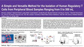 科学海报Isolation of Human Regulatory T Cells from Peripheral Blood Samples
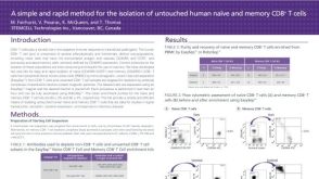 科学海报Isolation of Untouched Human Naive and Memory CD8 T Cells
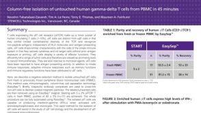 科学海报Immunomagnetic Cell Isolation of Human Gamma-Delta T Cells from PBMC
 沪公网安备31010102008431号