D. Duluc et al. ( 2014)
The Journal of Immunology 192 5776-88
Induction and activation of human Th17 by targeting antigens to dendritic cells via dectin-1
Recent compelling evidence indicates that Th17 confer host immunity against a variety of microbes,including extracellular and intracellular pathogens. Therefore,understanding mechanisms for the induction and activation of Ag-specific Th17 is important for the rational design of vaccines against pathogens. To study this,we employed an in vitro system in which influenza hemagglutinin (HA) 1 was delivered to dendritic cells (DCs) via Dectin-1 using anti-human Dectin-1 (hDectin-1)-HA1 recombinant fusion proteins. We found that healthy individuals maintained broad ranges of HA1-specific memory Th17 that were efficiently activated by DCs targeted with anti-hDectin-1-HA1. Nonetheless,these DCs were not able to induce a significant level of HA1-specific Th17 responses even in the presence of the Th17-promoting cytokines IL-1? and IL-6. We further found that the induction of surface IL-1R1 expression by signals via TCRs and common ?-chain receptors was essential for naive CD4(+) T cell differentiation into HA1-specific Th17. This process was dependent on MyD88,but not IL-1R-associated kinase 1/4. Thus,interruptions in STAT3 or MyD88 signaling led to substantially diminished HA1-specific Th17 induction. Taken together,the de novo generation of pathogen-specific human Th17 requires complex,but complementary,actions of multiple signals. Data from this study will help us design a new and effective vaccine strategy that can promote Th17-mediated immunity against microbial pathogens.
View Publication
Zeng J and Wang S (JAN 2014)
Stem cells translational medicine 3 1 69--80
Human dendritic cells derived from embryonic stem cells stably modified with CD1d efficiently stimulate antitumor invariant natural killer T cell response.
Invariant natural killer T (iNKT) cells are a unique lymphocyte subpopulation that mediates antitumor activities upon activation. A current strategy to harness iNKT cells for cancer treatment is endogenous iNKT cell activation using patient-derived dendritic cells (DCs). However,the limited number and functional defects of patient DCs are still the major challenges for this therapeutic approach. In this study,we investigated whether human embryonic stem cells (hESCs) with an ectopically expressed CD1d gene could be exploited to address this issue. Using a lentivector carrying an optimized expression cassette,we generated stably modified hESC lines that consistently overexpressed CD1d. These modified hESC lines were able to differentiate into DCs as efficiently as the parental line. Most importantly,more than 50% of such derived DCs were CD1d+. These CD1d-overexpressing DCs were more efficient in inducing iNKT cell response than those without modification,and their ability was comparable to that of DCs generated from monocytes of healthy donors. The iNKT cells expanded by the CD1d-overexpressing DCs were functional,as demonstrated by their ability to lyse iNKT cell-sensitive glioma cells. Therefore,hESCs stably modified with the CD1d gene may serve as a convenient,unlimited,and competent DC source for iNKT cell-based cancer immunotherapy.
View Publication
HIV-1 envelope protein binds to and signals through integrin alpha4beta7, the gut mucosal homing receptor for peripheral T cells.
Infection with human immunodeficiency virus 1 (HIV-1) results in the dissemination of virus to gut-associated lymphoid tissue. Subsequently,HIV-1 mediates massive depletion of gut CD4+ T cells,which contributes to HIV-1-induced immune dysfunction. The migration of lymphocytes to gut-associated lymphoid tissue is mediated by integrin alpha4beta7. We demonstrate here that the HIV-1 envelope protein gp120 bound to an activated form of alpha4beta7. This interaction was mediated by a tripeptide in the V2 loop of gp120,a peptide motif that mimics structures presented by the natural ligands of alpha4beta7. On CD4+ T cells,engagement of alpha4beta7 by gp120 resulted in rapid activation of LFA-1,the central integrin involved in the establishment of virological synapses,which facilitate efficient cell-to-cell spreading of HIV-1.
View Publication
产品类型:
产品号#:
19052
19052RF
19055
19055RF
产品名:
EasySep™人CD4+ T细胞富集试剂盒
RoboSep™ 人CD4+ T细胞富集试剂盒含滤芯吸头
EasySep™人NK细胞富集试剂盒
RoboSep™ 人NK细胞富集试剂盒含滤芯吸头
Poulin LF et al. (JUN 2010)
The Journal of experimental medicine 207 6 1261--71
Characterization of human DNGR-1+ BDCA3+ leukocytes as putative equivalents of mouse CD8alpha+ dendritic cells.
In mouse,a subset of dendritic cells (DCs) known as CD8alpha+ DCs has emerged as an important player in the regulation of T cell responses and a promising target in vaccination strategies. However,translation into clinical protocols has been hampered by the failure to identify CD8alpha+ DCs in humans. Here,we characterize a population of human DCs that expresses DNGR-1 (CLEC9A) and high levels of BDCA3 and resembles mouse CD8alpha+ DCs in phenotype and function. We describe the presence of such cells in the spleens of humans and humanized mice and report on a protocol to generate them in vitro. Like mouse CD8alpha+ DCs,human DNGR-1+ BDCA3hi DCs express Necl2,CD207,BATF3,IRF8,and TLR3,but not CD11b,IRF4,TLR7,or (unlike CD8alpha+ DCs) TLR9. DNGR-1+ BDCA3hi DCs respond to poly I:C and agonists of TLR8,but not of TLR7,and produce interleukin (IL)-12 when given innate and T cell-derived signals. Notably,DNGR-1+ BDCA3+ DCs from in vitro cultures efficiently internalize material from dead cells and can cross-present exogenous antigens to CD8+ T cells upon treatment with poly I:C. The characterization of human DNGR-1+ BDCA3hi DCs and the ability to grow them in vitro opens the door for exploiting this subset in immunotherapy.
View Publication
产品类型:
产品号#:
09600
09650
产品名:
StemSpan™ SFEM
StemSpan™ SFEM
Liu W et al. (JUL 2006)
The Journal of experimental medicine 203 7 1701--11
CD127 expression inversely correlates with FoxP3 and suppressive function of human CD4+ T reg cells.
Regulatory T (T reg) cells are critical regulators of immune tolerance. Most T reg cells are defined based on expression of CD4,CD25,and the transcription factor,FoxP3. However,these markers have proven problematic for uniquely defining this specialized T cell subset in humans. We found that the IL-7 receptor (CD127) is down-regulated on a subset of CD4(+) T cells in peripheral blood. We demonstrate that the majority of these cells are FoxP3(+),including those that express low levels or no CD25. A combination of CD4,CD25,and CD127 resulted in a highly purified population of T reg cells accounting for significantly more cells that previously identified based on other cell surface markers. These cells were highly suppressive in functional suppressor assays. In fact,cells separated based solely on CD4 and CD127 expression were anergic and,although representing at least three times the number of cells (including both CD25(+)CD4(+) and CD25(-)CD4(+) T cell subsets),were as suppressive as the classic" CD4(+)CD25(hi) T reg cell subset. Finally�
View Publication
产品类型:
产品号#:
15022
15062
15621
15661
产品名:
RosetteSep™人CD4+ T细胞富集抗体混合物
RosetteSep™人CD4+ T细胞富集抗体混合物
RosetteSep™人CD3去除抗体混合物
RosetteSep™人CD3去除抗体混合物
Frelin C et al. (JAN 2005)
Blood 105 2 804--11
Targeting NF-kappaB activation via pharmacologic inhibition of IKK2-induced apoptosis of human acute myeloid leukemia cells.
Acute myeloid leukemia (AML) cells are characterized by a constitutive and abnormal activation of the nuclear factor-kappaB (NF-kappaB) transcription factor. This study,conducted in vitro on 18 patients,shows that targeting the IKB kinase 2 (IKK2) kinase with the specific pharmacologic inhibitor AS602868 to block NF-kappaB activation led to apoptosis of human primary AML cells. Moreover,AS602868 potentiated the apoptotic response induced by the current chemotherapeutic drugs doxorubicin,cytarabine,or etoposide (VP16). AS602868-induced cell death was associated with rupture of the mitochondrial transmembrane potential and activation of cellular caspases. NF-kappaB inhibition did not affect normal CD34+ hematopoietic precursors,suggesting that it could represent a new adjuvant strategy for AML treatment.
View Publication
产品类型:
产品号#:
15026
15066
产品名:
RosetteSep™人造血祖细胞富集抗体混合物
RosetteSep™人造血祖细胞富集抗体混合物
Trotta R et al. (APR 2005)
Blood 105 8 3011--8
Differential expression of SHIP1 in CD56bright and CD56dim NK cells provides a molecular basis for distinct functional responses to monokine costimulation.
Monocyte cytokines (ie,monokines) induce natural killer (NK) cells to produce interferon-gamma (IFN-gamma),which is critical for monocyte clearance of infectious pathogens and tumor surveillance. Human CD56bright NK cells produce far more IFN-gamma in response to monokines than do CD56dim NK cells. The kinases and phosphatases involved in regulating IFN-gamma production by monokine-activated NK cells are not clearly identified. SHIP1 is a 5' inositol phosphatase that dephosphorylates the phosphatidylinositol-3 kinase (PI-3K) product PI3,4,5P3. Here,we show that constitutive expression of SHIP1 is distinctly lower in CD56bright NK cells compared with CD56dim NK cells,suggesting it could be an important negative regulator of IFN-gamma production in monokine-activated NK cells. Indeed,overexpression of SHIP1 in CD56bright NK cells followed by monokine activation substantially lowered IFN-gamma production. This effect was not seen when NK cells were infected with a SHIP1 mutant containing an inactive catalytic domain. Finally,NK cells in SHIP1-/- mice produced more IFN-gamma in response to monokines in vivo than did NK cells from wild-type mice. Collectively,these results demonstrate that SHIP1 negatively regulates monokine-induced NK cell IFN-gamma production in vitro and in vivo and provide the first molecular explanation for an important functional distinction observed between CD56bright and CD56dim human NK subsets.
View Publication
产品类型:
产品号#:
15025
15065
产品名:
RosetteSep™人NK细胞富集抗体混合物
RosetteSep™人NK细胞富集抗体混合物
Jeyanathan M et al. ( 2017)
Journal of immunology (Baltimore,Md. : 1950) 199 7 2555--2569
CXCR3 Signaling Is Required for Restricted Homing of Parenteral Tuberculosis Vaccine-Induced T Cells to Both the Lung Parenchyma and Airway.
Although most novel tuberculosis (TB) vaccines are designed for delivery via the muscle or skin for enhanced protection in the lung,it has remained poorly understood whether systemic vaccine-induced memory T cells can readily home to the lung mucosa prior to and shortly after pathogen exposure. We have investigated this issue by using a model of parenteral TB immunization and intravascular immunostaining. We find that systemically induced memory T cells are restricted to the blood vessels in the lung,unable to populate either the lung parenchymal tissue or the airway under homeostatic conditions. We further find that after pulmonary TB infection,it still takes many days before such T cells can enter the lung parenchymal tissue and airway. We have identified the acquisition of CXCR3 expression by circulating T cells to be critical for their entry to these lung mucosal compartments. Our findings offer new insights into mucosal T cell biology and have important implications in vaccine strategies against pulmonary TB and other intracellular infections in the lung.
View Publication
产品类型:
产品号#:
19853
19853RF
产品名:
EasySep™小鼠CD8+ T细胞分选试剂盒
RoboSep™ 小鼠CD8+ T细胞分选试剂盒
Wu X et al. (DEC 2008)
Blood 112 12 4675--82
Alternative splicing regulates activation-induced cytidine deaminase (AID): implications for suppression of AID mutagenic activity in normal and malignant B cells.
The mutagenic enzyme activation-induced cytidine deaminase (AID) is required for immunoglobulin class switch recombination (CSR) and somatic hypermutation (SHM) in germinal center (GC) B cells. Deregulated expression of AID is associated with various B-cell malignancies and,currently,it remains unclear how AID activity is extinguished to avoid illegitimate mutations. AID has also been shown to be alternatively spliced in malignant B cells,and there is limited evidence that this also occurs in normal blood B cells. The functional significance of these splice variants remains unknown. Here we show that normal GC human B cells and blood memory B cells similarly express AID splice variants and show for the first time that AID splicing variants are singly expressed in individual normal B cells as well as malignant B cells from chronic lymphocytic leukemia patients. We further demonstrate that the alternative AID splice variants display different activities ranging from inactivation of CSR to inactivation or heightened SHM activity. Our data therefore suggest that CSR and SHM are differentially switched off by varying the expression of splicing products of AID at the individual cell level. Most importantly,our findings suggest a novel tumor suppression mechanism by which unnecessary AID mutagenic activities are promptly contained for GC B cells.
View Publication
产品类型:
产品号#:
21000
20119
20155
19054
19054RF
19754
19754RF
产品名:
RoboSep™- S
RoboSep™ 吸头组件抛光剂
RoboSep™分选管套装(9个塑料管)
EasySep™人B细胞富集试剂盒
RoboSep™ 人B细胞富集试剂盒含滤芯吸头
Sø et al. (JUN 2014)
Molecular immunology 59 2 180--7
Natural mannosylation of HIV-1 gp120 imposes no immunoregulatory effects in primary human plasmacytoid dendritic cells.
Plasmacytoid dendritic cells (pDCs) play a vital role in activation of anti-HIV-1 immunity,and suppression of pDCs might mitigate immune responses against HIV-1. HIV-1 gp120 high-mannose has been attributed immunosuppressive roles in human myeloid DCs,but no receptors for high-mannose have so far been reported on human pDCs. Here we show that upon activation with HIV-1 or by a synthetic compound triggering the same receptor in human pDCs as single-stranded RNA,human pDCs upregulate the mannose receptor (MR,CD206). To examine the functional outcome of this upregulation,inactivated intact or viable HIV-1 particles with various degrees of mannosylation were cultured with pDCs. Activation of pDCs was determined by assaying secretion of IFN-alpha,viability,and upregulation of several pDC-activation markers: CD40,CD86,HLA-DR,CCR7,and PD-L1. The level of activation negatively correlated with degree of mannosylation,however,subsequent reduction in the original mannosylation level had no effect on the pDC phenotype. Furthermore,two of the infectious HIV-1 strains induced profound necrosis in pDCs,also in a mannose-independent manner. We therefore conclude that natural mannosylation of HIV-1 is not involved in HIV-1-mediated immune suppression of pDCs.
View Publication

 EasySep™小鼠TIL(CD45)正选试剂盒


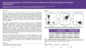 科学海报Fast and Easy Isolation of CD27-Positive Human Memory B Cells Using EasySep™ Releasable RapidSpheres™

 沪公网安备31010102008431号