Peters PJ et al. (JUL 2006)
Journal of virology 80 13 6324--32
Non-macrophage-tropic human immunodeficiency virus type 1 R5 envelopes predominate in blood, lymph nodes, and semen: implications for transmission and pathogenesis.
Human immunodeficiency virus type 1 (HIV-1) R5 isolates that predominantly use CCR5 as a coreceptor are frequently described as macrophage tropic. Here,we compare macrophage tropism conferred by HIV-1 R5 envelopes that were derived directly by PCR from patient tissue. This approach avoids potentially selective culture protocols used in virus isolation. Envelopes were amplified (i) from blood and semen of adult patients and (ii) from plasma of pediatric patients. The phenotypes of these envelopes were compared to those conferred by an extended panel of envelopes derived from brain and lymph node that we reported previously. Our results show that R5 envelopes vary by up to 1,000-fold in their capacity to confer infection of primary macrophages. Highly macrophage-tropic envelopes were predominate in brain but were infrequent in semen,blood,and lymph node samples. We also confirmed that the presence of N283 in the C2 CD4 binding site of gp120 is associated with HIV-1 envelopes from the brain but absent from macrophage-tropic envelopes amplified from blood and semen. Finally,we compared infection of macrophages,CD4(+) T cells,and peripheral blood mononuclear cells (PBMCs) conferred by macrophage-tropic and non-macrophage-tropic envelopes in the context of full-length replication competent viral clones. Non-macrophage-tropic envelopes conferred low-level infection of macrophages yet infected CD4(+) T cells and PBMCs as efficiently as highly macrophage-tropic brain envelopes. The lack of macrophage tropism for the majority of the envelopes amplified from lymph node,blood,and semen is striking and contrasts with the current consensus that R5 primary isolates are generally macrophage tropic. The extensive variation in R5 tropism reported here is likely to have an important impact on pathogenesis and on the capacity of HIV-1 to transmit.
View Publication
产品类型:
产品号#:
19052
19052RF
产品名:
EasySep™人CD4+ T细胞富集试剂盒
RoboSep™ 人CD4+ T细胞富集试剂盒含滤芯吸头
Balakrishnan K et al. (OCT 2006)
Blood 108 7 2392--8
Forodesine, an inhibitor of purine nucleoside phosphorylase, induces apoptosis in chronic lymphocytic leukemia cells.
Purine nucleoside phosphorylase (PNP) deficiency in humans results in T lymphocytopenia. Forodesine,a potent inhibitor of PNP,was designed based on the transition-state structure stabilized by the enzyme. Previous studies established that forodesine in the presence of deoxyguanosine (dGuo) inhibits the proliferation of T lymphocytes. A phase 1 clinical trial of forodesine in T-cell malignancies demonstrated significant antileukemic activity with an increase in intracellular dGuo triphosphate (dGTP). High accumulation of dGTP in T cells may be dependent on the levels of deoxynucleoside kinases. Because B-cell chronic lymphocytic leukemia (B-CLL) cells have high activity of deoxycytidine kinase (dCK),we hypothesized that these lymphocytes would respond to forodesine. This postulate was tested in primary lymphocytes during in vitro investigations. Lymphocytes from 12 patients with CLL were incubated with forodesine and dGuo. These CLL cells showed a wide variation in the accumulation of intracellular dGTP without any effect on other deoxynucleotides. This was associated with DNA damage-induced p53 stabilization,phosphorylation of p53 at Ser15,and activation of p21. The dGTP accumulation was related to induction of apoptosis measured by caspase activation,changes in mitochondrial membrane potential,and PARP cleavage. Based on these data,a phase 2 clinical trial of forodesine has been initiated for CLL patients.
View Publication
产品类型:
产品号#:
19051
19051RF
19054
19054RF
产品名:
EasySep™人T细胞富集试剂盒
RoboSep™ 人T细胞富集试剂盒含滤芯吸头
EasySep™人B细胞富集试剂盒
RoboSep™ 人B细胞富集试剂盒含滤芯吸头
Timm MM et al. (OCT 2006)
Leukemia 20 10 1863--9
Thymoglobulin targets multiple plasma cell antigens and has in vitro and in vivo activity in multiple myeloma.
Multiple myeloma is characterized by the proliferation of clonal plasma cells that have a heterogeneous expression of various cell surface markers,precluding successful use of monoclonal antibodies for therapeutic targeting of the tumor cell. Thymoglobulin (rabbit-derived polyclonal anti-thymocyte globulin),by virtue of its method of preparation,contains antibodies against several B-cell and plasma cell antigens and offers an attractive option for immunotherapy of myeloma. Here,we demonstrate potent anti-myeloma activity of the rabbit anti-thymocyte globulin preparation Thymoglobulin in vitro and in vivo in an animal model of myeloma. Thymoglobulin was able to induce dose- and time-dependent apoptosis of several myeloma cell lines,including those resistant to conventional anti-myeloma agents. Importantly,the anti-myeloma activity was preserved even when myeloma cells were grown with different cytokines demonstrating the ability to overcome microenvironment-mediated resistance. Thymoglobulin induced apoptosis of freshly isolated primary myeloma cells from patients. Using a competitive flow cytometric analysis,we were able to identify the potential antigen targets for Thymoglobulin preparation. Finally,in a plasmacytoma mouse model of myeloma,Thymoglobulin delayed the tumor growth in a dose-dependent manner providing convincing evidence for continued evaluation of this agent in the clinic in patients with myeloma,either alone or in combination with other agents.
View Publication
产品类型:
产品号#:
18357
18357RF
21000
20119
20155
产品名:
RoboSep™- S
RoboSep™ 吸头组件抛光剂
RoboSep™分选管套装(9个塑料管)
Cammenga J et al. (JAN 2007)
Cancer research 67 2 537--45
Mutations in the RUNX1 gene are found at high frequencies in minimally differentiated acute myelogenous leukemia. In addition to null mutations,many of the mutations generate Runx1 DNA-binding (RDB) mutants. To determine if these mutants antagonize wild-type protein activity,cDNAs were transduced into murine bone marrow or human cord blood cells using retroviral vectors. Significantly,the RDB mutants did not act in a transdominant fashion in vivo to disrupt Runx1 activity in either T-cell or platelet development,which are highly sensitive to Runx1 dosage. However,RDB mutant expression impaired expansion and differentiation of the erythroid compartment in which Runx1 expression is normally down-regulated,showing that a RDB-independent function is incompatible with erythroid differentiation. Significantly,both bone marrow progenitors expressing RDB mutants or deficient for Runx1 showed increased replating efficiencies in vitro,accompanied by the accumulation of myeloblasts and dysplastic progenitors,but the effect was more pronounced in RDB cultures. Disruption of the interface that binds CBFbeta,an important cofactor of Runx1,did not impair RDB mutant replating activity,arguing against inactivation of Runx1 function by CBFbeta sequestration. We propose that RDB mutants antagonize Runx1 function in early progenitors by disrupting a critical balance between DNA-binding-independent and DNA-binding-dependent signaling.
View Publication
产品类型:
产品号#:
03434
03444
09500
09600
09650
18096
18096RF
84434
84444
产品名:
MethoCult™GF M3434
MethoCult™GF M3434
BIT 9500血清替代物
StemSpan™ SFEM
StemSpan™ SFEM
Martin G et al. (JUN 2007)
Journal of virology 81 11 5872--81
Human immunodeficiency virus type 1-associated CD40 ligand transactivates B lymphocytes and promotes infection of CD4+ T cells.
Abnormal activation of B lymphocytes is a feature commonly seen in human immunodeficiency virus type 1 (HIV-1)-infected persons. However,the mechanism(s) responsible for this dysfunction is still poorly understood. Having recently shown that CD40L,the ligand for CD40,is inserted within emerging HIV-1 particles,we hypothesized that the contact between virus-anchored host CD40L and CD40 on the surface of B lymphocytes might result in the activation of this cell type. We report here that CD40L-bearing viruses,but not isogenic virions lacking host-derived CD40L,can induce immunoglobulin G and interleukin-6 production. Furthermore,such viral entities were found to induce B-cell homotypic adhesion. These effects were paralleled at the intracellular level by the nuclear translocation of the ubiquitous transcription factor NF-kappaB. The presence of host-derived CD40L within virions resulted in an increased virus attachment to B cells and a more-efficient B-cell-mediated transfer of HIV-1 to autologous CD4(+) T lymphocytes. All the above processes were independent of the virus-encoded envelope glycoproteins. Altogether,the data gathered from this series of investigations suggest that the incorporation of host-encoded CD40L in HIV-1 is likely to play a role in the B-cell abnormalities that are seen in infected individuals.
View Publication
产品类型:
产品号#:
19052
19052RF
19054
19054RF
产品名:
EasySep™人CD4+ T细胞富集试剂盒
RoboSep™ 人CD4+ T细胞富集试剂盒含滤芯吸头
EasySep™人B细胞富集试剂盒
RoboSep™ 人B细胞富集试剂盒含滤芯吸头
Gilbert C et al. (JUL 2007)
Journal of virology 81 14 7672--82
Human immunodeficiency virus type 1 replication in dendritic cell-T-cell cocultures is increased upon incorporation of host LFA-1 due to higher levels of virus production in immature dendritic cells.
Dendritic cells (DCs) act as a portal for invasion by human immunodeficiency virus type-1 (HIV-1). Here,we investigated whether virion-incorporated host cell membrane proteins can affect virus replication in DC-T-cell cocultures. Using isogenic viruses either devoid of or bearing host-derived leukocyte function-associated antigen 1 (LFA-1),we showed that HIV-1 production is augmented when LFA-1-bearing virions are used compared to that for viral entities lacking this adhesion molecule. This phenomenon was observed in immature monocyte-derived DCs (IM-MDDCs) only and not in DCs displaying a mature phenotype. The increase is not due to higher virus production in responder CD4(+) T cells but rather is linked with a more important productive infection of IM-MDDCs. We provided evidence that virus-associated host LFA-1 molecules do not affect a late event in the HIV-1 life cycle but rather exert an effect on an early step in virus replication. We demonstrated that the enhancement of productive infection of IM-MDDCs that is conferred by virus-anchored host LFA-1 involves the protein kinase A (PKA) and PKC signal transduction pathways. The biological significance of this phenomenon was established by performing experiments with virus stocks produced in primary human cells and anti-LFA-1 antibodies. Together,our results indicate that the association between some virus-bound host proteins and their natural cognate ligands can modulate de novo HIV-1 production by IM-MDDCs. Therefore,the additional interactions between virus-bound host cell membrane constituents and counter receptors on the surfaces of DCs can influence HIV-1 replication in IM-MDDC-T-cell cocultures.
View Publication
产品类型:
产品号#:
18058
18058RF
19052
19052RF
产品名:
EasySep™人CD4+ T细胞富集试剂盒
RoboSep™ 人CD4+ T细胞富集试剂盒含滤芯吸头
Arendt BK et al. (SEP 2008)
Blood 112 5 1931--41
Biologic and genetic characterization of the novel amyloidogenic lambda light chain-secreting human cell lines, ALMC-1 and ALMC-2.
Primary systemic amyloidosis (AL) is a rare monoclonal plasma cell (PC) disorder characterized by the deposition of misfolded immunoglobulin (Ig) light chains (LC) in vital organs throughout the body. To our knowledge,no cell lines have ever been established from AL patients. Here we describe the establishment of the ALMC-1 and ALMC-2 cell lines from an AL patient. Both cell lines exhibit a PC phenotype and display cytokine-dependent growth. Using a comprehensive genetic approach,we established the genetic relationship between the cell lines and the primary patient cells,and we were also able to identify new genetic changes accompanying tumor progression that may explain the natural history of this patient's disease. Importantly,we demonstrate that free lambda LC secreted by both cell lines contained a beta structure and formed amyloid fibrils. Despite absolute Ig LC variable gene sequence identity,the proteins show differences in amyloid formation kinetics that are abolished by the presence of Na(2)SO(4). The formation of amyloid fibrils from these naturally secreting human LC cell lines is unprecedented. Moreover,these cell lines will provide an invaluable tool to better understand AL,from the combined perspectives of amyloidogenic protein structure and amyloid formation,genetics,and cell biology.
View Publication
Engineering a stable and selective peptide blocker of the Kv1.3 channel in T lymphocytes.
Kv1.3 potassium channels maintain the membrane potential of effector memory (T(EM)) T cells that are important mediators of multiple sclerosis,type 1 diabetes mellitus,and rheumatoid arthritis. The polypeptide ShK-170 (ShK-L5),containing an N-terminal phosphotyrosine extension of the Stichodactyla helianthus ShK toxin,is a potent and selective blocker of these channels. However,a stability study of ShK-170 showed minor pH-related hydrolysis and oxidation byproducts that were exacerbated by increasing temperatures. We therefore engineered a series of analogs to minimize the formation of these byproducts. The analog with the greatest stability,ShK-192,contains a nonhydrolyzable phosphotyrosine surrogate,a methionine isostere,and a C-terminal amide. ShK-192 shows the same overall fold as ShK,and there is no evidence of any interaction between the N-terminal adduct and the rest of the peptide. The docking configuration of ShK-192 in Kv1.3 shows the N-terminal para-phosphonophenylalanine group lying at the junction of two channel monomers to form a salt bridge with Lys(411) of the channel. ShK-192 blocks Kv1.3 with an IC(50) of 140 pM and exhibits greater than 100-fold selectivity over closely related channels. After a single subcutaneous injection of 100 microg/kg,approximately 100 to 200 pM concentrations of active peptide is detectable in the blood of Lewis rats 24,48,and 72 h after the injection. ShK-192 effectively inhibits the proliferation of T(EM) cells and suppresses delayed type hypersensitivity when administered at 10 or 100 microg/kg by subcutaneous injection once daily. ShK-192 has potential as a therapeutic for autoimmune diseases mediated by T(EM) cells.
View Publication
产品类型:
产品号#:
19051
19051RF
产品名:
EasySep™人T细胞富集试剂盒
RoboSep™ 人T细胞富集试剂盒含滤芯吸头
Popovic R et al. (APR 2009)
Blood 113 14 3314--22
Regulation of mir-196b by MLL and its overexpression by MLL fusions contributes to immortalization.
Chromosomal translocations involving the Mixed Lineage Leukemia (MLL) gene produce chimeric proteins that cause abnormal expression of a subset of HOX genes and leukemia development. Here,we show that MLL normally regulates expression of mir-196b,a hematopoietic microRNA located within the HoxA cluster,in a pattern similar to that of the surrounding 5' Hox genes,Hoxa9 and Hoxa10,during embryonic stem (ES) cell differentiation. Within the hematopoietic lineage,mir-196b is most abundant in short-term hematopoietic stem cells and is down-regulated in more differentiated hematopoietic cells. Leukemogenic MLL fusion proteins cause overexpression of mir-196b,while treatment of MLL-AF9 transformed bone marrow cells with mir-196-specific antagomir abrogates their replating potential in methylcellulose. This demonstrates that mir-196b function is necessary for MLL fusion-mediated immortalization. Furthermore,overexpression of mir-196b was found specifically in patients with MLL associated leukemias as determined from analysis of 55 primary leukemia samples. Overexpression of mir-196b in bone marrow progenitor cells leads to increased proliferative capacity and survival,as well as a partial block in differentiation. Our results suggest a mechanism whereby increased expression of mir-196b by MLL fusion proteins significantly contributes to leukemia development.
View Publication
产品类型:
产品号#:
03534
19756
19756RF
产品名:
MethoCult™GF M3534
Maldonado RA et al. (APR 2009)
The Journal of experimental medicine 206 4 877--92
Control of T helper cell differentiation through cytokine receptor inclusion in the immunological synapse.
The antigen recognition interface formed by T helper precursors (Thps) and antigen-presenting cells (APCs),called the immunological synapse (IS),includes receptors and signaling molecules necessary for Thp activation and differentiation. We have recently shown that recruitment of the interferon-gamma receptor (IFNGR) into the IS correlates with the capacity of Thps to differentiate into Th1 effector cells,an event regulated by signaling through the functionally opposing receptor to interleukin-4 (IL4R). Here,we show that,similar to IFN-gamma ligation,TCR stimuli induce the translocation of signal transducer and activator of transcription 1 (STAT1) to IFNGR1-rich regions of the membrane. Unexpectedly,STAT1 is preferentially expressed,is constitutively serine (727) phosphorylated in Thp,and is recruited to the IS and the nucleus upon TCR signaling. IL4R engagement controls this process by interfering with both STAT1 recruitment and nuclear translocation. We also show that in cells with deficient Th1 or constitutive Th2 differentiation,the IL4R is recruited to the IS. This observation suggest that the IL4R is retained outside the IS,similar to the exclusion of IFNGR from the IS during IL4R signaling. This study provides new mechanistic cues for the regulation of lineage commitment by mutual immobilization of functionally antagonistic membrane receptors.
View Publication

 EasySep™小鼠TIL(CD45)正选试剂盒


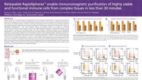 科学海报Releasable RapidSpheres Enable Immunomagnetic Purification of Highly Viable and Functional Immune Cells from Complex Tissues in Less Than 30 Minutes

 沪公网安备31010102008431号