Hassanzadeh-Kiabi N et al. (NOV 2016)
Journal of immunology (Baltimore,Md. : 1950)
Autocrine Type I IFN Signaling in Dendritic Cells Stimulated with Fungal β-Glucans or Lipopolysaccharide Promotes CD8 T Cell Activation.
Type I IFNs are key mediators of immune defense against viruses and bacteria. Type I IFNs were also previously implicated in protection against fungal infection,but their roles in antifungal immunity have not been thoroughly investigated. A recent study demonstrated that bacterial and fungal β-glucans stimulate IFN-β production by dendritic cells (DCs) following detection by the Dectin-1 receptor,but the effects of β-glucan-induced type I IFNs have not been defined. We investigated whether type I IFNs regulate CD8 T cell activation by fungal β-glucan particle-stimulated DCs. We demonstrate that β-glucan-stimulated DCs induce CD8 T cell proliferation,activation marker (CD44 and CD69) expression,and production of IFN-γ,IL-2,and granzyme B. Moreover,we show that type I IFNs support robust CD8 T cell activation (proliferation and IFN-γ and granzyme B production) by β-glucan-stimulated DCs in vitro and in vivo due to autocrine effects on the DCs. Specifically,type I IFNs promote Ag presentation on MHC I molecules,CD86 and CD40 expression,and the production of IL-12 p70,IL-2,IL-6,and TNF-α by β-glucan-stimulated DCs. We also demonstrate a role for autocrine type I IFN signaling in bacterial LPS-induced DC maturation,although,in the context of LPS stimulation,this mechanism is not so critical for CD8 T cell activation (promotes IFN-γ production but not proliferation or granzyme B production). This study provides insight into the mechanisms underlying CD8 T cell activation during infection,which may be useful in the rational design of vaccines directed against pathogens and tumors.
View Publication
产品类型:
产品号#:
19858
19858RF
产品名:
EasySep™ 小鼠Naïve CD8+ T细胞分选试剂盒
RoboSep™ 小鼠Naïve CD8+ T细胞分选试剂盒
Fu W et al. (DEC 2016)
Scientific reports 6 38162
Immune Activation Influences SAMHD1 Expression and Vpx-mediated SAMHD1 Degradation during Chronic HIV-1 Infection.
SAMHD1 restricts human immunodeficiency virus type 1 (HIV-1) replication in myeloid cells and CD4(+) T cells,while Vpx can mediate SAMHD1 degradation to promote HIV-1 replication. Although the restriction mechanisms of SAMHD1 have been well-described,SAMHD1 expression and Vpx-mediated SAMHD1 degradation during chronic HIV-1 infection were poorly understood. Flow cytometric analysis was used to directly visualize ex vivo,and after in vitro SIV-Vpx treatment,SAMHD1 expression in CD4(+) T cells and monocytes. Here we report activated CD4(+) T cells without SAMHD1 expression were severely reduced,and SAMHD1 in CD4(+) T cells became susceptible to SIV-Vpx mediated degradation during chronic HIV-1 infection,which was absent from uninfected donors. These alterations were irreversible,even after long-term fully suppressive antiretroviral treatment. Although SAMHD1 expression in CD4(+) T cells and monocytes was not found to correlate with plasma viral load,Vpx-mediated SAMHD1 degradation was associated with indicators of immune activation. In vitro assays further revealed that T-cell activation and an upregulated IFN-I pathway contributed to these altered SAMHD1 properties. These findings provide insight into how immune activation during HIV-1 infection leads to irreparable aberrations in restriction factors and in subsequent viral evasion from host antiviral defenses.
View Publication
产品类型:
产品号#:
17952
17952RF
19359
19359RF
100-0696
100-0697
产品名:
EasySep™人CD4+ T细胞分选试剂盒
RoboSep™ 人CD4+ T细胞分选试剂盒
EasySep™人单核细胞分选试剂盒
RoboSep™ 人单核细胞分选试剂盒
EasySep™人CD4+ T细胞分离试剂盒
EasySep™人单核细胞分选试剂盒
D. Xie et al. (MAY 2017)
Experimental cell research
The effects of activin A on the migration of human breast cancer cells and neutrophils and their migratory interaction.
Activin A belongs to the superfamily of transforming growth factor beta (TGF$\beta$) and is a critical regulatory cytokine in breast cancer and inflammation. However,the role of activin A in migration of breast cancer cells and immune cells was not well characterized. Here,a microfluidic device was used to examine the effect of activin A on the migration of human breast cancer cell line MDA-MB-231 cells and human blood neutrophils as well as their migratory interaction. We found that activin A promoted the basal migration but impaired epidermal growth factor (EGF)-induced migration of breast cancer cells. By contrast,activin A reduced neutrophil chemotaxis and transendothelial migration to N-Formyl-Met-Leu-Phe (fMLP). Finally,activin A promoted neutrophil chemotaxis to the supernatant from breast cancer cell culture. Collectively,our study revealed the different roles of activin A in regulating the migration of breast cancer cells and neutrophils and their migratory interaction. These findings suggested the potential of activin A as a therapeutic target for inflammation and breast cancers.
View Publication
产品类型:
产品号#:
19666
100-0404
产品名:
EasySep™ Direct人中性粒细胞分选试剂盒
RoboSep™ 人中性粒细胞分选试剂盒
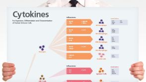
挂图
Human Immune Cytokines
Infographic of key cytokines for expansion, differentiation and characterization of major immune cell types
Abadier M et al. (DEC 2017)
Cell reports 21 13 3885--3899
Effector and Regulatory T Cells Roll at High Shear Stress by Inducible Tether and Sling Formation.
The adaptive immune response involves T cell differentiation and migration to sites of inflammation. T cell trafficking is initiated by rolling on inflamed endothelium. Tethers and slings,discovered in neutrophils,facilitate cell rolling at high shear stress. Here,we demonstrate that the ability to form tethers and slings during rolling is highly inducible in T helper 1 (Th1),Th17,and regulatory T (Treg) cells but less in Th2 cells. In vivo,endogenous Treg cells rolled stably in cremaster venules at physiological shear stress. Quantitative dynamic footprinting nanoscopy of Th1,Th17,and Treg cells uncovered the formation of multiple tethers per cell. Human Th1 cells also showed tethers and slings. RNA sequencing (RNA-seq) revealed the induction of cell migration and cytoskeletal genes in sling-forming cells. We conclude that differentiated CD4 T cells stabilize rolling by inducible tether and sling formation. These phenotypic changes approximate the adhesion phenotype of neutrophils and support CD4 T cell access to sites of inflammation.
View Publication
产品类型:
产品号#:
19762
19762RF
产品名:
EasySep™小鼠中性粒细胞富集试剂盒
RoboSep™ 小鼠中性粒细胞富集试剂盒含滤芯吸头
Kansy BA et al. (NOV 2017)
Cancer research 77 22 6353--6364
PD-1 Status in CD8+ T Cells Associates with Survival and Anti-PD-1 Therapeutic Outcomes in Head and Neck Cancer.
Improved understanding of expression of immune checkpoint receptors (ICR) on tumor-infiltrating lymphocytes (TIL) may facilitate more effective immunotherapy in head and neck cancer (HNC) patients. A higher frequency of PD-1+ TIL has been reported in human papillomavirus (HPV)+ HNC patients,despite the role of PD-1 in T-cell exhaustion. This discordance led us to hypothesize that the extent of PD-1 expression more accurately defines T-cell function and prognostic impact,because PD-1high T cells may be more exhausted than PD-1low T cells and may influence clinical outcome and response to anti-PD-1 immunotherapy. In this study,PD-1 expression was indeed upregulated on HNC patient TIL,and the frequency of these PD-1+ TIL was higher in HPV+ patients (P = 0.006),who nonetheless experienced significantly better clinical outcome. However,PD-1high CD8+ TILs were more frequent in HPV- patients and represented a more dysfunctional subset with compromised IFN-γ secretion. Moreover,HNC patients with higher frequencies of PD-1high CD8+ TIL showed significantly worse disease-free survival and higher hazard ratio for recurrence (P < 0.001),while higher fractions of PD-1low T cells associated with HPV positivity and better outcome. In a murine HPV+ HNC model,anti-PD-1 mAb therapy differentially modulated PD-1high/low populations,and tumor rejection associated with loss of dysfunctional PD-1high CD8+ T cells and a significant increase in PD-1low TIL. Thus,the extent of PD-1 expression on CD8+ TIL provides a potential biomarker for anti-PD-1-based immunotherapy. Cancer Res; 77(22); 6353-64. textcopyright2017 AACR.
View Publication
产品类型:
产品号#:
19051
19051RF
产品名:
EasySep™人T细胞富集试剂盒
RoboSep™ 人T细胞富集试剂盒含滤芯吸头
Marzaioli V et al. ( 2017)
Blood 130 15 1734--1745
NOX5 and p22phox are 2 novel regulators of human monocytic differentiation into dendritic cells.
Dendritic cells (DCs) are a heterogeneous population of professional antigen-presenting cells and are key cells of the immune system,acquiring different phenotypes in accordance with their localization during the immune response. A subset of inflammatory DCs is derived from circulating monocytes (Mo) and has a key role in inflammation and infection. The pathways controlling Mo-DC differentiation are not fully understood. Our objective was to investigate the possible role of nicotinamide adenine dinucleotide phosphate reduced form oxidases (NOXs) in Mo-DC differentiation. In this study,we revealed that Mo-DC differentiation was inhibited by NOX inhibitors and reactive oxygen species scavengers. We show that the Mo-DC differentiation was dependent on p22phox,and not on gp91phox/NOX2,as shown by the reduced Mo-DC differentiation observed in chronic granulomatous disease patients lacking p22phox. Moreover,we revealed that NOX5 expression was strongly increased during Mo-DC differentiation,but not during Mo-macrophage differentiation. NOX5 was expressed in circulating myeloid DC,and at a lower level in plasmacytoid DC. Interestingly,NOX5 was localized at the outer membrane of the mitochondria and interacted with p22phox in Mo-DC. Selective inhibitors and small interfering RNAs for NOX5 indicated that NOX5 controlled Mo-DC differentiation by regulating the JAK/STAT/MAPK and NFκB pathways. These data demonstrate that the NOX5-p22phox complex drives Mo-DC differentiation,and thus could be critical for immunity and inflammation.
View Publication
产品类型:
产品号#:
19061
19061RF
19062
19062RF
19359
19359RF
100-0697
产品名:
EasySep™人髓样DC富集试剂盒
RoboSep™ 人髓样DC富集试剂盒
EasySep™人浆细胞样DC富集试剂盒
RoboSep™ 人浆细胞样DC富集试剂盒含滤芯吸头
EasySep™人单核细胞分选试剂盒
RoboSep™ 人单核细胞分选试剂盒
EasySep™人单核细胞分选试剂盒
Tan Q et al. (JAN 2018)
JCI insight 3 1
Activation-induced cytidine deaminase deficiency accelerates autoimmune diabetes in NOD mice.
B cells play an important role in type 1 diabetes (T1D) development. However,the role of B cell activation-induced cytidine deaminase (AID) in diabetes development is not clear. We hypothesized that AID is important in the immunopathogenesis of T1D. To test this hypothesis,we generated AID-deficient (AID-/-) NOD mice. We found that AID-/-NOD mice developed accelerated T1D,with worse insulitis and high levels of anti-insulin autoantibody in the circulation. Interestingly,neither maternal IgG transferred through placenta,nor IgA transferred through milk affected the accelerated diabetes development. AID-/-NOD mice showed increased activation and proliferation of B and T cells. We found enhanced T-B cell interactions in AID-/-NOD mice,with increased T-bet and IFN-γ expression in CD4+ T cells in the presence of AID-/- B cells. Moreover,excessive lymphoid expansion was observed in AID-/-NOD mice. Importantly,antigen-specific BDC2.5 CD4+ T cells caused more rapid onset of diabetes when cotransferred with AID-/- B cells than when cotransferred with AID+/+ B cells. Thus,our study provides insights into the role of AID in T1D. Our data also suggest that AID is a negative regulator of immune tolerance and ablation of AID can lead to exacerbated islet autoimmunity and accelerated T1D development.
View Publication
产品类型:
产品号#:
19854
19854RF
产品名:
EasySep™小鼠B细胞分选试剂盒
RoboSep™ 小鼠B细胞分选试剂盒
Wu X et al. (JAN 2018)
Cell 172 3 423--438.e25
Intrinsic Immunity Shapes Viral Resistance of Stem Cells.
Stem cells are highly resistant to viral infection compared to their differentiated progeny; however,the mechanism is mysterious. Here,we analyzed gene expression in mammalian stem cells and cells at various stages of differentiation. We find that,conserved across species,stem cells express a subset of genes previously classified as interferon (IFN) stimulated genes (ISGs) but that expression is intrinsic,as stem cells are refractory to interferon. This intrinsic ISG expression varies in a cell-type-specific manner,and many ISGs decrease upon differentiation,at which time cells become IFN responsive,allowing induction of a broad spectrum of ISGs by IFN signaling. Importantly,we show that intrinsically expressed ISGs protect stem cells against viral infection. We demonstrate the in vivo importance of intrinsic ISG expression for protecting stem cells and their differentiation potential during viral infection. These findings have intriguing implications for understanding stem cell biology and the evolution of pathogen resistance.
View Publication
Dendritic Cells but Not Macrophages Sense Tumor Mitochondrial DNA for Cross-priming through Signal Regulatory Protein α Signaling.
Inhibition of cytosolic DNA sensing represents a strategy that tumor cells use for immune evasion,but the underlying mechanisms are unclear. Here we have shown that CD47-signal regulatory protein α (SIRPα) axis dictates the fate of ingested DNA in DCs for immune evasion. Although macrophages were more potent in uptaking tumor DNA,increase of DNA sensing by blocking the interaction of SIRPα with CD47 preferentially occurred in dendritic cells (DCs) but not in macrophages. Mechanistically,CD47 blockade enabled the activation of NADPH oxidase NOX2 in DCs,which in turn inhibited phagosomal acidification and reduced the degradation of tumor mitochondrial DNA (mtDNA) in DCs. mtDNA was recognized by cyclic-GMP-AMP synthase (cGAS) in the DC cytosol,contributing to type I interferon (IFN) production and antitumor adaptive immunity. Thus,our findings have demonstrated how tumor cells inhibit innate sensing in DCs and suggested that the CD47-SIRPα axis is critical for DC-driven antitumor immunity.
View Publication
产品类型:
产品号#:
18780
18780RF
18781
18781RF
19853
19853RF
70025
70025.1
70025.2
70025.3
产品名:
EasySep™ 小鼠CD11c正选试剂盒 II
RoboSep™ 小鼠CD11c正选试剂盒II
EasySep™小鼠CD11c正选试剂盒II及脾脏解离液
RoboSep™ 小鼠CD11c正选试剂盒II及脾脏解离液
EasySep™小鼠CD8+ T细胞分选试剂盒
RoboSep™ 小鼠CD8+ T细胞分选试剂盒
冻存的人外周血单个核细胞
冻存的人外周血单个核细胞
冻存的人外周血单个核细胞
冻存的人外周血单个核细胞
Chang C-F et al. (DEC 2017)
The Journal of clinical investigation
Erythrocyte efferocytosis modulates macrophages towards recovery after intracerebral hemorrhage.
Macrophages are a source of both proinflammatory and restorative functions in damaged tissue through complex dynamic phenotypic changes. Here,we sought to determine whether monocyte-derived macrophages (MDMs) contribute to recovery after acute sterile brain injury. By profiling the transcriptional dynamics of MDMs in the murine brain after experimental intracerebral hemorrhage (ICH),we found robust phenotypic changes in the infiltrating MDMs over time and demonstrated that MDMs are essential for optimal hematoma clearance and neurological recovery. Next,we identified the mechanism by which the engulfment of erythrocytes with exposed phosphatidylserine directly modulated the phenotype of both murine and human MDMs. In mice,loss of receptor tyrosine kinases AXL and MERTK reduced efferocytosis of eryptotic erythrocytes and hematoma clearance,worsened neurological recovery,exacerbated iron deposition,and decreased alternative activation of macrophages after ICH. Patients with higher circulating soluble AXL had poor 1-year outcomes after ICH onset,suggesting that therapeutically augmenting efferocytosis may improve functional outcomes by both reducing tissue injury and promoting the development of reparative macrophage responses. Thus,our results identify the efferocytosis of eryptotic erythrocytes through AXL/MERTK as a critical mechanism modulating macrophage phenotype and contributing to recovery from ICH.
View Publication
产品类型:
产品号#:
17858
17858RF
85450
85460
100-0694
产品名:
EasySep™人CD14正选试剂盒II
RoboSep™ 人CD14正选试剂盒II
SepMate™-50 (IVD)
SepMate™-50 (IVD)
EasySep™人CD14正选试剂盒II
Fan Y et al. (JAN 2018)
The Biochemical journal 475 1 23--44
Interrogating Parkinson's disease LRRK2 kinase pathway activity by assessing Rab10 phosphorylation in human neutrophils.
There is compelling evidence for the role of the leucine-rich repeat kinase 2 (LRRK2) and in particular its kinase function in Parkinson's disease. Orally bioavailable,brain penetrant and potent LRRK2 kinase inhibitors are in the later stages of clinical development. Here,we describe a facile and robust assay to quantify LRRK2 kinase pathway activity by measuring LRRK2-mediated phosphorylation of Rab10 in human peripheral blood neutrophils. We use the selective MJFF-pRab10 monoclonal antibody recognising the Rab10 Thr73 phospho-epitope that is phosphorylated by LRRK2. We highlight the feasibility and practicability of using our assay in the clinical setting by studying a few patients with G2019S LRRK2 associated and sporadic Parkinson's as well as healthy controls. We suggest that peripheral blood neutrophils are a valuable resource for LRRK2 research and should be considered for inclusion in Parkinson's bio-repository collections as they are abundant,homogenous and express relatively high levels of LRRK2 as well as Rab10. In contrast,the widely used peripheral blood mononuclear cells are heterogeneous and only a minority of cells (monocytes and contaminating neutrophils) express LRRK2. While our LRRK2 kinase pathway assay could assist in patient stratification based on LRRK2 kinase activity,we envision that it may find greater utility in pharmacodynamic and target engagement studies in future LRRK2 inhibitor trials.
View Publication

 EasySep™小鼠TIL(CD45)正选试剂盒


 挂图Human Immune Cytokines Infographic of key cytokines for expansion, differentiation and characterization of major immune cell types
 沪公网安备31010102008431号