Wulff H et al. (JUL 2004)
Journal of immunology (Baltimore,Md. : 1950) 173 2 776--86
K+ channel expression during B cell differentiation: implications for immunomodulation and autoimmunity.
Using whole-cell patch-clamp,fluorescence microscopy and flow cytometry,we demonstrate a switch in potassium channel expression during differentiation of human B cells from naive to memory cells. Naive and IgD(+)CD27(+) memory B cells express small numbers of the voltage-gated Kv1.3 and the Ca(2+)-activated intermediate-conductance IKCa1 channel when quiescent,and increase IKCa1 expression 45-fold upon activation with no change in Kv1.3 levels. In contrast,quiescent class-switched memory B cells express high levels of Kv1.3 ( approximately 2000 channels/cell) and maintain their Kv1.3(high) expression after activation. Consistent with their channel phenotypes,proliferation of naive and IgD(+)CD27(+) memory B cells is suppressed by the specific IKCa1 inhibitor TRAM-34 but not by the potent Kv1.3 blocker Stichodactyla helianthus toxin,whereas the proliferation of class-switched memory B cells is suppressed by Stichodactyla helianthus toxin but not TRAM-34. These changes parallel those reported for T cells. Therefore,specific Kv1.3 and IKCa1 inhibitors may have use in therapeutic manipulation of selective lymphocyte subsets in immunological disorders.
View Publication
Ramgolam VS et al. (OCT 2009)
Journal of immunology (Baltimore,Md. : 1950) 183 8 5418--27
IFN-beta inhibits human Th17 cell differentiation.
IFN-beta-1a has been used over the past 15 years as a primary therapy for relapsing-remitting multiple sclerosis (MS). However,the immunomodulatory mechanisms that provide a therapeutic effect against this CNS inflammatory disease are not yet completely elucidated. The effect of IFN-beta-1a on Th17 cells,which play a critical role in the development of the autoimmune response,has not been extensively studied in humans. We have investigated the effect of IFN-beta-1a on dendritic cells (DCs) and naive CD4(+)CD45RA(+) T cells derived from untreated MS patients and healthy controls in the context of Th17 cell differentiation. We report that IFN-beta-1a treatment down-regulated the expression of IL-1beta and IL-23p19 in DCs,whereas it induced the gene expression of IL-12p35 and IL-27p28. We propose that IFN-beta-1a-mediated up-regulation of the suppressor of cytokine signaling 3 expression,induced via STAT3 phosphorylation,mediates IL-1beta and IL-23 down-regulation,while IFN-beta-1a-induced STAT1 phosphorylation induces IL-27p28 expression. CD4(+)CD45RA(+) naive T cells cocultured with supernatants from IFN-beta-1a-treated DCs exhibited decreased gene expression of the Th17 cell markers retinoic acid-related orphan nuclear hormone receptor c (RORc),IL-17A,and IL-23R. A direct IFN-beta-1a treatment of CD45RA(+) T cells cultured in Th17-polarizing conditions also down-regulated RORc,IL-17A,and IL-23R,but up-regulated IL-10 gene expression. Studies of the mechanisms involved in the Th17 cell differentiation suggest that IFN-beta-1a inhibits IL-17 and induces IL-10 secretion via activated STAT1 and STAT3,respectively. IFN-beta's suppression of Th17 cell differentiation may represent its most relevant mechanism of selective suppression of the autoimmune response in MS.
View Publication
产品类型:
产品号#:
19059
19059RF
产品名:
EasySep™人单核细胞富集试剂盒
RoboSep™ 人单核细胞富集试剂盒含滤芯吸头
Ols ML et al. (OCT 2016)
Immunity
Dendritic Cells Regulate Extrafollicular Autoreactive B Cells via T Cells Expressing Fas and Fas Ligand.
The extrafollicular (EF) plasmablast response to self-antigens that contain Toll-like receptor (TLR) ligands is prominent in murine lupus models and some bacterial infections,but the inhibitors and activators involved have not been fully delineated. Here,we used two conventional dendritic cell (cDC) depletion systems to investigate the role of cDCs on a classical TLR-dependent autoreactive EF response elicited in rheumatoid-factor B cells by DNA-containing immune complexes. Contrary to our hypothesis,cDC depletion amplified rather than dampened the EF response in Fas-intact but not Fas-deficient mice. Further,we demonstrated that cDC-dependent regulation requires Fas and Fas ligand (FasL) expression by T cells,but not Fas expression by B cells. Thus,cDCs activate FasL-expressing T cells that regulate Fas-expressing extrafollicular helper T (Tefh) cells. These studies reveal a regulatory role for cDCs in B cell plasmablast responses and provide a mechanistic explanation for the excess autoantibody production observed in Fas deficiency.
View Publication
Aflaki E et al. (JUN 2014)
Science translational medicine 6 240 240ra73
Macrophage models of Gaucher disease for evaluating disease pathogenesis and candidate drugs.
Gaucher disease is caused by an inherited deficiency of glucocerebrosidase that manifests with storage of glycolipids in lysosomes,particularly in macrophages. Available cell lines modeling Gaucher disease do not demonstrate lysosomal storage of glycolipids; therefore,we set out to develop two macrophage models of Gaucher disease that exhibit appropriate substrate accumulation. We used these cellular models both to investigate altered macrophage biology in Gaucher disease and to evaluate candidate drugs for its treatment. We generated and characterized monocyte-derived macrophages from 20 patients carrying different Gaucher disease mutations. In addition,we created induced pluripotent stem cell (iPSC)-derived macrophages from five fibroblast lines taken from patients with type 1 or type 2 Gaucher disease. Macrophages derived from patient monocytes or iPSCs showed reduced glucocerebrosidase activity and increased storage of glucocerebroside and glucosylsphingosine in lysosomes. These macrophages showed efficient phagocytosis of bacteria but reduced production of intracellular reactive oxygen species and impaired chemotaxis. The disease phenotype was reversed with a noninhibitory small-molecule chaperone drug that enhanced glucocerebrosidase activity in the macrophages,reduced glycolipid storage,and normalized chemotaxis and production of reactive oxygen species. Macrophages differentiated from patient monocytes or patient-derived iPSCs provide cellular models that can be used to investigate disease pathogenesis and facilitate drug development.
View Publication
产品类型:
产品号#:
05850
05857
05870
05875
19059
19059RF
85850
85857
85870
85875
27845
27945
27840
27865
27940
27965
产品名:
EasySep™人单核细胞富集试剂盒
RoboSep™ 人单核细胞富集试剂盒含滤芯吸头
mTeSR™1
mTeSR™1
Marwali MR et al. (SEP 2004)
Journal of immunology (Baltimore,Md. : 1950) 173 5 2960--7
Lipid rafts mediate association of LFA-1 and CD3 and formation of the immunological synapse of CTL.
Lipid rafts accumulate in the immunological synapse formed by an organized assembly of the TCR/CD3,LFA-1,and signaling molecules. However,the precise role of lipid rafts in the formation of the immunological synapse is unclear. In this study,we show that LFA-1 on CTL is constitutively active and mediates Ag-independent binding of CTL to target cells expressing its ligands. LFA-1 and CD3 on CTL,but not resting T cells,colocalize in lipid rafts. Binding of LFA-1 on CTL to targets initiates the formation of the immunological synapse,which is formed by LFA-1,CD3,and ganglioside GM1 distributed in the periphery of the cell contact site and cholesterol is more widely distributed. The formation of this synapse is Ag independent,but the recognition of Ag by the TCR induces accumulation of tyrosine phosphorylated proteins in the synapse as well as redistribution of the microtubule organization center toward the cell contact site. Our results suggest that LFA-1 recruits lipid rafts and the TCR/CD3 to the synapse,and facilitates efficient and rapid activation of CTL.
View Publication

 EasySep™小鼠TIL(CD45)正选试剂盒


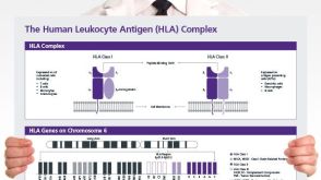 挂图The Human Leukocyte Antigen (HLA) Complex Provides an overview of the human leukocyte antigen (HLA) complex and nomenclature of HLA alleles
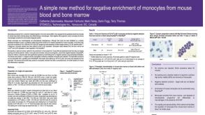 科学海报Method for Negative Enrichment of Monocytes from Mouse Blood and Bone Marrow
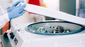 实验方案How to Process Leukocyte Reduction System (LRS) Cones/Chambers for Downstream Cell Isolation
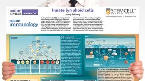 挂图Innate Lymphoid Cells Overview of innate lymphoid cells (ILCs) development, classification, plasticity and functional diversity
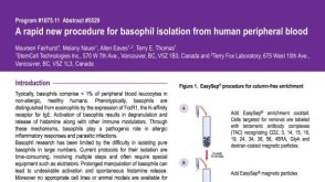 科学海报Basophil Isolation from Human Peripheral Blood
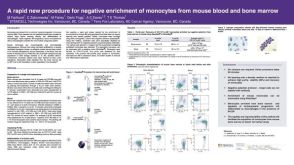 科学海报Procedure for Negative Enrichment of Monocytes from Mouse Blood and Bone Marrow

 沪公网安备31010102008431号