Mace EM et al. (NOV 2016)
The Journal of clinical investigation
Biallelic mutations in IRF8 impair human NK cell maturation and function.
Human NK cell deficiencies are rare yet result in severe and often fatal disease,particularly as a result of viral susceptibility. NK cells develop from hematopoietic stem cells,and few monogenic errors that specifically interrupt NK cell development have been reported. Here we have described biallelic mutations in IRF8,which encodes an interferon regulatory factor,as a cause of familial NK cell deficiency that results in fatal and severe viral disease. Compound heterozygous or homozygous mutations in IRF8 in 3 unrelated families resulted in a paucity of mature CD56dim NK cells and an increase in the frequency of the immature CD56bright NK cells,and this impairment in terminal maturation was also observed in Irf8-/-,but not Irf8+/-,mice. We then determined that impaired maturation was NK cell intrinsic,and gene expression analysis of human NK cell developmental subsets showed that multiple genes were dysregulated by IRF8 mutation. The phenotype was accompanied by deficient NK cell function and was stable over time. Together,these data indicate that human NK cells require IRF8 for development and functional maturation and that dysregulation of this function results in severe human disease,thereby emphasizing a critical role for NK cells in human antiviral defense.
View Publication
Role for the conserved N-terminal cysteines in the anti-chemokine activities by the chemokine-like protein MC148R1 encoded by Molluscum contagiosum virus.
Molluscum contagiosum poxvirus (MCV) type 1 and type 2 encode two chemokine-like proteins MC148R1 and MC148R2. It is believed that MC148R proteins function by blocking the inflammatory response. However,the mechanism of the proposed biological activities of MC148R proteins and the role of the additional C-terminal cysteines that do not exist in other chemokines are not understood. Here,we demonstrated in two different assay systems that His-tagged MC148R1 displaces the interaction between CXCL12α and CXCR4. The N-terminal cysteines but not the additional C-terminal cysteines modulate this displacement. His-tagged MC148R1 blocked both CXCL12α-mediated and MIP-1α-mediated chemotaxis. In contrast,MC148R2 blocked MIP-1α-mediated but not CXCL12α-mediated chemotaxis. Immunoprecipitation by antibodies to MC148R1 or CXCL12α followed by immunoblotting and detection by antibodies to the other protein demonstrated physical interaction of His-tagged CXCL12α and His-tagged MC148R1. Interaction with chemokines might mask the receptor interaction site resulting in decreased binding and impairment of the biological activities.
View Publication
产品类型:
产品号#:
70025
70025.1
70025.2
70025.3
70047
70047.1
70047.2
70048
70048.1
70048.2
产品名:
冻存的人外周血单个核细胞
冻存的人外周血单个核细胞
冻存的人外周血单个核细胞
冻存的人外周血单个核细胞
Mitchell WB et al. (MAY 2007)
Blood 109 9 3725--32
Mapping early conformational changes in alphaIIb and beta3 during biogenesis reveals a potential mechanism for alphaIIbbeta3 adopting its bent conformation.
Current evidence supports a model in which the low-affinity state of the platelet integrin alphaIIbbeta3 results from alphaIIbbeta3 adopting a bent conformation. To assess alphaIIbbeta3 biogenesis and how alphaIIbbeta3 initially adopts the bent conformation,we mapped the conformational states occupied by alphaIIb and beta3 during biogenesis using conformation-specific monoclonal antibodies (mAbs). We found that alphaIIbbeta3 complex formation was not limited by the availability of either free pro-alphaIIb or free beta3,suggesting that other molecules,perhaps chaperones,control complex formation. Five beta3-specific,ligand-induced binding site (LIBS) mAbs reacted with much or all free beta3 but not with beta3 when in complex with mature alphaIIb,suggesting that beta3 adopts its mature conformation only after complex formation. Conversely,2 alphaIIb-specific LIBS mAbs directed against the alphaIIb Calf-2 region adjacent to the membrane reacted with only minor fractions of free pro-alphaIIb,raising the possibility that pro-alphaIIb adopts a bent conformation early in biogenesis. Our data suggest a working model in which pro-alphaIIb adopts a bent conformation soon after synthesis,and then beta3 assumes its bent conformation by virtue of its interaction with the bent pro-alphaIIb.
View Publication
Inoue S et al. (AUG 2006)
Cancer research 66 15 7741--7
Inhibitory effects of B cells on antitumor immunity.
B-cell functions in antitumor immunity are not well understood. In this study,we evaluated the role of B cells in the development of antitumor immunity using Friend murine leukemia virus gag-expressing mouse EL-4 (EL-4 gag),D5 mouse melanoma,or MCA304 mouse sarcoma cells. To screen tumors for susceptibility to B-cell-deficient immune environments,spleen cells from naive C57BL/6 [wild-type (WT)] and B-cell knockout (BKO) mice were cultured with irradiated tumor cells in vitro. When cells were stimulated with EL-4 gag or D5 (but not MCA304 tumors),IFN-gamma production from CD8 T cells and natural killer cells was markedly decreased in WT compared with BKO cultures. IFN-gamma production was correlated with CD40 ligand expression on the tumor and inversely with interleukin-10 (IL-10) production by B cells. Sorted WT B cells produced more IL-10 than CD40 knockout (CD40KO) B cells when cocultured with EL-4 gag or D5 (but not MCA304). IFN-gamma production by BKO cells was reduced by the addition of sorted naive WT B cells (partially by CD40KO B cells) or recombinant mouse IL-10. In vivo tumor progression mirrored in vitro studies in that WT mice were unable to control tumor growth whereas EL-4 gag and D5 tumors (but not MCA304) were eliminated in BKO mice. Robust in vivo antitumor CTLs developed only in BKO tumor-challenged mice. Our studies provide the first mechanistic basis for the concept that B-cell depletion could therapeutically enhance antitumor immune responses to certain tumors by decreasing IL-10 production from B cells.
View Publication
产品类型:
产品号#:
18754
18754RF
产品名:
Suto A et al. (JUN 2008)
The Journal of experimental medicine 205 6 1369--79
Development and characterization of IL-21-producing CD4+ T cells.
It has recently been shown that interleukin (IL)-21 is produced by Th17 cells,functions as an autocrine growth factor for Th17 cells,and plays critical roles in autoimmune diseases. In this study,we investigated the differentiation and characteristics of IL-21-producing CD4(+) T cells by intracellular staining. Unexpectedly,we found that under Th17-polarizing conditions,the majority of IL-21-producing CD4(+) T cells did not produce IL-17A and -17F. We also found that IL-6 and -21 potently induced the development of IL-21-producing CD4(+) T cells without the induction of IL-4,IFN-gamma,IL-17A,or IL-17F production. On the other hand,TGF-beta inhibited IL-6- and IL-21-induced development of IL-21-producing CD4(+) T cells. IL-2 enhanced the development of IL-21-producing CD4(+) T cells under Th17-polarizing conditions. Finally,IL-21-producing CD4(+) T cells exhibited a stable phenotype of IL-21 production in the presence of IL-6,but retained the potential to produce IL-4 under Th2-polarizing conditions and IL-17A under Th17-polarizing conditions. These results suggest that IL-21-producing CD4(+) T cells exhibit distinct characteristics from Th17 cells and develop preferentially in an IL-6-rich environment devoid of TGF-beta,and that IL-21 functions as an autocrine growth factor for IL-21-producing CD4(+) T cells.
View Publication
Comparison of gene expression profiles between human and mouse monocyte subsets.
Blood of both humans and mice contains 2 main monocyte subsets. Here,we investigated the extent of their similarity using a microarray approach. Approximately 270 genes in humans and 550 genes in mice were differentially expressed between subsets by 2-fold or more. More than 130 of these gene expression differences were conserved between mouse and human monocyte subsets. We confirmed numerous of these differences at the cell surface protein level. Despite overall conservation,some molecules were conversely expressed between the 2 species' subsets,including CD36,CD9,and TREM-1. Other differences included a prominent peroxisome proliferator-activated receptor gamma (PPARgamma) signature in mouse monocytes,which is absent in humans,and strikingly opposed patterns of receptors involved in uptake of apoptotic cells and other phagocytic cargo between human and mouse monocyte subsets. Thus,whereas human and mouse monocyte subsets are far more broadly conserved than currently recognized,important differences between the species deserve consideration when models of human disease are studied in mice.
View Publication
产品类型:
产品号#:
15028
15068
产品名:
RosetteSep™人单核细胞富集抗体混合物
RosetteSep™人单核细胞富集抗体混合物
Wu X et al. (JAN 2018)
Cell 172 3 423--438.e25
Intrinsic Immunity Shapes Viral Resistance of Stem Cells.
Stem cells are highly resistant to viral infection compared to their differentiated progeny; however,the mechanism is mysterious. Here,we analyzed gene expression in mammalian stem cells and cells at various stages of differentiation. We find that,conserved across species,stem cells express a subset of genes previously classified as interferon (IFN) stimulated genes (ISGs) but that expression is intrinsic,as stem cells are refractory to interferon. This intrinsic ISG expression varies in a cell-type-specific manner,and many ISGs decrease upon differentiation,at which time cells become IFN responsive,allowing induction of a broad spectrum of ISGs by IFN signaling. Importantly,we show that intrinsically expressed ISGs protect stem cells against viral infection. We demonstrate the in vivo importance of intrinsic ISG expression for protecting stem cells and their differentiation potential during viral infection. These findings have intriguing implications for understanding stem cell biology and the evolution of pathogen resistance.
View Publication

 EasySep™小鼠TIL(CD45)正选试剂盒


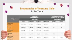 挂图Frequencies of Immune Cells in Rat Tissue Lists the estimated frequencies of more than 15 immune cell types in Sprague Dawley rats
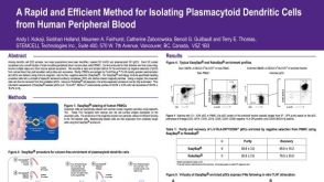 科学海报Isolation of Plasmacytoid Dendritic Cells from Peripheral Blood
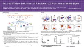 科学海报Fast and Efficient Enrichment of Functional ILC2 From Human Whole Blood


 沪公网安备31010102008431号