
 EasySep™小鼠TIL(CD45)正选试剂盒


搜索结果: 'EasySep Direct'
-

产品类型:
产品号#:
05150
产品名:
MyeloCult™H5100
-
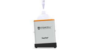
 科学海报Easy 250 EasySep™ Magnet: A Novel Magnetic Platform for Large-Volume Cell Isolation
产品类型:
产品号#:
产品名:
-
 3:04
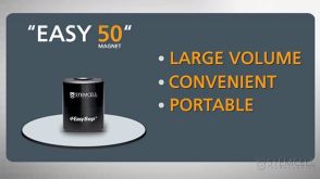
视频The Easy 50 EasySep™ Magnet: Portable Large-Volume Cell Separation In 25 Minutes发布日期: 10/10/2010
3:04
视频The Easy 50 EasySep™ Magnet: Portable Large-Volume Cell Separation In 25 Minutes发布日期: 10/10/2010 -
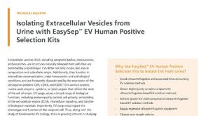 技术公告Isolating Extracellular Vesicles from Urine with EasySep™ EV Human Positive Selection Kits
产品类型:
细胞类型:
其他细胞系
产品号#:
产品名:
发布日期: 01/01/2024


 沪公网安备31010102008431号