S. Bhatia et al. (may 2019)
Cancer research 79 10 2722--2735
Inhibition of EphB4-Ephrin-B2 Signaling Reprograms the Tumor Immune Microenvironment in Head and Neck Cancers.
Identifying targets present in the tumor microenvironment that contribute to immune evasion has become an important area of research. In this study,we identified EphB4-ephrin-B2 signaling as a regulator of both innate and adaptive components of the immune system. EphB4 belongs to receptor tyrosine kinase family that interacts with ephrin-B2 ligand at sites of cell-cell contact,resulting in bidirectional signaling. We found that EphB4-ephrin-B2 inhibition alone or in combination with radiation (RT) reduced intratumoral regulatory T cells (Tregs) and increased activation of both CD8+ and CD4+Foxp3- T cells compared with the control group in an orthotopic head and neck squamous cell carcinoma (HNSCC) model. We also compared the effect of EphB4-ephrin-B2 inhibition combined with RT with combined anti-PDL1 and RT and observed similar tumor growth suppression,particularly at early time-points. A patient-derived xenograft model showed reduction of tumor-associated M2 macrophages and favored polarization towards an antitumoral M1 phenotype following EphB4-ephrin-B2 inhibition with RT. In vitro,EphB4 signaling inhibition decreased Ki67-expressing Tregs and Treg activation compared with the control group. Overall,our study is the first to implicate the role of EphB4-ephrin-B2 in tumor immune response. Moreover,our findings suggest that EphB4-ephrin-B2 inhibition combined with RT represents a potential alternative for patients with HNSCC and could be particularly beneficial for patients who are ineligible to receive or cannot tolerate anti-PDL1 therapy. SIGNIFICANCE: These findings present EphB4-ephrin-B2 inhibition as an alternative to anti-PDL1 therapeutics that can be used in combination with radiation to induce an effective antitumor immune response in patients with HNSCC.
View Publication
产品类型:
产品号#:
17952
17952RF
100-0696
产品名:
EasySep™人CD4+ T细胞分选试剂盒
RoboSep™ 人CD4+ T细胞分选试剂盒
EasySep™人CD4+ T细胞分离试剂盒
B. L. Jamison et al. (jul 2019)
Journal of immunology (Baltimore,Md. : 1950) 203 1 48--57
Nanoparticles Containing an Insulin-ChgA Hybrid Peptide Protect from Transfer of Autoimmune Diabetes by Shifting the Balance between Effector T Cells and Regulatory T Cells.
CD4 T cells play a critical role in promoting the development of autoimmunity in type 1 diabetes. The diabetogenic CD4 T cell clone BDC-2.5,originally isolated from a NOD mouse,has been widely used to study the contribution of autoreactive CD4 T cells and relevant Ags to autoimmune diabetes. Recent work from our laboratory has shown that the Ag for BDC-2.5 T cells is a hybrid insulin peptide (2.5HIP) consisting of an insulin C-peptide fragment fused to a peptide from chromogranin A (ChgA) and that endogenous 2.5HIP-reactive T cells are major contributors to autoimmune pathology in NOD mice. The objective of this study was to determine if poly(lactide-co-glycolide) (PLG) nanoparticles (NPs) loaded with the 2.5HIP Ag (2.5HIP-coupled PLG NPs) can tolerize BDC-2.5 T cells. Infusion of 2.5HIP-coupled PLG NPs was found to prevent diabetes in an adoptive transfer model by impairing the ability of BDC-2.5 T cells to produce proinflammatory cytokines through induction of anergy,leading to an increase in the ratio of Foxp3+ regulatory T cells to IFN-gamma+ effector T cells. To our knowledge,this work is the first to use a hybrid insulin peptide,or any neoepitope,to re-educate diabetogenic T cells and may have significant implications for the development of an Ag-specific therapy for type 1 diabetes patients.
View Publication
产品类型:
产品号#:
19852
19852RF
18783
18783RF
18765
18765RF
产品名:
EasySep™小鼠CD4+ T细胞分选试剂盒
RoboSep™ 小鼠CD4+ T细胞分选试剂盒
EasySep™ 小鼠CD4+CD25+调节性T细胞分选试剂盒 II
RoboSep™ 小鼠CD4+CD25+调节性T细胞分选试剂盒II
EasySep™ 小鼠CD4+CD62L+ T细胞分选试剂盒
RoboSep™ 小鼠CD4+ CD62L+ T细胞分选试剂盒
R. J. Komban et al. ( 2019)
Nature communications 10 1 2423
Activated Peyer's patch B cells sample antigen directly from M cells in the subepithelial dome.
The germinal center (GC) reaction in Peyer's patches (PP) requires continuous access to antigens,but how this is achieved is not known. Here we show that activated antigen-specific CCR6+CCR1+GL7- B cells make close contact with M cells in the subepithelial dome (SED). Using in situ photoactivation analysis of antigen-specific SED B cells,we find migration of cells towards the GC. Following antigen injection into ligated intestinal loops containing PPs,40{\%} of antigen-specific SED B cells bind antigen within 2 h,whereas unspecifc cells do not,indicating B cell-receptor involvment. Antigen-loading is not observed in M cell-deficient mice,but is unperturbed in mice depleted of classical dendritic cells (DC). Thus,we report a M cell-B cell antigen-specific transporting pathway in PP that is independent of DC. We propose that this antigen transporting pathway has a critical role in gut IgA responses,and should be taken into account when developing mucosal vaccines.
View Publication
产品类型:
产品号#:
19854
19854RF
产品名:
EasySep™小鼠B细胞分选试剂盒
RoboSep™ 小鼠B细胞分选试剂盒
S. Omenetti et al. (jun 2019)
Immunity
The Intestine Harbors Functionally Distinct Homeostatic Tissue-Resident and Inflammatory Th17 Cells.
T helper 17 (Th17) cells are pathogenic in many inflammatory diseases,but also support the integrity of the intestinal barrier in a non-inflammatory manner. It is unclear what distinguishes inflammatory Th17 cells elicited by pathogens and tissue-resident homeostatic Th17 cells elicited by commensals. Here,we compared the characteristics of Th17 cells differentiating in response to commensal bacteria (SFB) to those differentiating in response to a pathogen (Citrobacter rodentium). Homeostatic Th17 cells exhibited little plasticity towards expression of inflammatory cytokines,were characterized by a metabolism typical of quiescent or memory T cells,and did not participate in inflammatory processes. In contrast,infection-induced Th17 cells showed extensive plasticity towards pro-inflammatory cytokines,disseminated widely into the periphery,and engaged aerobic glycolysis in addition to oxidative phosphorylation typical for inflammatory effector cells. These findings will help ensure that future therapies directed against inflammatory Th17 cells do not inadvertently damage the resident gut population.
View Publication
产品类型:
产品号#:
19752
19752RF
19052
19052RF
产品名:
EasySep™人CD4+ T细胞富集试剂盒
RoboSep™ 人CD4+ T细胞富集试剂盒含滤芯吸头
C. Onyilagha et al. (jun 2019)
Journal of immunology (Baltimore,Md. : 1950)
NK Cells Are Critical for Optimal Immunity to Experimental Trypanosoma congolense Infection.
NK cells are key innate immune cells that play critical roles in host defense. Although NK cells have been shown to regulate immunity to some infectious diseases,their role in immunity to Trypanosoma congolense has not been investigated. NK cells are vital sources of IFN-gamma and TNF-alpha; two key cytokines that are known to play important roles in resistance to African trypanosomes. In this article,we show that infection with T. congolense leads to increased levels of activated and functional NK cells in multiple tissue compartments. Systemic depletion of NK cells with anti-NK1.1 mAb led to increased parasitemia,which was accompanied by significant reduction in IFN-gamma production by immune cells in the spleens and liver of infected mice. Strikingly,infected NFIL3-/- mice (which genetically lack NK cell development and function) on the normally resistant background were highly susceptible to T. congolense infection. These mice developed fulminating and uncontrolled parasitemia and died significantly earlier (13 ± 1 d) than their wild-type control mice (106 ± 26 d). The enhanced susceptibility of NFIL3-/- mice to infection was accompanied by significantly impaired cytokine (IFN-gamma and TNF-alpha) response by CD3+ T cells in the spleens and liver. Adoptive transfer of NK cells into NFIL3-/- mice before infection rescued them from acute death in a perforin-dependent manner. Collectively,these studies show that NK cells are critical for optimal resistance to T. congolense,and its deficiency leads to enhanced susceptibility in infected mice.
View Publication
产品类型:
产品号#:
19855
19855RF
产品名:
EasySep™小鼠NK细胞分选试剂盒
RoboSep™ 小鼠NK细胞分选试剂盒
Z. Yan et al. (apr 2019)
JCI insight 5
Deficiency of Socs3 leads to brain-targeted EAE via enhanced neutrophil activation and ROS production.
Dysregulation of the JAK/STAT signaling pathway is associated with Multiple Sclerosis (MS) and its mouse model,Experimental Autoimmune Encephalomyelitis (EAE). Suppressors Of Cytokine Signaling (SOCS) negatively regulate the JAK/STAT pathway. We previously reported a severe,brain-targeted,atypical form of EAE in mice lacking Socs3 in myeloid cells (Socs3DeltaLysM),which is associated with cerebellar neutrophil infiltration. There is emerging evidence that neutrophils are detrimental in the pathology of MS/EAE,however,their exact function is unclear. Here we demonstrate that neutrophils from the cerebellum of Socs3DeltaLysM mice show a hyper-activated phenotype with excessive production of reactive oxygen species (ROS) at the peak of EAE. Neutralization of ROS in vivo delayed the onset and reduced severity of atypical EAE. Mechanistically,Socs3-deficient neutrophils exhibit enhanced STAT3 activation,a hyper-activated phenotype in response to G-CSF,and upon G-CSF priming,increased ROS production. Neutralization of G-CSF in vivo significantly reduced the incidence and severity of the atypical EAE phenotype. Overall,our work elucidates that hypersensitivity of G-CSF/STAT3 signaling in Socs3DeltaLysM mice leads to atypical EAE by enhanced neutrophil activation and increased oxidative stress,which may explain the detrimental role of G-CSF in MS patients.
View Publication
产品类型:
产品号#:
19762
19762RF
产品名:
EasySep™小鼠中性粒细胞富集试剂盒
RoboSep™ 小鼠中性粒细胞富集试剂盒含滤芯吸头
C. Yang et al. (may 2019)
The Journal of experimental medicine 216 5 1182--1198
Thyrotropin aggravates atherosclerosis by promoting macrophage inflammation in plaques.
Subclinical hypothyroidism is associated with cardiovascular diseases,yet the underlying mechanism remains largely unknown. Herein,in a common population (n = 1,103),TSH level was found to be independently correlated with both carotid plaque prevalence and intima-media thickness. Consistently,TSH receptor ablation in ApoE-/- mice attenuated atherogenesis,accompanied by decreased vascular inflammation and macrophage burden in atherosclerotic plaques. These results were also observed in myeloid-specific Tshr-deficient ApoE-/- mice,which indicated macrophages to be a critical target of the proinflammatory and atherogenic effects of TSH. In vitro experiments further revealed that TSH activated MAPKs (ERK1/2,p38alpha,and JNK) and IkappaB/p65 pathways in macrophages and increased inflammatory cytokine production and their recruitment of monocytes. Thus,the present study has elucidated the new mechanisms by which TSH,as an independent risk factor of atherosclerosis,aggravates vascular inflammation and contributes to atherogenesis.
View Publication
Liu J et al. (NOV 2015)
Nature Protocols 10 11 1842--59
Efficient delivery of nuclease proteins for genome editing in human stem cells and primary cells.
Targeted nucleases,including zinc-finger nucleases (ZFNs),transcription activator-like (TAL) effector nucleases (TALENs) and clustered regularly interspaced short palindromic repeat (CRISPR)/CRISPR-associated protein 9 (Cas9),have provided researchers with the ability to manipulate nearly any genomic sequence in human cells and model organisms. However,realizing the full potential of these genome-modifying technologies requires their safe and efficient delivery into relevant cell types. Unlike methods that rely on expression from nucleic acids,the direct delivery of nuclease proteins to cells provides rapid action and fast turnover,leading to fewer off-target effects while maintaining high rates of targeted modification. These features make nuclease protein delivery particularly well suited for precision genome engineering. Here we describe procedures for implementing protein-based genome editing in human embryonic stem cells and primary cells. Protocols for the expression,purification and delivery of ZFN proteins,which are intrinsically cell-permeable; TALEN proteins,which can be internalized via conjugation with cell-penetrating peptide moieties; and Cas9 ribonucleoprotein,whose nucleofection into cells facilitates rapid induction of multiplexed modifications,are described,along with procedures for evaluating nuclease protein activity. Once they are constructed,nuclease proteins can be expressed and purified within 6 d,and they can be used to induce genomic modifications in human cells within 2 d.
View Publication
产品类型:
产品号#:
05850
05857
05870
05875
07920
17952
17952RF
19052
19052RF
18000
15470
15450
15420
15460
15425
15465
15430
15415
85850
85857
85870
85875
100-0696
07922
产品名:
ACCUTASE™
EasySep™人CD4+ T细胞分选试剂盒
RoboSep™ 人CD4+ T细胞分选试剂盒
EasySep™人CD4+ T细胞富集试剂盒
RoboSep™ 人CD4+ T细胞富集试剂盒含滤芯吸头
EasySep™磁极
mTeSR™1
mTeSR™1
EasySep™人CD4+ T细胞分离试剂盒
ACCUTASE™
Baker RL et al. (JAN 2016)
Journal of Immunology 196 1 39--43
Cutting Edge: Nonobese Diabetic Mice Deficient in Chromogranin A Are Protected from Autoimmune Diabetes.
T cells reactive to β cell Ags are critical players in the development of autoimmune type 1 diabetes. Using a panel of diabetogenic CD4 T cell clones derived from the NOD mouse,we recently identified the β cell secretory granule protein,chromogranin A (ChgA),as a new autoantigen in type 1 diabetes. CD4 T cells reactive to ChgA are pathogenic and rapidly transfer diabetes into young NOD recipients. We report in this article that NOD.ChgA(-/-) mice do not develop diabetes and show little evidence of autoimmunity in the pancreatic islets. Using tetramer analysis,we demonstrate that ChgA-reactive T cells are present in these mice but remain naive. In contrast,in NOD.ChgA(+/+) mice,a majority of the ChgA-reactive T cells are Ag experienced. Our results suggest that the presence of ChgA and subsequent activation of ChgA-reactive T cells are essential for the initiation and development of autoimmune diabetes in NOD mice.
View Publication
产品类型:
产品号#:
19852
19852RF
产品名:
EasySep™小鼠CD4+ T细胞分选试剂盒
RoboSep™ 小鼠CD4+ T细胞分选试剂盒
Cao Y et al. (MAR 2016)
Journal of Immunology 196 5 2075--84
Autoreactive T Cells from Patients with Myasthenia Gravis Are Characterized by Elevated IL-17, IFN-γ, and GM-CSF and Diminished IL-10 Production.
Myasthenia gravis (MG) is a prototypical autoimmune disease that is among the few for which the target Ag and the pathogenic autoantibodies are clearly defined. The pathology of the disease is affected by autoantibodies directed toward the acetylcholine receptor (AChR). Mature,Ag-experienced B cells rely on the action of Th cells to produce these pathogenic Abs. The phenotype of the MG Ag-reactive T cell compartment is not well defined; thus,we sought to determine whether such cells exhibit both a proinflammatory and a pathogenic phenotype. A novel T cell library assay that affords multiparameter interrogation of rare Ag-reactive CD4(+) T cells was applied. Proliferation and cytokine production in response to both AChR and control Ags were measured from 3120 T cell libraries derived from 11 MG patients and paired healthy control subjects. The frequency of CCR6(+) memory T cells from MG patients proliferating in response to AChR-derived peptides was significantly higher than that of healthy control subjects. Production of both IFN-γ and IL-17,in response to AChR,was also restricted to the CCR6(+) memory T cell compartment in the MG cohort,indicating a proinflammatory phenotype. These T cells also included an elevated expression of GM-CSF and absence of IL-10 expression,indicating a proinflammatory and pathogenic phenotype. This component of the autoimmune response in MG is of particular importance when considering the durability of MG treatment strategies that eliminate B cells,because the autoreactive T cells could renew autoimmunity in the reconstituted B cell compartment with ensuing clinical manifestations.
View Publication
产品类型:
产品号#:
17952
17952RF
100-0696
产品名:
EasySep™人CD4+ T细胞分选试剂盒
RoboSep™ 人CD4+ T细胞分选试剂盒
EasySep™人CD4+ T细胞分离试剂盒
Krummey SM et al. (MAR 2016)
Journal of Immunology 196 6 2838--46
Low-Affinity Memory CD8+ T Cells Mediate Robust Heterologous Immunity.
Heterologous immunity is recognized as a significant barrier to transplant tolerance. Whereas it has been established that pathogen-elicited memory T cells can have high or low affinity for cross-reactive allogeneic peptide-MHC,the role of TCR affinity during heterologous immunity has not been explored. We established a model with which to investigate the impact of TCR-priming affinity on memory T cell populations following a graft rechallenge. In contrast to high-affinity priming,low-affinity priming elicited fully differentiated memory T cells with a CD45RB(hi) status. High CD45RB status enabled robust secondary responses in vivo,as demonstrated by faster graft rejection kinetics and greater proliferative responses. CD45RB blockade prolonged graft survival in low affinity-primed mice,but not in high affinity-primed mice. Mechanistically,low affinity-primed memory CD8(+) T cells produced more IL-2 and significantly upregulated IL-2Rα expression during rechallenge. We found that CD45RB(hi) status was also a stable marker of priming affinity within polyclonal CD8(+) T cell populations. Following high-affinity rechallenge,low affinity-primed CD45RB(hi) cells became CD45RB(lo),demonstrating that CD45RB status acts as an affinity-based differentiation switch on CD8(+) T cells. Thus,these data establish a novel mechanism by which CD45 isoforms tune low affinity-primed memory CD8(+) T cells to become potent secondary effectors following heterologous rechallenge. These findings have direct implications for allogeneic heterologous immunity by demonstrating that despite a lower precursor frequency,low-affinity priming is sufficient to generate memory cells that mediate potent secondary responses against a cross-reactive graft challenge.
View Publication

 EasySep™小鼠TIL(CD45)正选试剂盒


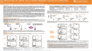 科学海报Depletion of Dead Cells from Primary Tissue in 6 Minutes
 沪公网安备31010102008431号