Inhibition of angiotensin I-converting enzyme induces radioprotection by preserving murine hematopoietic short-term reconstituting cells.
Angiotensin I-converting enzyme (ACE) inhibitors can affect hematopoiesis by several mechanisms including inhibition of angiotensin II formation and increasing plasma concentrations of AcSDKP (acetyl-N-Ser-Asp-Lys-Pro),an ACE substrate and a negative regulator of hematopoiesis. We tested whether ACE inhibition could decrease the hematopoietic toxicity of lethal or sublethal irradiation protocols. In all cases,short treatment with the ACE inhibitor perindopril protected against irradiation-induced death. ACE inhibition accelerated hematopoietic recovery and led to a significant increase in platelet and red cell counts. Pretreatment with perindopril increased bone marrow cellularity and the number of hematopoietic progenitors (granulocyte macrophage colony-forming unit [CFU-GM],erythroid burst-forming unit [BFU-E],and megakaryocyte colony-forming unit [CFU-MK]) from day 7 to 28 after irradiation. Perindopril also increased the number of hematopoietic stem cells with at least a short-term reconstitutive activity in animals that recovered from irradiation. To determine the mechanism of action involved,we evaluated the effects of increasing AcSDKP plasma concentrations and of an angiotensin II type 1 (AT1) receptor antagonist (telmisartan) on radioprotection. We found that the AT1-receptor antagonism mediated similar radioprotection as the ACE inhibitor. These results suggest that ACE inhibitors and AT1-receptor antagonists could be used to decrease the hematopoietic toxicity of irradiation.
View Publication
产品类型:
产品号#:
03134
产品名:
MethoCult™ M3134
Drowley L et al. (FEB 2016)
Stem cells translational medicine 5 2 164--74
Human Induced Pluripotent Stem Cell-Derived Cardiac Progenitor Cells in Phenotypic Screening: A Transforming Growth Factor-β Type 1 Receptor Kinase Inhibitor Induces Efficient Cardiac Differentiation.
Several progenitor cell populations have been reported to exist in hearts that play a role in cardiac turnover and/or repair. Despite the presence of cardiac stem and progenitor cells within the myocardium,functional repair of the heart after injury is inadequate. Identification of the signaling pathways involved in the expansion and differentiation of cardiac progenitor cells (CPCs) will broaden insight into the fundamental mechanisms playing a role in cardiac homeostasis and disease and might provide strategies for in vivo regenerative therapies. To understand and exploit cardiac ontogeny for drug discovery efforts,we developed an in vitro human induced pluripotent stem cell-derived CPC model system using a highly enriched population of KDR(pos)/CKIT(neg)/NKX2.5(pos) CPCs. Using this model system,these CPCs were capable of generating highly enriched cultures of cardiomyocytes under directed differentiation conditions. In order to facilitate the identification of pathways and targets involved in proliferation and differentiation of resident CPCs,we developed phenotypic screening assays. Screening paradigms for therapeutic applications require a robust,scalable,and consistent methodology. In the present study,we have demonstrated the suitability of these cells for medium to high-throughput screens to assess both proliferation and multilineage differentiation. Using this CPC model system and a small directed compound set,we identified activin-like kinase 5 (transforming growth factor-β type 1 receptor kinase) inhibitors as novel and potent inducers of human CPC differentiation to cardiomyocytes. Significance: Cardiac disease is a leading cause of morbidity and mortality,with no treatment available that can result in functional repair. This study demonstrates how differentiation of induced pluripotent stem cells can be used to identify and isolate cell populations of interest that can translate to the adult human heart. Two separate examples of phenotypic screens are discussed,demonstrating the value of this biologically relevant and reproducible technology. In addition,this assay system was able to identify novel and potent inducers of differentiation and proliferation of induced pluripotent stem cell-derived cardiac progenitor cells.
View Publication
产品类型:
产品号#:
70919
产品名:
Ma Z et al. (JUL 2015)
Nature communications 6 May 7413
Self-organizing human cardiac microchambers mediated by geometric confinement.
Tissue morphogenesis and organ formation are the consequences of biochemical and biophysical cues that lead to cellular spatial patterning in development. To model such events in vitro,we use PEG-patterned substrates to geometrically confine human pluripotent stem cell colonies and spatially present mechanical stress. Modulation of the WNT/β-catenin pathway promotes spatial patterning via geometric confinement of the cell condensation process during epithelial-mesenchymal transition,forcing cells at the perimeter to express an OCT4+ annulus,which is coincident with a region of higher cell density and E-cadherin expression. The biochemical and biophysical cues synergistically induce self-organizing lineage specification and creation of a beating human cardiac microchamber confined by the pattern geometry. These highly defined human cardiac microchambers can be used to study aspects of embryonic spatial patterning,early cardiac development and drug-induced developmental toxicity.
View Publication

 EasySep™小鼠TIL(CD45)正选试剂盒


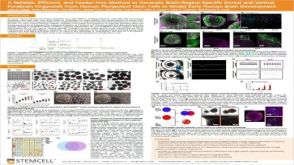 科学海报A Reliable, Efficient, and Feeder-Free Method to Generate Brain-Region-Specific Dorsal and Ventral Forebrain Organoids From Human Pluripotent Stem Cells to Model Early Human Brain Development
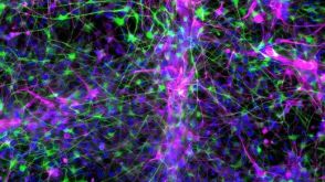 实验方案How to Co-Culture Astrocytes and NGN2 mRNA-Driven Induced Forebrain Neurons Derived from Human Pluripotent Stem Cells
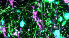 实验方案How to Tri-Culture Astrocytes, Microglia, and NGN2 mRNA-LNP-Induced Forebrain Neurons Derived from Human Pluripotent Stem Cells
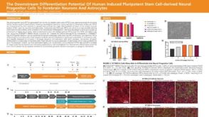 科学海报The Downstream Differentiation Potential of Human Induced Pluripotent Stem Cell-Derived Neural Progenitor Cells to Forebrain Neurons and Astrocytes
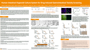 科学海报Human Intestinal Organoid Culture System for Drug-Induced Gastrointestinal Toxicity Screening
 31:49
线上讲座Using Human Pluripotent Stem Cell-Derived Neural Organoids for Disease Modeling发布日期: 08/01/2024
31:49
线上讲座Using Human Pluripotent Stem Cell-Derived Neural Organoids for Disease Modeling发布日期: 08/01/2024


 沪公网安备31010102008431号