Tareen SU et al. (MAR 2014)
Molecular therapy : the journal of the American Society of Gene Therapy 22 3 575--87
Design of a novel integration-deficient lentivector technology that incorporates genetic and posttranslational elements to target human dendritic cells.
As sentinels of the immune system,dendritic cells (DCs) play an essential role in regulating cellular immune responses. One of the main challenges of developing DC-targeted therapies includes the delivery of antigen to DCs in order to promote the activation of antigen-specific effector CD8 T cells. With the goal of creating antigen-directed immunotherapeutics that can be safely administered directly to patients,Immune Design has developed a platform of novel integration-deficient lentiviral vectors that target and deliver antigen-encoding nucleic acids to human DCs. This platform,termed ID-VP02,utilizes a novel genetic variant of a Sindbis virus envelope glycoprotein with posttranslational carbohydrate modifications in combination with Vpx,a SIVmac viral accessory protein,to achieve efficient targeting and transduction of human DCs. In addition,ID-VP02 incorporates safety features in its design that include two redundant mechanisms to render ID-VP02 integration-deficient. Here,we describe the characteristics that allow ID-VP02 to specifically transduce human DCs,and the advances that ID-VP02 brings to conventional third-generation lentiviral vector design as well as demonstrate upstream production yields that will enable manufacturing feasibility studies to be conducted.
View Publication
Vieillard V et al. (AUG 2005)
Proceedings of the National Academy of Sciences 102 31 10981--86
NK cytotoxicity against CD4+ T cells during HIV-1 infection: A gp41 peptide induces the expression of an NKp44 ligand
HIV infection leads to a state of chronic immune activation and progressive deterioration in immune function,manifested most recognizably by the progressive depletion of CD4+ T cells. A substantial percentage of natural killer (NK) cells from patients with HIV infection are activated and express the natural cytotoxicity receptor (NCR) NKp44. Here we show that a cellular ligand for NKp44 (NKp44L) is expressed during HIV-1 infection and is correlated with both the progression of CD4+ T cell depletion and the increase of viral load. CD4+ T cells expressing this ligand are highly sensitive to the NK lysis activity mediated by NKp44+ NK cells. The expression of NKp44L is induced by the linear motif NH2-SWSNKS-COOH of the HIV-1 envelope gp41 protein. This highly conserved motif appears critical to the sharp increase in NK lysis of CD4+ T cells from HIV-infected patients. These studies strongly suggest that induction of NKp44L plays a key role in the lysis of CD4+ T cells by activated NK cells in HIV infection and consequently provide a framework for considering how HIV-1 may use NK cell immune surveillance to trigger CD4+ T cells. Understanding this mechanism may help to develop future therapeutic strategies and vaccines against HIV-1 infection.
View Publication
产品类型:
产品号#:
03800
03801
03802
03803
03804
03805
03806
05150
15021
15061
产品名:
ClonaCell™-HY杂交瘤试剂盒
ClonaCell™-HY培养基A
ClonaCell™-HY 培养基 B
ClonaCell™-HY 培养基 C
ClonaCell™-HY 培养基 D
ClonaCell™-HY 培养基 E
ClonaCell™-HY PEG
MyeloCult™ H5100
RosetteSep™人T细胞富集抗体混合物
RosetteSep™人T细胞富集抗体混合物
Schiedlmeier B et al. (MAR 2003)
Blood 101 5 1759--68
High-level ectopic HOXB4 expression confers a profound in vivo competitive growth advantage on human cord blood CD34+ cells, but impairs lymphomyeloid differentiation.
Ectopic retroviral expression of homeobox B4 (HOXB4) causes an accelerated and enhanced regeneration of murine hematopoietic stem cells (HSCs) and is not known to compromise any program of lineage differentiation. However,HOXB4 expression levels for expansion of human stem cells have still to be established. To test the proposed hypothesis that HOXB4 could become a prime tool for in vivo expansion of genetically modified human HSCs,we retrovirally overexpressed HOXB4 in purified cord blood (CB) CD34+ cells together with green fluorescent protein (GFP) as a reporter protein,and evaluated the impact of ectopic HOXB4 expression on proliferation and differentiation in vitro and in vivo. When injected separately into nonobese diabetic-severe combined immunodeficient (NOD/SCID) mice or in competition with control vector-transduced cells,HOXB4-overexpressing cord blood CD34+ cells had a selective growth advantage in vivo,which resulted in a marked enhancement of the primitive CD34+ subpopulation (P =.01). However,high HOXB4 expression substantially impaired the myeloerythroid differentiation program,and this was reflected in a severe reduction of erythroid and myeloid progenitors in vitro (P textless.03) and in vivo (P =.01). Furthermore,HOXB4 overexpression also significantly reduced B-cell output (P textless.01). These results show for the first time unwanted side effects of ectopic HOXB4 expression and therefore underscore the need to carefully determine the therapeutic window of HOXB4 expression levels before initializing clinical trials.
View Publication
Ketola K et al. (DEC 2010)
Molecular cancer therapeutics 9 12 3175--85
Monensin is a potent inducer of oxidative stress and inhibitor of androgen signaling leading to apoptosis in prostate cancer cells.
Current treatment options for advanced and hormone refractory prostate cancer are limited and responses to commonly used androgen pathway inhibitors are often unsatisfactory. Our recent results indicated that sodium ionophore monensin is one of the most potent and cancer-specific inhibitors in a systematic sensitivity testing of most known drugs and drug-like molecules in a panel of prostate cancer cell models. Because monensin has been extensively used in veterinary applications to build muscle mass in cattle,the link to prostate cancer and androgen signaling was particularly interesting. Here,we showed that monensin effects at nanomolar concentrations are linked to induction of apoptosis and potent reduction of androgen receptor mRNA and protein in prostate cancer cells. Monensin also elevated intracellular oxidative stress in prostate cancer cells as evidenced by increased generation of intracellular reactive oxygen species and by induction of a transcriptional profile characteristic of an oxidative stress response. Importantly,the antiproliferative effects of monensin were potentiated by combinatorial treatment with the antiandrogens and antagonized by antioxidant vitamin C. Taken together,our results suggest monensin as a potential well-tolerated,in vivo compatible drug with strong proapoptotic effects in prostate cancer cells,and synergistic effects with antiandrogens. Moreover,our data suggest a general strategy by which the effects of antiandrogens could be enhanced by combinatorial administration with agents that increase oxidative stress in prostate cancer cells.
View Publication
产品类型:
产品号#:
01700
01705
01702
产品名:
ALDEFLUOR™ 试剂盒
ALDEFLUOR™ DEAB试剂, 1.5 mM, 1 mL
ALDEFLUOR™检测缓冲液
Aichberger KJ et al. (DEC 2009)
Blood 114 26 5342--51
Identification of proapoptotic Bim as a tumor suppressor in neoplastic mast cells: role of KIT D816V and effects of various targeted drugs.
Systemic mastocytosis (SM) is a myeloid neoplasm involving mast cells (MCs) and their progenitors. In most cases,neoplastic cells display the D816V-mutated variant of KIT. KIT D816V exhibits constitutive tyrosine kinase (TK) activity and has been implicated in increased survival and growth of neoplastic MCs. Recent data suggest that the proapoptotic BH3-only death regulator Bim plays a role as a tumor suppressor in various myeloid neoplasms. We found that KIT D816V suppresses expression of Bim in Ba/F3 cells. The KIT D816-induced down-regulation of Bim was rescued by the KIT-targeting drug PKC412/midostaurin. Both PKC412 and the proteasome-inhibitor bortezomib were found to decrease growth and promote expression of Bim in MC leukemia cell lines HMC-1.1 (D816V negative) and HMC-1.2 (D816V positive). Both drugs were also found to counteract growth of primary neoplastic MCs. Furthermore,midostaurin was found to cooperate with bortezomib and with the BH3-mimetic obatoclax in producing growth inhibition in both HMC-1 subclones. Finally,a Bim-specific siRNA was found to rescue HMC-1 cells from PKC412-induced cell death. Our data show that KIT D816V suppresses expression of proapoptotic Bim in neoplastic MCs. Targeting of Bcl-2 family members by drugs promoting Bim (re)-expression,or by BH3-mimetics such as obatoclax,may be an attractive therapy concept in SM.
View Publication
产品类型:
产品号#:
09600
09650
产品名:
StemSpan™ SFEM
StemSpan™ SFEM
Secchiero P et al. (MAY 2006)
Blood 107 10 4122--9
Functional integrity of the p53-mediated apoptotic pathway induced by the nongenotoxic agent nutlin-3 in B-cell chronic lymphocytic leukemia (B-CLL).
Deletions and/or mutations of p53 are relatively rare and late events in the natural history of B-cell chronic lymphocytic leukemia (B-CLL). However,it is unknown whether p53 signaling is functional in B-CLL and if targeted nongenotoxic activation of the p53 pathway by using nutlin-3,a small molecule inhibitor of the p53/MDM2 interaction,is sufficient to kill B-CLL cells. In vitro treatment with nutlin-3 induced a significant cytotoxicity on primary CD19(+) B-CLL cells,but not on normal CD19(+) B lymphocytes,peripheral-blood mononuclear cells,or bone marrow hematopoietic progenitors. Among 29 B-CLL samples examined,only one was resistant to nutlin-3-mediated cytotoxicity. The induction of p53 by nutlin-3 in B-CLL samples was accompanied by alterations of the mitochondrial potential and activation of the caspase-dependent apoptotic pathway. Among several genes related to the p53 pathway,nutlin-3 up-regulated the steady-state mRNA levels of PCNA,CDKN1A/p21,GDF15,TNFRSF10B/TRAIL-R2,TP53I3/PIG3,and GADD45. This profile of gene activation showed a partial overlapping with that induced by the genotoxic drug fludarabine. Moreover,nutlin-3 synergized with both fludarabine and chlorambucil in inducing B-CLL apoptosis. Our data strongly suggest that nutlin-3 should be further investigated for clinical applications in the treatment of B-CLL.
View Publication
Clendening JW et al. (AUG 2010)
Proceedings of the National Academy of Sciences of the United States of America 107 34 15051--6
Dysregulation of the mevalonate pathway promotes transformation.
The importance of cancer metabolism has been appreciated for many years,but the intricacies of how metabolic pathways interconnect with oncogenic signaling are not fully understood. With a clear understanding of how metabolism contributes to tumorigenesis,we will be better able to integrate the targeting of these fundamental biochemical pathways into patient care. The mevalonate (MVA) pathway,paced by its rate-limiting enzyme,hydroxymethylglutaryl coenzyme A reductase (HMGCR),is required for the generation of several fundamental end-products including cholesterol and isoprenoids. Despite years of extensive research from the perspective of cardiovascular disease,the contribution of a dysregulated MVA pathway to human cancer remains largely unexplored. We address this issue directly by showing that dysregulation of the MVA pathway,achieved by ectopic expression of either full-length HMGCR or its novel splice variant,promotes transformation. Ectopic HMGCR accentuates growth of transformed and nontransformed cells under anchorage-independent conditions or as xenografts in immunocompromised mice and,importantly,cooperates with RAS to drive the transformation of primary mouse embryonic fibroblasts cells. We further explore whether the MVA pathway may play a role in the etiology of human cancers and show that high mRNA levels of HMGCR and additional MVA pathway genes correlate with poor prognosis in a meta-analysis of six microarray datasets of primary breast cancer. Taken together,our results suggest that HMGCR is a candidate metabolic oncogene and provide a molecular rationale for further exploring the statin family of HMGCR inhibitors as anticancer agents.
View Publication
Adaptive secretion of granulocyte-macrophage colony-stimulating factor (GM-CSF) mediates imatinib and nilotinib resistance in BCR/ABL+ progenitors via JAK-2/STAT-5 pathway activation.
Overcoming imatinib mesylate (IM) resistance and disease persistence in patients with chronic myeloid leukemia (CML) is of considerable importance to the issue of potential cure. Here we asked whether autocrine signaling contributes to survival of BCR/ABL+ cells in the presence of IM and nilotinib (NI; AMN107),a novel,more selective Abl inhibitor. Conditioned media (CM) of IM-resistant LAMA84 cell clones (R-CM) was found to substantially protect IM-naive LAMA cells and primary CML progenitors from IM- or NI-induced cell death. This was due to an increased secretion of the granulocyte-macrophage colony-stimulating factor (GM-CSF),which was identified as the causative factor mediating IM resistance in R-CM. GM-CSF elicited IM and NI drug resistance via a BCR/ABL-independent activation of the janus kinases 2 (JAK-2)/signal transducer and activator of transcription 5 (STAT-5) signaling pathway in GM-CSF receptor alpha receptor (CD116)-expressing cells,including primary CD34+/CD116+ GM progenitors (GMPs). Elevated mRNA and protein levels of GM-CSF were detected in IM-resistant patient samples,suggesting a contribution of GM-CSF secretion for IM and NI resistance in vivo. Importantly,inhibition of JAK-2 with AG490 abrogated GM-CSF-mediated STAT-5 phosphorylation and NI resistance in vitro. Together,adaptive autocrine secretion of GM-CSF mediates BCR/ABL-independent IM and NI resistance via activation of the antiapoptotic JAK-2/STAT-5 pathway. Inhibition of JAK-2 overcomes GM-CSF-induced IM and NI progenitor cell resistance,providing a rationale for the application of JAK-2 inhibitors to eradicate residual disease in CML.
View Publication

 EasySep™小鼠TIL(CD45)正选试剂盒


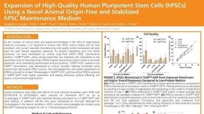 科学海报Expansion of High-Quality Human Pluripotent Stem Cells (hPSCs) Using a Novel Animal Origin-Free and Stabilized hPSC Maintenance Medium
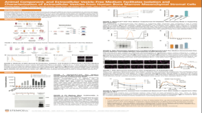 科学海报Animal Component- and Extracellular Vesicle-Free Medium Facilitates Isolation and Characterization of Extracellular Vesicles from Human Bone Marrow Mesenchymal Stromal Cells

 沪公网安备31010102008431号