Watson CL et al. (NOV 2014)
Nature Medicine 20 11 1310--4
An in vivo model of human small intestine using pluripotent stem cells.
Differentiation of human pluripotent stem cells (hPSCs) into organ-specific subtypes offers an exciting avenue for the study of embryonic development and disease processes,for pharmacologic studies and as a potential resource for therapeutic transplant. To date,limited in vivo models exist for human intestine,all of which are dependent upon primary epithelial cultures or digested tissue from surgical biopsies that include mesenchymal cells transplanted on biodegradable scaffolds. Here,we generated human intestinal organoids (HIOs) produced in vitro from human embryonic stem cells (ESCs) or induced pluripotent stem cells (iPSCs) that can engraft in vivo. These HIOs form mature human intestinal epithelium with intestinal stem cells contributing to the crypt-villus architecture and a laminated human mesenchyme,both supported by mouse vasculature ingrowth. In vivo transplantation resulted in marked expansion and maturation of the epithelium and mesenchyme,as demonstrated by differentiated intestinal cell lineages (enterocytes,goblet cells,Paneth cells,tuft cells and enteroendocrine cells),presence of functional brush-border enzymes (lactase,sucrase-isomaltase and dipeptidyl peptidase 4) and visible subepithelial and smooth muscle layers when compared with HIOs in vitro. Transplanted intestinal tissues demonstrated digestive functions as shown by permeability and peptide uptake studies. Furthermore,transplanted HIO-derived tissue was responsive to systemic signals from the host mouse following ileocecal resection,suggesting a role for circulating factors in the intestinal adaptive response. This model of the human small intestine may pave the way for studies of intestinal physiology,disease and translational studies.
View Publication
产品类型:
产品号#:
05854
05855
05850
05857
05870
05875
85850
85857
85870
85875
产品名:
mFreSR™
mFreSR™
mTeSR™1
mTeSR™1
Zielske SP et al. (NOV 2003)
The Journal of clinical investigation 112 10 1561--70
In vivo selection of MGMT(P140K) lentivirus-transduced human NOD/SCID repopulating cells without pretransplant irradiation conditioning.
Infusion of transduced hematopoietic stem cells into nonmyeloablated hosts results in ineffective in vivo levels of transduced cells. To increase the proportion of transduced cells in vivo,selection based on P140K O6-methylguanine-DNA-methyltransferase (MGMT[P140K]) gene transduction and O6-benzylguanine/1,3-bis(2-chloroethyl)-1-nitrosourea (BG/BCNU) treatment has been devised. In this study,we transduced human NOD/SCID repopulating cells (SRCs) with MGMT(P140K) using a lentiviral vector and infused them into BG/BCNU-conditioned NOD/SCID mice before rounds of BG/BCNU treatment as a model for in vivo selection. Engraftment was not observed until the second round of BG/BCNU treatment,at which time human cells emerged to compose up to 20% of the bone marrow. Furthermore,99% of human CFCs derived from NOD/SCID mice were positive for provirus as measured by PCR,compared with 35% before transplant and 11% in untreated irradiation-preconditioned mice,demonstrating selection. Bone marrow showed BG-resistant O6-alkylguanine-DNA-alkyltransferase (AGT) activity,and CFUs were stained intensely for AGT protein,indicating high transgene expression. Real-time PCR estimates of the number of proviral insertions in individual CFUs ranged from 3 to 22. Selection resulted in expansion of one or more SRC clones containing similar numbers of proviral copies per mouse. To our knowledge,these results provide the first evidence of potent in vivo selection of MGMT(P140K) lentivirus-transduced human SRCs following BG/BCNU treatment.
View Publication
Adherent cells generated during long-term culture of human umbilical cord blood CD34+ cells have characteristics of endothelial cells and beneficial effect on cord blood ex vivo expansion.
Hematopoiesis depends on the association of hematopoietic stem cells with stromal cells that constitute the hematopoietic microenvironment. The in vitro development of the endothelial cell from umbilical cord blood (UCB) is not well established and has met very limited success. In this study,UCB CD34(+) cells were cultured for 5 weeks in a stroma-free liquid culture system using thrombopoietin,flt3 ligand,and granulocyte-colony stimulating factor. By week 4-5,we found that firmly adherent fibroblast-like cells were established. These cells showed characteristics of endothelial cells expressing von Willebrand factor,human vascular cell adhesion molecule-1,human intracellular adhesion molecule-1,human CD31,E-selectin,and human macrophage. Furthermore,when comparing an ex vivo system without an established endothelial monolayer to an ex vivo system with an established endothelial monolayer,better expansion of total nucleated cells,CD34(+) cells,and colony-forming units (CFUs)-granulocyte-macrophage and CFUs-granulocyte-erythroid-megakaryocyte-macrophage were found during culture. This phenomenon was in part due to the fact that a significant reduction of apoptotic fractions was found in the CD34(+) cells,which were cultured on the adherent monolayer for up to 5 weeks. To gather quantitative data on the number of endothelial cells derived from a given number of CD34 cells,we performed limiting dilution assay by using Poisson distribution: the number of tested cells (linear scale) producing a 37% negative culture (logarithmic scale) is the number of cells containing one endothelial cell. By this method,one endothelial cell may be found from 314 CD34(+) cells after 5 weeks of culture. These results suggest that the UCB CD34(+) cell fraction contains endothelial cell precursors,establishing the hematopoietic microenvironment and providing the beneficial effects through downregulating apoptosis on UCB expansion protocols. These observations may provide insight for future cellular therapy or graft engineering.
View Publication
产品类型:
产品号#:
04434
04444
产品名:
MethoCult™H4434经典
MethoCult™H4434经典
Gilpin SE et al. (NOV 2014)
The Annals of thoracic surgery 98 5 1721--------9; discussion 1729
Enhanced lung epithelial specification of human induced pluripotent stem cells on decellularized lung matrix.
BACKGROUND Whole-lung scaffolds can be created by perfusion decellularization of cadaveric donor lungs. The resulting matrices can then be recellularized to regenerate functional organs. This study evaluated the capacity of acellular lung scaffolds to support recellularization with lung progenitors derived from human induced pluripotent stem cells (iPSCs). METHODS Whole rat and human lungs were decellularized by constant-pressure perfusion with 0.1% sodium dodecyl sulfate solution. Resulting lung scaffolds were cryosectioned into slices or left intact. Human iPSCs were differentiated to definitive endoderm,anteriorized to a foregut fate,and then ventralized to a population expressing NK2 homeobox 1 (Nkx2.1). Cells were seeded onto slices and whole lungs,which were maintained under constant perfusion biomimetic culture. Lineage specification was assessed by quantitative polymerase chain reaction and immunofluorescent staining. Regenerated left lungs were transplanted in an orthotopic position. RESULTS Activin-A treatment,followed by transforming growth factor-$\$,induced differentiation of human iPSCs to anterior foregut endoderm as confirmed by forkhead box protein A2 (FOXA2),SRY (Sex Determining Region Y)-Box 17 (SOX17),and SOX2 expression. Cells cultured on decellularized lung slices demonstrated proliferation and lineage commitment after 5 days. Cells expressing Nkx2.1 were identified at 40% to 60% efficiency. Within whole-lung scaffolds and under perfusion culture,cells further upregulated Nkx2.1 expression. After orthotopic transplantation,grafts were perfused and ventilated by host vasculature and airways. CONCLUSIONS Decellularized lung matrix supports the culture and lineage commitment of human iPSC-derived lung progenitor cells. Whole-organ scaffolds and biomimetic culture enable coseeding of iPSC-derived endothelial and epithelial progenitors and enhance early lung fate. Orthotopic transplantation may enable further in vivo graft maturation.
View Publication
Generation of GFAP::GFP astrocyte reporter lines from human adult fibroblast-derived iPS cells using zinc-finger nuclease technology.
Astrocytes are instrumental to major brain functions,including metabolic support,extracellular ion regulation,the shaping of excitatory signaling events and maintenance of synaptic glutamate homeostasis. Astrocyte dysfunction contributes to numerous developmental,psychiatric and neurodegenerative disorders. The generation of adult human fibroblast-derived induced pluripotent stem cells (iPSCs) has provided novel opportunities to study mechanisms of astrocyte dysfunction in human-derived cells. To overcome the difficulties of cell type heterogeneity during the differentiation process from iPSCs to astroglial cells (iPS astrocytes),we generated homogenous populations of iPS astrocytes using zinc-finger nuclease (ZFN) technology. Enhanced green fluorescent protein (eGFP) driven by the astrocyte-specific glial fibrillary acidic protein (GFAP) promoter was inserted into the safe harbor adeno-associated virus integration site 1 (AAVS1) locus in disease and control-derived iPSCs. Astrocyte populations were enriched using Fluorescence Activated Cell Sorting (FACS) and after enrichment more than 99% of iPS astrocytes expressed mature astrocyte markers including GFAP,S100$\$,NFIA and ALDH1L1. In addition,mature pure GFP-iPS astrocytes exhibited a well-described functional astrocytic activity in vitro characterized by neuron-dependent regulation of glutamate transporters to regulate extracellular glutamate concentrations. Engraftment of GFP-iPS astrocytes into rat spinal cord grey matter confirmed in vivo cell survival and continued astrocytic maturation. In conclusion,the generation of GFAP::GFP-iPS astrocytes provides a powerful in vitro and in vivo tool for studying astrocyte biology and astrocyte-driven disease pathogenesis and therapy.
View Publication
产品类型:
产品号#:
05850
05857
05870
05875
85850
85857
85870
85875
05835
05839
08581
08582
产品名:
mTeSR™1
mTeSR™1
STEMdiff™ 神经诱导培养基
STEMdiff™ 神经诱导培养基
STEMdiff™SMADi神经诱导试剂盒
STEMdiff™SMADi神经诱导试剂盒,2套
Seeger FH et al. (MAR 2007)
European heart journal 28 6 766--72
Cell isolation procedures matter: a comparison of different isolation protocols of bone marrow mononuclear cells used for cell therapy in patients with acute myocardial infarction.
AIM: The recently published REPAIR-AMI and ASTAMI trial showed differences in contractile recovery of left ventricular function after infusion of bone marrow-derived cells in acute myocardial infarction. Since the trials used different protocols for cell isolation and storage (REPAIR-AMI: Ficoll,storage in X-vivo 10 medium plus serum; ASTAMI: Lymphoprep,storage in NaCl plus plasma),we compared the functional activity of BMC isolated by the two different protocols. METHODS AND RESULTS: The recovery of total cell number,colony-forming units (CFU),and the number of mesenchymal stem cells were significantly reduced to 77 +/- 4%,83 +/- 16%,and 65 +/- 15%,respectively,when using the ASTAMI protocol compared with the REPAIR protocol. The capacity of the isolated BMC to migrate in response to stromal cell-derived factor 1 (SDF-1) was profoundly reduced when using the ASTAMI cell isolation procedure (42 +/- 8% and 78 +/- 3% reduction in healthy and CAD-patient cells,respectively). Finally,infusion of BMC into a hindlimb ischaemia model demonstrated a significantly blunted blood-flow-recovery by BMC isolated with the ASTAMI protocol (54 +/- 6% of the effect obtained by REPAIR cells). Comparison of the individual steps identified the use of NaCl and plasma for cell storage as major factors for functional impairment of the BMC. CONCLUSION: Cell isolation protocols have a major impact on the functional activity of bone marrow-derived progenitor cells. The assessment of cell number and viability may not entirely reflect the functional capacity of cells in vivo. Additional functional testing appears to be mandatory to assure proper cell function before embarking on clinical cell therapy trials.
View Publication
Anti-leukemia activity of alloreactive NK cells in KIR ligand-mismatched haploidentical HSCT for pediatric patients: evaluation of the functional role of activating KIR and redefinition of inhibitory KIR specificity.
We analyzed 21 children with leukemia receiving haploidentical hematopoietic stem cell transplantation (haplo-HSCT) from killer immunoglobulin (Ig)-like receptors (KIR) ligand-mismatched donors. We showed that,in most transplantation patients,variable proportions of donor-derived alloreactive natural killer (NK) cells displaying anti-leukemia activity were generated and maintained even late after transplantation. This was assessed through analysis of donor KIR genotype,as well as through phenotypic and functional analyses of NK cells,both at the polyclonal and clonal level. Donor-derived KIR2DL1(+) NK cells isolated from the recipient displayed the expected capability of selectively killing C1/C1 target cells,including patient leukemia blasts. Differently,KIR2DL2/3(+) NK cells displayed poor alloreactivity against leukemia cells carrying human leukocyte antigen (HLA) alleles belonging to C2 group. Unexpectedly,this was due to recognition of C2 by KIR2DL2/3,as revealed by receptor blocking experiments and by binding assays of soluble KIR to HLA-C transfectants. Remarkably,however,C2/C2 leukemia blasts were killed by KIR2DL2/3(+) (or by NKG2A(+)) NK cells that coexpressed KIR2DS1. This could be explained by the ability of KIR2DS1 to directly recognize C2 on leukemia cells. A role of the KIR2DS2 activating receptor in leukemia cell lysis could not be demonstrated. Altogether,these results may have important clinical implications for the selection of optimal donors for haplo-HSCT.
View Publication
Kang YK et al. (MAR 2016)
Blood research 51 1 31--6
Humanizing NOD/SCID/IL-2Rγnull (NSG) mice using busulfan and retro-orbital injection of umbilical cord blood-derived CD34(+) cells.
BACKGROUND Humanized mouse models are still under development,and various protocols exist to improve human cell engraftment and function. METHODS Fourteen NOD/SCID/IL-2Rγnull (NSG) mice (4‒5 wk old) were conditioned with busulfan and injected with human umbilical cord blood (hUCB)-derived CD34(+) hematopoietic stem cells (HSC) via retro-orbital sinuses. The bone marrow (BM),spleen,and peripheral blood (PB) were analyzed 8 and 12 weeks after HSC transplantation. RESULTS Most of the NSG mice tolerated the regimen well. The percentage of hCD45(+) and CD19(+) cells rose significantly in a time-dependent manner. The median percentage of hCD45(+)cells in the BM was 55.5% at week 8,and 67.2% at week 12. The median percentage of hCD45(+) cells in the spleen at weeks 8 and 12 was 42% and 51%,respectively. The median percentage of hCD19(+) cells in BM at weeks 8 and 12 was 21.5% and 39%,respectively (P=0.04). Similarly,the median percentage of hCD19(+) cells in the spleen at weeks 8 and 12 was 10% and 24%,respectively (P=0.04). The percentage of hCD19(+) B cells in PB was 23% at week 12. At week 8,hCD3(+) T cells were barely detectable,while hCD7(+) was detected in the BM and spleen. The percentage of hCD3(+) T cells was 2‒3% at week 12 in the BM,spleen,and PB of humanized NSG mice. CONCLUSION We adopted a simplified protocol for establishing humanized NSG mice. We observed a higher engraftment rate of human CD45(+) cells than earlier studies without any significant toxicity. And human CD45(+) cell engraftment at week 8 was comparable to that of week 12.
View Publication

 EasySep™小鼠TIL(CD45)正选试剂盒


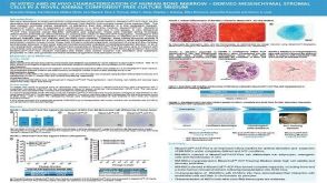 科学海报In Vitro and in Vivo Characterization of Human Bone Marrow-Derived Mesenchymal Stromal Cells in a Novel Animal Component-Free Culture Medium

 沪公网安备31010102008431号