High levels of lymphoid expression of enhanced green fluorescent protein in nonhuman primates transplanted with cytokine-mobilized peripheral blood CD34(+) cells.
We have used a murine retrovirus vector containing an enhanced green fluorescent protein complimentary DNA (EGFP cDNA) to dynamically follow vector-expressing cells in the peripheral blood (PB) of transplanted rhesus macaques. Cytokine mobilized CD34(+) cells were transduced with an amphotropic vector that expressed EGFP and a dihydrofolate reductase cDNA under control of the murine stem cell virus promoter. The transduction protocol used the CH-296 recombinant human fibronectin fragment and relatively high concentrations of the flt-3 ligand and stem cell factor. Following transplantation of the transduced cells,up to 55% EGFP-expressing granulocytes were obtained in the peripheral circulation during the early posttransplant period. This level of myeloid marking,however,decreased to 0.1% or lower within 2 weeks. In contrast,EGFP expression in PB lymphocytes rose from 2%-5% shortly following transplantation to 10% or greater by week 5. After 10 weeks,the level of expression in PB lymphocytes continued to remain at 3%-5% as measured by both flow cytometry and Southern blot analysis,and EGFP expression was observed in CD4(+),CD8(+),CD20(+),and CD16/56(+) lymphocyte subsets. EGFP expression was only transiently detected in red blood cells and platelets soon after transplantation. Such sustained levels of lymphocyte marking may be therapeutic in a number of human gene therapy applications that require targeting of the lymphoid compartment. The transient appearance of EGFP(+) myeloid cells suggests that transduction of a lineage-restricted myeloid progenitor capable of short-term engraftment was obtained with this protocol. (Blood. 2000;95:445-452)
View Publication
Pharmacologic modulation of the calcium-sensing receptor enhances hematopoietic stem cell lodgment in the adult bone marrow.
The ability of hematopoietic stem cells (HSCs) to undergo self-renewal is partly regulated by external signals originating from the stem cell niche. Our previous studies with HSCs obtained from fetal liver of mice deficient for the calcium-sensing receptor (CaR) have shown the crucial role of this receptor in HSC lodgment and engraftment in the bone marrow (BM) endosteal niche. Using a CaR agonist,Cinacalcet,we assessed the effects of stimulating the CaR on the function of murine HSCs. Our results show that CaR stimulation increases primitive hematopoietic cell activity in vitro,including growth in stromal cell cocultures,adhesion to extracellular matrix molecules such as collagen I and fibronectin,and migration toward the chemotactic stimulus,stromal cell-derived factor 1α. Receptor stimulation also led to augmented in vivo homing,CXCR4-mediated lodgment at the endosteal niche,and engraftment capabilities. These mechanisms by which stimulating the CaR dictates preferential localization of HSCs in the BM endosteal niche provide additional insights into the fundamental interrelationship between the stem cell and its niche. These studies also have implications in the area of clinical stem cell transplantation,where ex vivo modulation of the CaR may be envisioned as a strategy to enhance HSC engraftment in the BM.
View Publication
Thirukkumaran CM et al. (JUL 2003)
Blood 102 1 377--87
Reovirus oncolysis as a novel purging strategy for autologous stem cell transplantation.
Hematologic stem cell rescue after high-dose cytotoxic therapy is extensively used for the treatment of many hematopoietic and solid cancers. Gene marking studies suggest that occult tumor cells within the autograft may contribute to clinical relapse. To date purging of autografts contaminated with cancer cells has been unsuccessful. The selective oncolytic property of reovirus against myriad malignant histologies in in vitro,in vivo,and ex vivo systems has been previously demonstrated. In the present study we have shown that reovirus can successfully purge cancer cells within autografts. Human monocytic and myeloma cell lines as well as enriched ex vivo lymphoma,myeloma,and Waldenström macroglobulinemia patient tumor specimens were used in an experimental purging model. Viability of the cell lines or purified ex vivo tumor cells of diffuse large B-cell lymphoma,chronic lymphocytic leukemia,Waldenström macroglobulinemia,and small lymphocytic lymphoma was significantly reduced after reovirus treatment. Further,[35S]-methionine labeling and sodium dodecyl sulfate-polyacrylamide gel electrophoresis (SDS-PAGE) of cellular proteins demonstrated reovirus protein synthesis and disruption of host cell protein synthesis as early as 24 hours. Admixtures of apheresis product with the abovementioned tumor cells and cell lines treated with reovirus showed complete purging of disease. In contrast,reovirus purging of enriched ex vivo multiple myeloma,Burkitt lymphoma,and follicular lymphoma was incomplete. The oncolytic action of reovirus did not affect CD34+ stem cells or their long-term colony-forming assays even after granulocyte colony-stimulating factor (G-CSF) stimulation. Our results indicate the ex vivo use of an unattenuated oncolytic virus as an attractive purging strategy for autologous stem cell transplantations.
View Publication
Progenitor cell dose determines the pace and completeness of engraftment in a xenograft model for cord blood transplantation.
Two critical concerns in clinical cord blood transplantation are the initial time to engraftment and the subsequent restoration of immune function. These studies measured the impact of progenitor cell dose on both the pace and strength of hematopoietic reconstitution by transplanting nonobese diabetic/severe combined immunodeficiency/interleukin-2 receptor-gamma-null (NSγ) mice with lineage-depleted aldehyde dehydrogenase-bright CD34(+) human cord blood progenitors. The progress of each transplant was monitored over an extended time course by repeatedly analyzing the peripheral blood for human hematopoietic cells. In vivo human hematopoietic development was complete. After long-term transplantation assays (≥ 19 weeks),human T-cell development was documented within multiple tissues in 16 of 32 NSγ mice. Human T-cell differentiation was active within NSγ thymuses,as documented by the presence of CD4(+) CD8(+) T-cell progenitors as well as T-cell receptor excision circles. It is important to note that although myeloid and B-cell engraftment was detected as early as 4 weeks after transplantation,human T-cell development was exclusively late onset. High progenitor cell doses were associated with a robust human hematopoietic chimerism that accelerated both initial time to engraftment and subsequent T-cell development. At lower progenitor cell doses,the chimerism was weak and the human hematopoietic lineage development was frequently incomplete.
View Publication

 EasySep™小鼠TIL(CD45)正选试剂盒


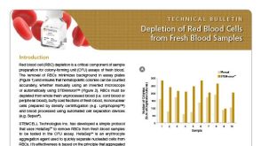 技术公告Depletion of Red Blood Cells from Fresh Blood Samples


 沪公网安备31010102008431号