High levels of lymphoid expression of enhanced green fluorescent protein in nonhuman primates transplanted with cytokine-mobilized peripheral blood CD34(+) cells.
We have used a murine retrovirus vector containing an enhanced green fluorescent protein complimentary DNA (EGFP cDNA) to dynamically follow vector-expressing cells in the peripheral blood (PB) of transplanted rhesus macaques. Cytokine mobilized CD34(+) cells were transduced with an amphotropic vector that expressed EGFP and a dihydrofolate reductase cDNA under control of the murine stem cell virus promoter. The transduction protocol used the CH-296 recombinant human fibronectin fragment and relatively high concentrations of the flt-3 ligand and stem cell factor. Following transplantation of the transduced cells,up to 55% EGFP-expressing granulocytes were obtained in the peripheral circulation during the early posttransplant period. This level of myeloid marking,however,decreased to 0.1% or lower within 2 weeks. In contrast,EGFP expression in PB lymphocytes rose from 2%-5% shortly following transplantation to 10% or greater by week 5. After 10 weeks,the level of expression in PB lymphocytes continued to remain at 3%-5% as measured by both flow cytometry and Southern blot analysis,and EGFP expression was observed in CD4(+),CD8(+),CD20(+),and CD16/56(+) lymphocyte subsets. EGFP expression was only transiently detected in red blood cells and platelets soon after transplantation. Such sustained levels of lymphocyte marking may be therapeutic in a number of human gene therapy applications that require targeting of the lymphoid compartment. The transient appearance of EGFP(+) myeloid cells suggests that transduction of a lineage-restricted myeloid progenitor capable of short-term engraftment was obtained with this protocol. (Blood. 2000;95:445-452)
View Publication
产品类型:
产品号#:
04436
04064
04100
04230
04236
04431
04434
04444
04464
04531
04535
04545
04536
04564
04035
04330
04034
04044
04435
04445
04534
04544
产品名:
MethoCult™ SF H4436
MethoCult™ H4034 Optimum 入门试剂盒
MethoCult™ H4100
MethoCult™ H4230
MethoCult™ SF H4236
MethoCult™ H4431
MethoCult™ H4434 Classic
MethoCult™ H4434 Classic
MethoCult™ H4434 Classic 套装
MethoCult™ H4531
MethoCult™ H4535 Enriched,不含EPO
MethoCult™ H4535 Enriched,不含EPO
MethoCult™ SF H4536
MethoCult™ H4534 Classic 无 EPO 入门试剂盒
MethoCult™ 不含EPO的H4035 Optimum
MethoCult™ H4330
MethoCult™ H4034 Optimum
MethoCult™ H4034 Optimum
MethoCult™ H4435 Enriched
MethoCult™ H4435 Enriched
MethoCult™ H4534 Classic(不含 EPO)
MethoCult™ H4534 Classic(不含 EPO)
Penicka M et al. (JUL 2007)
Heart (British Cardiac Society) 93 7 837--41
One-day kinetics of myocardial engraftment after intracoronary injection of bone marrow mononuclear cells in patients with acute and chronic myocardial infarction.
OBJECTIVE: To investigate the kinetics of myocardial engraftment of bone marrow-derived mononuclear cells (BMNCs) after intracoronary injection using 99mTc-d,l-hexamethylpropylene amine oxime (99mTc-HMPAO) nuclear imaging in patients with acute and chronic anterior myocardial infarction. DESIGN: Nuclear imaging-derived tracking of BMNCs at 2 and 20 h after injection in the left anterior descending (LAD) coronary artery. SETTING: Academical cardiocentre. PATIENTS: Five patients with acute (mean (SD) age 58 (11) years; ejection fraction range 33-45%) and five patients with chronic (mean (SD) age 50 (6) years; ejection fraction range 28-34%) anterior myocardial infarction. INTERVENTIONS: A total of 24.2 x 10(8)-57.0 x 10(8) BMNCs (20% labelled with 700-1000 MBq 99mTc-HMPAO) were injected in the LAD coronary artery. RESULTS: At 2 h after BMNC injection,myocardial activity was observed in all patients with acute (range 1.31-5.10%) and in all but one patient with chronic infarction (range 1.10-3.0%). At 20 h,myocardial engraftment was noted only in three patients with acute myocardial infarction,whereas no myocardial activity was noted in any patient with chronic infarction. CONCLUSIONS: Engraftment of BMNCs shows dynamic changes within the first 20 h after intracoronary injection. Persistent myocardial engraftment was noted only in a subset of patients with acute myocardial infarction.
View Publication
Anti-CD45-mediated cytoreduction to facilitate allogeneic stem cell transplantation.
The CD45 antigen is present on all cells of the hematopoietic lineage. Using a murine model,we have determined whether a lytic CD45 monoclonal antibody can produce persistent aplasia and whether it could facilitate syngeneic or allogeneic stem cell engraftment. After its systemic administration,we found saturating quantities of the antibody on all cells expressing the CD45 antigen,both in marrow and in lymphoid organs. All leukocyte subsets in peripheral blood were markedly diminished during or soon after anti-CD45 treatment,but only the effect on the lymphoid compartment was sustained. In contrast to the prolonged depletion of T and B lymphocytes from the thymus and spleen,peripheral blood neutrophils began to recover within 24 hours after the first anti-CD45 injection and marrow progenitor cells were spared from destruction,despite being coated with saturating quantities of anti-CD45. Given the transient effects of the monoclonal antibody on myelopoiesis and the more persistent effects on lymphopoiesis,we asked whether this agent could contribute to donor hematopoietic engraftment following nonmyeloablative transplantation. Treatment with anti-CD45 alone did not enhance syngeneic engraftment,consistent with its inability to destroy progenitor cells and permit competitive repopulation with syngeneic donor stem cells. By contrast,the combination of anti-CD45 and an otherwise inactive dose of total-body irradiation allowed engraftment of H2 fully allogeneic donor stem cells. We attribute this result to the recipient immunosuppression produced by depletion of CD45(+) lymphocytes. Monoclonal antibodies of this type may therefore have an adjunctive role in nonmyeloablative conditioning regimens for allogeneic stem cell transplantation.
View Publication
Effects of MLN518, a dual FLT3 and KIT inhibitor, on normal and malignant hematopoiesis.
Internal tandem duplications (ITDs) of the FMS-like tyrosine kinase 3 (FLT3) receptor tyrosine kinase are found in approximately 30% of patients with acute myelogenous leukemia (AML) and are associated with a poor prognosis. FLT3 ITD mutations result in constitutive kinase activation and are thought to be pathogenetically relevant,implicating FLT3 as a plausible therapeutic target. MLN518 (formerly CT53518) is a small molecule inhibitor of the FLT3,KIT,and platelet-derived growth-factor receptor (PDGFR) tyrosine kinases with significant activity in murine models of FLT3 ITD-positive leukemia. Given the importance of FLT3 and KIT for normal hematopoietic progenitor cells,we analyzed the effect of MLN518 on murine hematopoiesis under steady-state conditions,after chemotherapy-induced myelosuppression,and during bone marrow transplantation. In these assays,we show that MLN518 has mild toxicity toward normal hematopoiesis at concentrations that are effective in treating FLT3 ITD-positive leukemia in mice. We also demonstrate that MLN518 preferentially inhibits the growth of blast colonies from FLT3 ITD-positive compared with ITD-negative patients with AML,at concentrations that do not significantly affect colony formation by normal human progenitor cells. In analogy to imatinib mesylate in BCR-ABL-positive acute leukemia,MLN518-induced remissions may not be durable. Our studies provide the basis for integrating this compound into chemotherapy and transplantation protocols.
View Publication

 EasySep™小鼠TIL(CD45)正选试剂盒


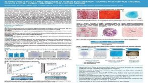 科学海报In Vitro and in Vivo Characterization of Human Bone Marrow-Derived Mesenchymal Stromal Cells in a Novel Animal Component-Free Culture Medium

 沪公网安备31010102008431号