Downregulation of miRNA-200c links breast cancer stem cells with normal stem cells.
Human breast tumors contain a breast cancer stem cell (BCSC) population with properties reminiscent of normal stem cells. We found 37 microRNAs that were differentially expressed between human BCSCs and nontumorigenic cancer cells. Three clusters,miR-200c-141,miR-200b-200a-429,and miR-183-96-182 were downregulated in human BCSCs,normal human and murine mammary stem/progenitor cells,and embryonal carcinoma cells. Expression of BMI1,a known regulator of stem cell self-renewal,was modulated by miR-200c. miR-200c inhibited the clonal expansion of breast cancer cells and suppressed the growth of embryonal carcinoma cells in vitro. Most importantly,miR-200c strongly suppressed the ability of normal mammary stem cells to form mammary ducts and tumor formation driven by human BCSCs in vivo. The coordinated downregulation of three microRNA clusters and the similar functional regulation of clonal expansion by miR-200c provide a molecular link that connects BCSCs with normal stem cells.
View Publication
Liu S et al. (JAN 2011)
Cancer research 71 2 614--24
Breast cancer stem cells are regulated by mesenchymal stem cells through cytokine networks.
We have used in vitro and mouse xenograft models to examine the interaction between breast cancer stem cells (CSC) and bone marrow-derived mesenchymal stem cells (MSC). We show that both of these cell populations are organized in a cellular hierarchy in which primitive aldehyde dehydrogenase expressing mesenchymal cells regulate breast CSCs through cytokine loops involving IL6 and CXCL7. In NOD/SCID mice,labeled MSCs introduced into the tibia traffic to sites of growing breast tumor xenografts where they accelerated tumor growth by increasing the breast CSC population. With immunochemistry,we identified MSC-CSC niches in these tumor xenografts as well as in frozen sections from primary human breast cancers. Bone marrow-derived MSCs may accelerate human breast tumor growth by generating cytokine networks that regulate the CSC population.
View Publication
Speen AM et al. ( 2016)
Journal of Biological Chemistry 291 48 25192--25206
Ozone-derived oxysterols affect liver X receptor (LXR) signaling: A potential role for lipid-protein adducts
When inhaled,ozone (O3) interacts with cholesterols of airway epithelial cell membranes or the lung lining fluid,generating chemically reactive oxysterols. The mechanism by which O3-derived oxysterols affect molecular function is unknown. Our data show that in vitro exposure of human bronchial epithelial cells to O3 results in the formation of oxysterols,epoxycholesterol-α and β (α-EpCh,β-EpCh) and Secosterol A and B (Seco A,SecoB),in cell lysates and apical washes. Similarly,bronchoalveolar lavage fluid obtained from human volunteers exposed to O3 contained elevated levels of these oxysterol species. As expected,O3-derived oxysterols have a pro-inflammatory effect and increase NF-κB activity. Interestingly,expression of the cholesterol efflux pump ATP-binding cassette transporter 1 (ABCA1),which is regulated by activation of the liver X receptor (LXR),was suppressed in epithelial cells exposed to O3. Additionally,exposure of LXR knockout mice to O3 enhanced pro-inflammatory cytokine production in the lung,suggesting LXR inhibits O3-induced inflammation. Using alkynyl surrogates of O3-derived oxysterols,our data demonstrate adduction of LXR with Seco A. Similarly,supplementation of epithelial cells with alkynyl-tagged cholesterol followed by O3 exposure causes observable lipid-LXR adduct formation. Experiments using Seco A and the LXR agonist T0901317 (T09) showed reduced expression of ABCA1 as compared to stimulation with T09 alone,indicating that Seco A-LXR protein adduct formation inhibits LXR activation by traditional agonists. Overall,these data demonstrate that O3-derived oxysterols have pro-inflammatory functions and form lipid-protein adducts with LXR,thus leading to suppressed cholesterol regulatory gene expression and providing a biochemical mechanism mediating O3-derived formation of oxidized lipids in the airways and subsequent adverse health effects.
View Publication
产品类型:
产品号#:
05001
05021
05022
产品名:
PneumaCult™-ALI 培养基
PneumaCult™-ALI 培养基含12 mm Transwell®插件
PneumaCult™-ALI 培养基含6.5 mm Transwell®插件
Aladegbami B et al. (JUL 2017)
Scientific reports 7 1 5580
Epithelial cell specific Raptor is required for initiation of type 2 mucosal immunity in small intestine.
Intestinal tuft cells are one of 4 secretory cell linages in the small intestine and the source of IL-25,a critical initiator of the type 2 immune response to parasite infection. When Raptor,a critical scaffold protein for mammalian target of rapamycin complex 1 (mTORC1),was acutely deleted in intestinal epithelium via Tamoxifen injection in Tritrichomonas muris (Tm) infected mice,tuft cells,IL-25 in epithelium and IL-13 in the mesenchyme were significantly reduced,but Tm burden was not affected. When Tm infected mice were treated with rapamycin,DCLK1 and IL-25 expression in enterocytes and IL-13 expression in mesenchyme were diminished. After massive small bowel resection,tuft cells and Tm were diminished due to the diet used postoperatively. The elimination of Tm and subsequent re-infection of mice with Tm led to type 2 immune response only in WT,but Tm colonization in both WT and Raptor deficient mice. When intestinal organoids were stimulated with IL-4,tuft cells and IL-25 were induced in both WT and Raptor deficient organoids. In summary,our study reveals that enterocyte specific Raptor is required for initiating a type 2 immune response which appears to function through the regulation of mTORC1 activity.
View Publication
产品类型:
产品号#:
06005
产品名:
IntestiCult™ 类器官生长培养基 (小鼠)
Pond AC et al. ( 2013)
Stem cells (Dayton,Ohio) 31 1 10.1002/stem.1266
Fibroblast Growth Factor Receptor Signaling Is Essential for Normal Mammary Gland Development and Stem Cell Function
Fibroblast growth factor (FGF) signaling plays an important role in embryonic stem cells and adult tissue homeostasis,but the function of FGFs in mammary gland stem cells is less well defined. Both FGFR1 and FGFR2 are expressed in basal and luminal mammary epithelial cells (MECs),suggesting that together they might play a role in mammary gland development and stem cell dynamics. Previous studies have demonstrated that the deletion of FGFR2 resulted only in transient developmental defects in branching morphogenesis. Using a conditional deletion strategy,we investigated the consequences of FGFR1 deletion alone and then the simultaneous deletion of both FGFR1 and FGFR2 in the mammary epithelium. FGFR1 deletion using a keratin 14 promoter-driven Cre-recombinase resulted in an early,yet transient delay in development. However,no reduction in functional outgrowth potential was observed following limiting dilution transplantation analysis. In contrast,a significant reduction in outgrowth potential was observed upon the deletion of both FGFR1 and FGFR2 in MECs using adenovirus-Cre. Additionally,using a fluorescent reporter mouse model to monitor Cre-mediated recombination,we observed a competitive disadvantage following transplantation of both FGFR1/R2-null MECs,most prominently in the basal epithelial cells. This correlated with the complete loss of the mammary stem cell repopulating population in the FGFR1/R2-attenuated epithelium. FGFR1/R2-null MECs were partially rescued in chimeric outgrowths containing wild-type MECs,suggesting the potential importance of paracrine mechanisms involved in the maintenance of the basal epithelial stem cells. These studies document the requirement for functional FGFR signaling in mammary stem cells during development.
View Publication
产品类型:
产品号#:
19758
60099
60099.1
60099AD
60099AD.1
60099AZ
60099AZ.1
60099BT
60099BT.1
60099FI
60099FI.1
60099PE
60099PE.1
60099PS
60099PS.1
60037
60037AD
60037AD.1
60037AZ
60037AZ.1
60037BT
60037BT.1
60037FI
60037FI.1
60037PE
60037PE.1
60037PB
60037PB.1
产品名:
抗小鼠CD24抗体,clone M1/69
抗小鼠CD24抗体,clone M1/69
抗小鼠CD24抗体,clone M1/69,Alexa Fluor® 488
抗小鼠CD24抗体,clone M1/69,Alexa Fluor® 488
抗小鼠CD24抗体,clone M1/69,APC
抗小鼠CD24抗体,clone M1/69,Biotin
抗小鼠CD24抗体,clone M1/69,Biotin
抗小鼠CD24抗体,clone M1/69,PE
抗小鼠CD24抗体,clone M1/69,PE
抗小鼠CD24抗体,clone M1/69,PerCP-Cy5.5
抗小鼠CD24抗体,clone M1/69,PerCP-Cy5.5
抗小鼠CD49f抗体,clone GoH3
抗小鼠CD49f抗体,clone GoH3,Alexa Fluor® 488
抗小鼠CD49f抗体,clone GoH3,Alexa Fluor® 488
抗小鼠CD49f抗体,clone GoH3,APC
抗小鼠CD49f抗体,clone GoH3,Biotin
抗小鼠CD49f抗体,clone GoH3,FITC
抗小鼠CD49f抗体,clone GoH3,PE
抗小鼠CD49f抗体,clone GoH3,PE
抗小鼠CD49f抗体,clone GoH3,Pacific Blue™
抗小鼠CD49f抗体,clone GoH3,Pacific Blue™
E. A. Davis et al. (JUN 2018)
Physiological reports 6 12 e13745
Evidence for a direct effect of the autonomic nervous system on intestinal epithelial stem cell proliferation.
The sympathetic (SNS) and parasympathetic (PNS) branches of the autonomic nervous system have been implicated in the modulation of the renewal of many tissues,including the intestinal epithelium. However,it is not known whether these mechanisms are direct,requiring an interaction between autonomic neurotransmitters and receptors on proliferating epithelial cells. To evaluate the existence of a molecular framework for a direct effect of the SNS or PNS on intestinal epithelial renewal,we measured gene expression for the main autonomic neurotransmitter receptors in this tissue. We separately evaluated intestinal epithelial regions comprised of the stem,progenitor,and mature cells,which allowed us to investigate the distinct contributions of each cell population to this proposed autonomic effect. Notably,we found that the stem cells expressed the receptors for the SNS-associated alpha2A adrenoreceptor and the PNS-associated muscarinic acetylcholine receptors (M1 and M3). In a separate experiment,we found that the application of norepinephrine or acetylcholine decreases the expression of cyclin D1,a gene necessary for cell cycle progression,in intestinal epithelial organoids compared with controls (P {\textless} 0.05). Together,these results provide evidence of a direct mechanism for the autonomic nervous system influence on intestinal epithelial stem cell proliferation.
View Publication
产品类型:
产品号#:
06005
产品名:
IntestiCult™ 类器官生长培养基 (小鼠)
Liu S and Wicha MS (SEP 2010)
Journal of clinical oncology : official journal of the American Society of Clinical Oncology 28 25 4006--12
Targeting breast cancer stem cells.
There is increasing evidence that many cancers,including breast cancer,contain populations of cells that display stem-cell properties. These breast cancer stem cells,by virtue of their relative resistance to radiation and cytotoxic chemotherapy,may contribute to treatment resistance and relapse. The elucidation of pathways that regulate these cells has led to the identification of potential therapeutic targets. A number of agents capable of targeting breast cancer stem cells in preclinical models are currently entering clinical trials. Assessment of the efficacy of the agents will require development of innovative clinical trial designs with appropriate biologic and clinical end points. The effective targeting of breast cancer stem cells has the potential to significantly improve outcome for women with both early-stage and advanced breast cancer.
View Publication

 EasySep™小鼠TIL(CD45)正选试剂盒


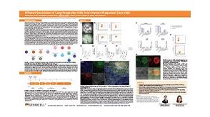 科学海报Efficient Generation of Lung Progenitor Cells From Human Pluripotent Stem Cells
 实验方案How to Dissociate and Plate Human Pluripotent Stem Cell-Derived Cardiomyocytes for Microelectrode Array (MEA) Assay
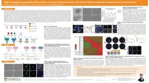 科学海报High-Throughput-Compatible Differentiation of Human Pluripotent Stem Cell Lines to Kidney Organoids for Nephrotoxic Drug Screening

 沪公网安备31010102008431号