Eden JA (JUL 2010)
Menopause (New York,N.Y.) 17 4 801--10
Human breast cancer stem cells and sex hormones--a narrative review.
OBJECTIVE: The aim of this narrative review was to evaluate the role of cancer stem cells (CSCs) and sex steroids in the pathophysiology of human breast cancer. METHODS: A key-word search was performed using the Scopus database. Preference was given to studies using human cells and tissues. RESULTS: Long-term estrogen-progestin hormone therapy is known to increase breast cancer risk,although the mechanisms are poorly understood. In the last few years,it has become clear that many human breast cancers contain CSCs,which may be responsible for much of the tumor's malignant behavior. Very recently,the impact of estrogen,progesterone,and progestins on breast CSCs and their progeny has been studied and clarified. Most breast CSCs are estrogen receptor negative and progesterone receptor negative,although some intermediary progenitor forms have hormone receptors,especially progesterone receptor. Most mature human breast cancer cellsare estrogen receptor positive and can thus be stimulated by estrogen. Breast CSCs usually elaborate CD44+,CD24j/low and/or ALDEFLUOR+ cell markers and are lineage markers negative. One of the main roles of progesterone and progestin seems to be on certain breast cancer stem intermediate forms,inducing them to revert back to a more primitive breast CSC form. CONCLUSIONS: As the pathophysiology of human breast CSC is clarified,it is probable that this will lead to novel,effective breast cancer treatments and,perhaps,new breast cancer preventive agents. This research may also lead to safer hormone therapy regimens.
View Publication
产品类型:
产品号#:
01700
01705
01702
产品名:
ALDEFLUOR™ 试剂盒
ALDEFLUOR™ DEAB试剂, 1.5 mM, 1 mL
ALDEFLUOR™检测缓冲液
Zhu S et al. (JUN 2017)
Nature 546 7660 667--670
Nlrp9b inflammasome restricts rotavirus infection in intestinal epithelial cells.
Rotavirus,a leading cause of severe gastroenteritis and diarrhoea in young children,accounts for around 215,000 deaths annually worldwide. Rotavirus specifically infects the intestinal epithelial cells in the host small intestine and has evolved strategies to antagonize interferon and NF-κB signalling,raising the question as to whether other host factors participate in antiviral responses in intestinal mucosa. The mechanism by which enteric viruses are sensed and restricted in vivo,especially by NOD-like receptor (NLR) inflammasomes,is largely unknown. Here we uncover and mechanistically characterize the NLR Nlrp9b that is specifically expressed in intestinal epithelial cells and restricts rotavirus infection. Our data show that,via RNA helicase Dhx9,Nlrp9b recognizes short double-stranded RNA stretches and forms inflammasome complexes with the adaptor proteins Asc and caspase-1 to promote the maturation of interleukin (Il)-18 and gasdermin D (Gsdmd)-induced pyroptosis. Conditional depletion of Nlrp9b or other inflammasome components in the intestine in vivo resulted in enhanced susceptibility of mice to rotavirus replication. Our study highlights an important innate immune signalling pathway that functions in intestinal epithelial cells and may present useful targets in the modulation of host defences against viral pathogens.
View Publication
产品类型:
产品号#:
06005
产品名:
IntestiCult™ 类器官生长培养基 (小鼠)
Gazdhar A et al. ( 2017)
Frontiers in immunology 8 April 447
Human Bronchial Epithelial Cells Induce CD141/CD123/DC-SIGN/FLT3Monocytes That Promote Allogeneic Th17 Differentiation.
Little is known about monocyte differentiation in the lung mucosal environment and about how the epithelium shapes monocyte function. We studied the role of the soluble component of bronchial epithelial cells (BECs) obtained under basal culture conditions in innate and adaptive monocyte responses. Monocytes cultured in bronchial epithelial cell-conditioned media (BEC-CM) specifically upregulate CD141,CD123,and DC-SIGN surface levels andFLT3expression,as well as the release of IL-1β,IL-6,and IL-10. BEC-conditioned monocytes stimulate naive T cells to produce IL-17 through IL-1β mechanism and also trigger IL-10 production by memory T cells. Furthermore,monocytes cultured in an inflammatory environment induced by the cytokines IL-6,IL-8,IL-1β,IL-15,TNF-α,and GM-CSF also upregulate CD123 and DC-SIGN expression. However,only inflammatory cytokines in the epithelial environment boost the expression of CD141. Interestingly,we identified a CD141/CD123/DC-SIGN triple positive population in the bronchoalveolar lavage fluid (BALF) from patients with different inflammatory conditions,demonstrating that this monocyte population existsin vivo. The frequency of this monocyte population was significantly increased in patients with sarcoidosis,suggesting a role in inflammatory mechanisms. Overall,these data highlight the specific role that the epithelium plays in shaping monocyte responses. Therefore,the unraveling of these mechanisms contributes to the understanding of the function that the epithelium may playin vivo.
View Publication
产品类型:
产品号#:
05001
05021
05022
产品名:
PneumaCult™-ALI 培养基
PneumaCult™-ALI 培养基含12 mm Transwell®插件
PneumaCult™-ALI 培养基含6.5 mm Transwell®插件
Gilpin SE et al. ( 2016)
Biomaterials 108 111--119
Regenerative potential of human airway stem cells in lung epithelial engineering
Bio-engineered organs for transplantation may ultimately provide a personalized solution for end-stage organ failure,without the risk of rejection. Building upon the process of whole organ perfusion decellularization,we aimed to develop novel,translational methods for the recellularization and regeneration of transplantable lung constructs. We first isolated a proliferative KRT5+TP63+ basal epithelial stem cell population from human lung tissue and demonstrated expansion capacity in conventional 2D culture. We then repopulated acellular rat scaffolds in ex vivo whole organ culture and observed continued cell proliferation,in combination with primary pulmonary endothelial cells. To show clinical scalability,and to test the regenerative capacity of the basal cell population in a human context,we then recellularized and cultured isolated human lung scaffolds under biomimetic conditions. Analysis of the regenerated tissue constructs confirmed cell viability and sustained metabolic activity over 7 days of culture. Tissue analysis revealed extensive recellularization with organized tissue architecture and morphology,and preserved basal epithelial cell phenotype. The recellularized lung constructs displayed dynamic compliance and rudimentary gas exchange capacity. Our results underline the regenerative potential of patient-derived human airway stem cells in lung tissue engineering. We anticipate these advances to have clinically relevant implications for whole lung bioengineering and ex vivo organ repair.
View Publication
Ibiza S et al. (JUL 2016)
Nature 535 7612 440--443
Glial-cell-derived neuroregulators control type 3 innate lymphoid cells and gut defence.
Group 3 innate lymphoid cells (ILC3) are major regulators of inflammation and infection at mucosal barriers. ILC3 development is thought to be programmed,but how ILC3 perceive,integrate and respond to local environmental signals remains unclear. Here we show that ILC3 in mice sense their environment and control gut defence as part of a glial"ILC3"epithelial cell unit orchestrated by neurotrophic factors. We found that enteric ILC3 express the neuroregulatory receptor RET. ILC3-autonomous Ret ablation led to decreased innate interleukin-22 (IL-22),impaired epithelial reactivity,dysbiosis and increased susceptibility to bowel inflammation and infection. Neurotrophic factors directly controlled innate Il22 downstream of the p38 MAPK/ERK-AKT cascade and STAT3 activation. Notably,ILC3 were adjacent to neurotrophic-factor-expressing glial cells that exhibited stellate-shaped projections into ILC3 aggregates. Glial cells sensed microenvironmental cues in a MYD88-dependent manner to control neurotrophic factors and innate IL-22. Accordingly,glial-intrinsic Myd88 deletion led to impaired production of ILC3-derived IL-22 and a pronounced propensity towards gut inflammation and infection. Our work sheds light on a novel multi-tissue defence unit,revealing that glial cells are central hubs of neuron and innate immune regulation by neurotrophic factor signals.
View Publication
产品类型:
产品号#:
06005
产品名:
IntestiCult™ 类器官生长培养基 (小鼠)
Diehn M et al. (APR 2009)
Nature 458 7239 780--3
Association of reactive oxygen species levels and radioresistance in cancer stem cells.
The metabolism of oxygen,although central to life,produces reactive oxygen species (ROS) that have been implicated in processes as diverse as cancer,cardiovascular disease and ageing. It has recently been shown that central nervous system stem cells and haematopoietic stem cells and early progenitors contain lower levels of ROS than their more mature progeny,and that these differences are critical for maintaining stem cell function. We proposed that epithelial tissue stem cells and their cancer stem cell (CSC) counterparts may also share this property. Here we show that normal mammary epithelial stem cells contain lower concentrations of ROS than their more mature progeny cells. Notably,subsets of CSCs in some human and murine breast tumours contain lower ROS levels than corresponding non-tumorigenic cells (NTCs). Consistent with ROS being critical mediators of ionizing-radiation-induced cell killing,CSCs in these tumours develop less DNA damage and are preferentially spared after irradiation compared to NTCs. Lower ROS levels in CSCs are associated with increased expression of free radical scavenging systems. Pharmacological depletion of ROS scavengers in CSCs markedly decreases their clonogenicity and results in radiosensitization. These results indicate that,similar to normal tissue stem cells,subsets of CSCs in some tumours contain lower ROS levels and enhanced ROS defences compared to their non-tumorigenic progeny,which may contribute to tumour radioresistance.
View Publication
产品类型:
产品号#:
05601
产品名:
EpiCult™-B 人培养基
Zhao X et al. (AUG 2010)
Proceedings of the National Academy of Sciences of the United States of America 107 32 14146--51
Telomerase-immortalized human mammary stem/progenitor cells with ability to self-renew and differentiate.
There is increasing evidence that breast and other cancers originate from and are maintained by a small fraction of stem/progenitor cells with self-renewal properties. Whether such cancer stem/progenitor cells originate from normal stem cells based on initiation of a de novo stem cell program,by reprogramming of a more differentiated cell type by oncogenic insults,or both remains unresolved. A major hurdle in addressing these issues is lack of immortal human stem/progenitor cells that can be deliberately manipulated in vitro. We present evidence that normal and human telomerase reverse transcriptase (hTERT)-immortalized human mammary epithelial cells (hMECs) isolated and maintained in Dana-Farber Cancer Institute 1 (DFCI-1) medium retain a fraction with progenitor cell properties. These cells coexpress basal (K5,K14,and vimentin),luminal (E-cadherin,K8,K18,or K19),and stem/progenitor (CD49f,CD29,CD44,and p63) cell markers. Clonal derivatives of progenitors coexpressing these markers fall into two distinct types--a K5(+)/K19(-) type and a K5(+)/K19(+) type. We show that both types of progenitor cells have self-renewal and differentiation ability. Microarray analyses confirmed the differential expression of components of stem/progenitor-associated pathways,such as Notch,Wnt,Hedgehog,and LIF,in progenitor cells compared with differentiated cells. Given the emerging evidence that stem/progenitor cells serve as precursors for cancers,these cellular reagents represent a timely and invaluable resource to explore unresolved questions related to stem/progenitor origin of breast cancer.
View Publication
Ma I and Allan AL (JUN 2011)
Stem cell reviews 7 2 292--306
The role of human aldehyde dehydrogenase in normal and cancer stem cells.
Normal stem cells and cancer stem cells (CSCs) share similar properties,in that both have the capacity to self-renew and differentiate into multiple cell types. In both the normal stem cell and cancer stem cell fields,there has been a great need for a universal marker that can effectively identify and isolate these rare populations of cells in order to characterize them and use this information for research and therapeutic purposes. Currently,it would appear that certain isoenzymes of the aldehyde dehydrogenase (ALDH) superfamily may be able to fulfill this role as a marker for both normal and cancer stem cells. ALDH has been identified as an important enzyme in the protection of normal hematopoietic stem cells,and is now also widely used as a marker to identify and isolate various types of normal stem cells and CSCs. In addition,emerging evidence suggests that ALDH1 is not only a marker for stem cells,but may also play important functional roles related to self-protection,differentiation,and expansion. This comprehensive review discusses the role that ALDH plays in normal stem cells and CSCs,with focus on ALDH1 and ALDH3A1. Discrepancies in the functional themes between cell types and future perspectives for therapeutic applications will also be discussed.
View Publication

 EasySep™小鼠TIL(CD45)正选试剂盒


 56:31
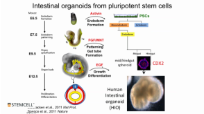
线上讲座Modeling Human Gastrointestinal Development and Disease Using Pluripotent Stem Cells发布日期: 03/06/2017
56:31
线上讲座Modeling Human Gastrointestinal Development and Disease Using Pluripotent Stem Cells发布日期: 03/06/2017 科学海报A Human Pluripotent Stem Cell-Derived Organoid Model for Recapitulation of Central Nervous System (CNS) Barrier and Fluid Secretion Functions of the Choroid Plexus
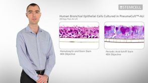

 沪公网安备31010102008431号