Liu C et al. (MAY 2012)
Molecular biology reports 39 5 5875--81
Co-expression of Oct-4 and Nestin in human breast cancers.
The aim is to investigate the clinical implications of the Oct-4 and Nestin protein in human breast cancers. A total of 346 cases including 26 fresh and 320 paraffin-embedded tumor tissues were selected for characterizing the frequency of CD44(+)CD24(-) tumor cells by flow cytometry and the differential expression of the stem cell-related genes between CD44(+)CD24(-) and non-CD44(+)CD24(-) tumor cells was analyzed by PCR Array and immunofluorescence. In comparison with the non-CD44(+)CD24(-) tumor cells,the CD44(+)CD24(-),particularly for those with high percentage of Oct-4(+) and Nestin(+),tumor cells had higher tumorigenicity by forming mammospheres in vitro. More importantly,42 (13.125%) out of 320 tumor tissues were positive for Oct-4 and Nestin staining. Universal analysis and multivariate analysis revealed that the expression of Oct-4 and Nestin was associated significantly with younger age,pathogenic degrees,lymph node metastasis and triple-negative breast cancer independently (P textless 0.05) as well as shorter survival (P = 0.001). Oct-4 and Nestin were important regulators of the development of breast cancer,and Oct-4 and Nestin may be used as predictors for the prognosis of breast cancers.
View Publication
Yu C et al. ( )
In vivo (Athens,Greece) 25 1 69--76
ALDH activity indicates increased tumorigenic cells, but not cancer stem cells, in prostate cancer cell lines.
BACKGROUND: Cancer stem cells (CSCs) have been shown to be a small stem cell-like cell population which appears to drive tumorigenesis,tumor recurrence and metastasis. Thus,identification and characterization of CSCs may be critical to defining effective anticancer therapies. In prostate cancer (PCa),the CD44(+) cell population appears to have stem cell-like properties including being tumorigenic. The enzyme aldehyde dehydrogenase (ALDH) has been found to identify hematopoietic stem cells and our aim was to determine the utility of ALDH activity and CD44 in identifying PCa stem cell-like cells in PCa cell lines. MATERIALS AND METHODS: LNCaP cells and PC-3 cells were sorted based on their expression of CD44 and ALDH activity. The cell populations were investigated using colony-forming assays,invasion assays,sphere formation experiments in a non-adherent environment and 3-D Matrigel matrix culture to observe the in vitro stem-cell like properties. Different sorted cell populations were injected subcutaneously into NOD/SCID mice to determine the corresponding tumorigenic capacities. RESULTS: ALDH(hi) CD44(+) cells exhibit a higher proliferative,clonogenic and metastatic capacity in vitro and demonstrate higher tumorigenicity capacity in vivo than did ALDH(lo) CD44(-) cells. The tumors recapitulated the population of the original cell line. However,ALDHlo CD44(-) cells were able to develop tumors,albeit with longer latency periods. CONCLUSION: ALDH activity and CD44 do not appear to identify PCa stem cells; however,they do indicate increased tumorigenic and metastatic potential,indicating their potential importance for further exploration.
View Publication
Griggs TF et al. ( 2017)
Respiratory research 18 1 84
Rhinovirus C targets ciliated airway epithelial cells.
BACKGROUND The Rhinovirus C (RV-C),first identified in 2006,produce high symptom burdens in children and asthmatics,however,their primary target host cell in the airways remains unknown. Our primary hypotheses were that RV-C target ciliated airway epithelial cells (AECs),and that cell specificity is determined by restricted and high expression of the only known RV-C cell-entry factor,cadherin related family member 3 (CDHR3). METHODS RV-C15 (C15) infection in differentiated human bronchial epithelial cell (HBEC) cultures was assessed using immunofluorescent and time-lapse epifluorescent imaging. Morphology of C15-infected differentiated AECs was assessed by immunohistochemistry. RESULTS C15 produced a scattered pattern of infection,and infected cells were shed from the epithelium. The percentage of cells infected with C15 varied from 1.4 to 14.7% depending on cell culture conditions. Infected cells had increased staining for markers of ciliated cells (acetylated-alpha-tubulin [aat],p < 0.001) but not markers of goblet cells (wheat germ agglutinin or Muc5AC,p = ns). CDHR3 expression was increased on ciliated epithelial cells,but not other epithelial cells (p < 0.01). C15 infection caused a 27.4% reduction of ciliated cells expressing CDHR3 (p < 0.01). During differentiation of AECs,CDHR3 expression progressively increased and correlated with both RV-C binding and replication. CONCLUSIONS The RV-C only replicate in ciliated AECs in vitro,leading to infected cell shedding. CDHR3 expression positively correlates with RV-C binding and replication,and is largely confined to ciliated AECs. Our data imply that factors regulating differentiation and CDHR3 production may be important determinants of RV-C illness severity.
View Publication
Development of a primary human co-culture model of inflamed airway mucosa
Neutrophil breach of the mucosal surface is a common pathological consequence of infection. We present an advanced co-culture model to explore neutrophil transepithelial migration utilizing airway mucosal barriers differentiated from primary human airway basal cells and examined by advanced imaging. Human airway basal cells were differentiated and cultured at air-liquid interface (ALI) on the underside of 3 μm pore-sized transwells,compatible with the study of transmigrating neutrophils. Inverted ALIs exhibit beating cilia and mucus production,consistent with conventional ALIs,as visualized by micro-optical coherence tomography (μOCT). μOCT is a recently developed imaging modality with the capacity for real time two- A nd three-dimensional analysis of cellular events in marked detail,including neutrophil transmigratory dynamics. Further,the newly devised and imaged primary co-culture model recapitulates key molecular mechanisms that underlie bacteria-induced neutrophil transepithelial migration previously characterized using cell line-based models. Neutrophils respond to imposed chemotactic gradients,and migrate in response to Pseudomonas aeruginosa infection of primary ALI barriers through a hepoxilin A3-directed mechanism. This primary cell-based co-culture system combined with μOCT imaging offers significant opportunity to probe,in great detail,micro-anatomical and mechanistic features of bacteria-induced neutrophil transepithelial migration and other important immunological and physiological processes at the mucosal surface.
View Publication
产品类型:
产品号#:
05001
05021
05022
产品名:
PneumaCult™-ALI 培养基
PneumaCult™-ALI 培养基含12 mm Transwell®插件
PneumaCult™-ALI 培养基含6.5 mm Transwell®插件
Smith GH (JAN 1996)
Breast cancer research and treatment 39 1 21--31
Experimental mammary epithelial morphogenesis in an in vivo model: evidence for distinct cellular progenitors of the ductal and lobular phenotype.
An in vivo transplantation system has been used to evaluate the developmental capacities of specific mouse mammary epithelial cell populations. Specifically,mouse mammary epithelial cells with distinctly limited developmental potentials have been identified using this procedure. Two distinct epithelial cell progenitors have been identified by experiments designed to determine whether basal lobular and ductal phenotypes could develop independently under conditions imposed by a limiting dilution. The prediction that these separate epithelial progenitors must exist was based upon the results from transplantation experiments carried out in epithelium-divested mammary fat pads of syngeneic mice with mammary epithelium from two different transgenic mouse models. The results presented here demonstrate the following points: 1) lobular,i.e. secretory,progenitor cells are present as distinct entities among the mammary epithelial cells found in immature virgin female mice; 2) similarly,ductal epithelial progenitors are present within the same population; 3) lobular progenitors are present in greater numbers,although both cell populations are extremely small; 4) as expected,some inocula produce outgrowths with simultaneous development of both lobular and ductal phenotypes--it is not known whether this indicates cooperative interaction between the two epithelial progenitors or signals the presence of a third progenitor type capable of producing both ductular and lobular committed daughters; 5) these findings have important consequences in the design of experiments aimed at testing the effects of known and putative mammary oncogenes and tumor suppressor genes,using techniques which include cellular transformation in vitro followed by in vivo cultivation and evaluation.
View Publication

 EasySep™小鼠TIL(CD45)正选试剂盒


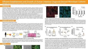 科学海报Efficient Establishment and Growth of Human Intestinal Organoid-Derived Monolayers
 15:48
线上讲座Generating Pancreatic Organoids from Healthy Murine and Human Pancreatic Ducts发布日期: 07/07/2023
15:48
线上讲座Generating Pancreatic Organoids from Healthy Murine and Human Pancreatic Ducts发布日期: 07/07/2023 实验方案Detection of CYP3A4 Activity in Human Hepatic Organoids by LC-MS
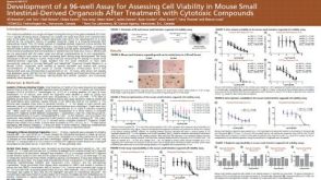 科学海报Development of a 96-well Assay for Assessing Cell Viability in Mouse Small Intestinal-Derived Organoids after Treatment with Cytotoxic Compounds
 实验方案Cryopreserving Hepatic Organoids Expanded in HepatiCult™ Organoid Growth Medium (Human)
 实验方案How to Perform Albumin Detection Assays in Human Hepatic Organoids
 沪公网安备31010102008431号