Crook JM et al. (MAR 2015)
Expert review of neurotherapeutics 15 3 295--304
The potential of induced pluripotent stem cells in models of neurological disorders: implications on future therapy.
There is an urgent need for new and advanced approaches to modeling the pathological mechanisms of complex human neurological disorders. This is underscored by the decline in pharmaceutical research and development efficiency resulting in a relative decrease in new drug launches in the last several decades. Induced pluripotent stem cells represent a new tool to overcome many of the shortcomings of conventional methods,enabling live human neural cell modeling of complex conditions relating to aberrant neurodevelopment,such as schizophrenia,epilepsy and autism as well as age-associated neurodegeneration. This review considers the current status of induced pluripotent stem cell-based modeling of neurological disorders,canvassing proven and putative advantages,current constraints,and future prospects of next-generation culture systems for biomedical research and translation.
View Publication
产品类型:
产品号#:
85850
85857
产品名:
mTeSR™1
mTeSR™1
Chen Y-W et al. (NOV 2010)
Molecular cancer therapeutics 9 11 2879--92
Cucurbitacin I suppressed stem-like property and enhanced radiation-induced apoptosis in head and neck squamous carcinoma--derived CD44(+)ALDH1(+) cells.
Head and neck squamous cell carcinoma (HNSCC) is a prevalent cancer worldwide. Signal transducers and activators of transcription 3 (STAT3) signaling is reported to promote tumor malignancy and recurrence in HNSCC. Cucurbitacins,triterpenoid derivatives,are strong STAT3 inhibitors with anticancer properties. Recent studies have shown aldehyde dehydrogenase 1 (ALDH1) to be a marker of cancer stem cells (CSC) in HNSCC. The aim of this study was to investigate the therapeutic effect of cucurbitacin I in HNSCC-derived CSCs. Using immunohistochemical analysis,we firstly showed that CD44,ALDH1,and phosphorylated STAT3 (p-STAT3) were higher in high-grade HNSCCs,and that triple positivity for CD44/ALDH1/p-STAT3 indicated a worse prognosis for HNSCC patients. Secondly,CD44(+)ALDH1(+) cells isolated from seven HNSCC patients showed greater tumorigenicity,radioresistance,and high expression of stemness (Bmi-1/Oct-4/Nanog) and epithelial-mesenchymal-transitional (Snail/Twist) genes as p-STAT3 level increased. Furthermore,we found that cucurbitacin I (JSI-124) can effectively inhibit the expression of p-STAT3 and capacities for tumorigenicity,sphere formation,and radioresistance in HNSCC-CD44(+)ALDH1(+). Notably,150 nmol/L cucurbitacin I effectively blocked STAT3 signaling and downstream survivin and Bcl-2 expression,and it induced apoptosis in HNSCC-CD44(+)ALDH1(+). Moreover,microarray data indicated that 100 nmol/L cucurbitacin I facilitated CD44(+)ALDH1(+) cells to differentiate into CD44�?�ALDH1�?� and enhanced the radiosensitivity of HNSCC-CD44(+)ALDH1(+). Xenotransplant experiments revealed that cucurbitacin I combined with radiotherapy significantly suppressed tumorigenesis and lung metastasis and further improved the survival rate in HNSCC-CD44(+)ALDH1(+)-transplanted immunocompromised mice. Taken together,our data show that cucurbitacin I,STAT3 inhibitor,reduces radioresistant,distant-metastatic,and CSC-like properties of HNSCC-CD44(+)ALDH1(+) cells. The potential of cucurbitacin I as a radiosensitizer should be verified in future anti-CSC therapy.
View Publication
Puissant A et al. (FEB 2010)
Cancer research 70 3 1042--52
Resveratrol promotes autophagic cell death in chronic myelogenous leukemia cells via JNK-mediated p62/SQSTM1 expression and AMPK activation.
Autophagy that is induced by starvation or cellular stress can enable cancer cell survival by sustaining energy homeostasis and eliminating damaged organelles and proteins. In response to stress,cancer cells have been reported to accumulate the protein p62/SQSTM1 (p62),but its role in the regulation of autophagy is controversial. Here,we report that the plant phytoalexin resveratrol (RSV) triggers autophagy in imatinib-sensitive and imatinib-resistant chronic myelogenous leukemia (CML) cells via JNK-dependent accumulation of p62. JNK inhibition or p62 knockdown prevented RSV-mediated autophagy and antileukemic effects. RSV also stimulated AMPK,thereby inhibiting the mTOR pathway. AMPK knockdown or mTOR overexpression impaired RSV-induced autophagy but not JNK activation. Lastly,p62 expression and autophagy in CD34+ progenitors from patients with CML was induced by RSV,and disrupting autophagy protected CD34+ CML cells from RSV-mediated cell death. We concluded that RSV triggered autophagic cell death in CML cells via both JNK-mediated p62 overexpression and AMPK activation. Our findings show that the JNK and AMPK pathways can cooperate to eliminate CML cells via autophagy.
View Publication
产品类型:
产品号#:
09600
09650
产品名:
StemSpan™ SFEM
StemSpan™ SFEM
Tareen SU et al. (MAR 2014)
Molecular therapy : the journal of the American Society of Gene Therapy 22 3 575--87
Design of a novel integration-deficient lentivector technology that incorporates genetic and posttranslational elements to target human dendritic cells.
As sentinels of the immune system,dendritic cells (DCs) play an essential role in regulating cellular immune responses. One of the main challenges of developing DC-targeted therapies includes the delivery of antigen to DCs in order to promote the activation of antigen-specific effector CD8 T cells. With the goal of creating antigen-directed immunotherapeutics that can be safely administered directly to patients,Immune Design has developed a platform of novel integration-deficient lentiviral vectors that target and deliver antigen-encoding nucleic acids to human DCs. This platform,termed ID-VP02,utilizes a novel genetic variant of a Sindbis virus envelope glycoprotein with posttranslational carbohydrate modifications in combination with Vpx,a SIVmac viral accessory protein,to achieve efficient targeting and transduction of human DCs. In addition,ID-VP02 incorporates safety features in its design that include two redundant mechanisms to render ID-VP02 integration-deficient. Here,we describe the characteristics that allow ID-VP02 to specifically transduce human DCs,and the advances that ID-VP02 brings to conventional third-generation lentiviral vector design as well as demonstrate upstream production yields that will enable manufacturing feasibility studies to be conducted.
View Publication
Jä et al. (SEP 2010)
Proceedings of the National Academy of Sciences of the United States of America 107 37 16280--5
Isolation and killing of candidate chronic myeloid leukemia stem cells by antibody targeting of IL-1 receptor accessory protein.
Chronic myeloid leukemia (CML) is genetically characterized by the Philadelphia (Ph) chromosome,formed through a reciprocal translocation between chromosomes 9 and 22 and giving rise to the constitutively active tyrosine kinase P210 BCR/ABL1. Therapeutic strategies aiming for a cure of CML will require full eradication of Ph chromosome-positive (Ph(+)) CML stem cells. Here we used gene-expression profiling to identify IL-1 receptor accessory protein (IL1RAP) as up-regulated in CML CD34(+) cells and also in cord blood CD34(+) cells as a consequence of retroviral BCR/ABL1 expression. To test whether IL1RAP expression distinguishes normal (Ph(-)) and leukemic (Ph(+)) cells within the CML CD34(+)CD38(-) cell compartment,we established a unique protocol for conducting FISH on small numbers of sorted cells. By using this method,we sorted cells directly into drops on slides to investigate their Ph-chromosome status. Interestingly,we found that the CML CD34(+)CD38(-)IL1RAP(+) cells were Ph(+),whereas CML CD34(+)CD38(-)IL1RAP(-) cells were almost exclusively Ph(-). By performing long-term culture-initiating cell assays on the two cell populations,we found that Ph(+) and Ph(-) candidate CML stem cells could be prospectively separated. In addition,by generating an anti-IL1RAP antibody,we provide proof of concept that IL1RAP can be used as a target on CML CD34(+)CD38(-) cells to induce antibody-dependent cell-mediated cytotoxicity. This study thus identifies IL1RAP as a unique cell surface biomarker distinguishing Ph(+) from Ph(-) candidate CML stem cells and opens up a previously unexplored avenue for therapy of CML.
View Publication
产品类型:
产品号#:
09600
09650
04435
04445
产品名:
StemSpan™ SFEM
StemSpan™ SFEM
MethoCult™H4435富集
MethoCult™H4435富集
Vieillard V et al. (AUG 2005)
Proceedings of the National Academy of Sciences 102 31 10981--86
NK cytotoxicity against CD4+ T cells during HIV-1 infection: A gp41 peptide induces the expression of an NKp44 ligand
HIV infection leads to a state of chronic immune activation and progressive deterioration in immune function,manifested most recognizably by the progressive depletion of CD4+ T cells. A substantial percentage of natural killer (NK) cells from patients with HIV infection are activated and express the natural cytotoxicity receptor (NCR) NKp44. Here we show that a cellular ligand for NKp44 (NKp44L) is expressed during HIV-1 infection and is correlated with both the progression of CD4+ T cell depletion and the increase of viral load. CD4+ T cells expressing this ligand are highly sensitive to the NK lysis activity mediated by NKp44+ NK cells. The expression of NKp44L is induced by the linear motif NH2-SWSNKS-COOH of the HIV-1 envelope gp41 protein. This highly conserved motif appears critical to the sharp increase in NK lysis of CD4+ T cells from HIV-infected patients. These studies strongly suggest that induction of NKp44L plays a key role in the lysis of CD4+ T cells by activated NK cells in HIV infection and consequently provide a framework for considering how HIV-1 may use NK cell immune surveillance to trigger CD4+ T cells. Understanding this mechanism may help to develop future therapeutic strategies and vaccines against HIV-1 infection.
View Publication
产品类型:
产品号#:
03800
03801
03802
03803
03804
03805
03806
05150
15021
15061
产品名:
ClonaCell™-HY 杂交瘤试剂盒
ClonaCell™-HY Medium
ClonaCell™-HY Medium
ClonaCell™-HY Medium
ClonaCell™-HY Medium
ClonaCell™-HY Medium
ClonaCell™-HY PEG (融合)
MyeloCult™H5100
RosetteSep™人T细胞富集抗体混合物
RosetteSep™人T细胞富集抗体混合物
Fakler M et al. (FEB 2009)
Blood 113 8 1710--22
Small molecule XIAP inhibitors cooperate with TRAIL to induce apoptosis in childhood acute leukemia cells and overcome Bcl-2-mediated resistance.
Defects in apoptosis contribute to poor outcome in pediatric acute lymphoblastic leukemia (ALL),calling for novel strategies that counter apoptosis resistance. Here,we demonstrate for the first time that small molecule inhibitors of the antiapoptotic protein XIAP cooperate with TRAIL to induce apoptosis in childhood acute leukemia cells. XIAP inhibitors at subtoxic concentrations,but not a structurally related control compound,synergize with TRAIL to trigger apoptosis and to inhibit clonogenic survival of acute leukemia cells,whereas they do not affect viability of normal peripheral blood lymphocytes,suggesting some tumor selectivity. Analysis of signaling pathways reveals that XIAP inhibitors enhance TRAIL-induced activation of caspases,loss of mitochondrial membrane potential,and cytochrome c release in a caspase-dependent manner,indicating that they promote a caspase-dependent feedback mitochondrial amplification loop. Of note,XIAP inhibitors even overcome Bcl-2-mediated resistance to TRAIL by enhancing Bcl-2 cleavage and Bak conformational change. Importantly,XIAP inhibitors kill leukemic blasts from children with ALL ex vivo and cooperate with TRAIL to induce apoptosis. In vivo,they significantly reduce leukemic burden in a mouse model of pediatric ALL engrafted in non-obese diabetic/severe combined immunodeficient (NOD/SCID) mice. Thus,XIAP inhibitors present a promising novel approach for apoptosis-based therapy of childhood ALL.
View Publication

 EasySep™小鼠TIL(CD45)正选试剂盒


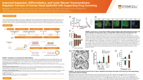 科学海报Improved Expansion, Differentiation, and Cystic Fibrosis Transmembrane Regulator Function of Human Nasal Epithelial Cells Supporting Drug Screening
 科学海报Routine Monitoring of Common Genetic Abnormalities in Human Pluripotent Stem Cells Using the hPSC Genetic Analysis Kit
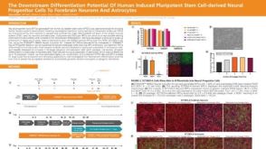 科学海报The Downstream Differentiation Potential of Human Induced Pluripotent Stem Cell-Derived Neural Progenitor Cells to Forebrain Neurons and Astrocytes
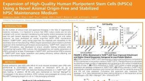 科学海报Expansion of High-Quality Human Pluripotent Stem Cells (hPSCs) Using a Novel Animal Origin-Free and Stabilized hPSC Maintenance Medium

 沪公网安备31010102008431号