Siatskas C et al. (OCT 2005)
FASEB journal : official publication of the Federation of American Societies for Experimental Biology 19 12 1752--4
Specific pharmacological dimerization of KDR in lentivirally transduced human hematopoietic cells activates anti-apoptotic and proliferative mechanisms.
Selective and regulatable expansion of transduced cells could augment gene therapy for many disorders. The activation of modified growth factor receptors via synthetic chemical inducers of dimerization allows for the coordinated growth of transduced cells. This system can also provide information on specific receptor-mediated signaling without interference from other family members. Although several receptor subunits have been investigated in this context,little is known about the precise molecular events associated with dimerizer-initiated signaling. We have constructed and expressed an AP20187-regulated KDR chimeric receptor in human TF1 cells and analyzed activation of this gene switch using functional,biochemical,and microarray analyses. When deprived of natural ligands,GM-CSF,interleukin-3,or erythropoietin,AP20187 prevented apoptosis of transduced TF1 cells,induced dose-dependent proliferation,and supported long-term growth. In addition,AP20187 stimulation activated the signaling molecules associated with mitogen-activated protein kinase and phosphatidyl-inositol 3-kinase/Akt pathways. Microarray analysis determined that a number of transcripts involved in a variety of cellular processes were differentially expressed. Notably,mRNAs affiliated with heat stress,including Hsp70 and Hsp105,were up-regulated. Functional assays showed that Hsp70 and Hsp105 protected transduced TF1 cells from apoptosis and premature senescence,in part through regulation of Akt. These observations delineate specific roles for kinase insert domain-containing receptor,or KDR,signaling and suggest strategies to endow genetically modified cells with a survival advantage enabling the generation of adequate cell numbers for therapeutic outcomes.
View Publication
产品类型:
产品号#:
04230
84434
84444
产品名:
MethoCult™H4230
Su YR et al. (AUG 2008)
Arteriosclerosis,thrombosis,and vascular biology 28 8 1439--46
Lentiviral transduction of apoAI into hematopoietic progenitor cells and macrophages: applications to cell therapy of atherosclerosis.
OBJECTIVE: We used genetically engineered mouse hematopoietic progenitor cells (HPCs) to investigate the therapeutic effects of human apoAI on atherosclerosis in apoE(-/-) mice. METHODS AND RESULTS: Lentiviral constructs expressing either human apoAI (LV-apoAI) or green fluorescent protein (LV-GFP) cDNA under a macrophage specific promoter (CD68) were generated and used for ex vivo transduction of mouse HPCs and macrophages. The transduction efficiency was textgreater25% for HPCs and textgreater70% for macrophages. ApoAI was found in the macrophage culture media,mostly associated with the HDL fraction. Interestingly,a significant increase in mRNA and protein levels for ATP binding cassette A1 (ABCA1) and ABCG1 were found in apoAI-expressing macrophages after acLDL loading. Expression of apoAI significantly increased cholesterol efflux in wild-type and apoE(-/-) macrophages. HPCs transduced with LV-apoAI ex vivo and then transplanted into apoE(-/-) mice caused a 50% reduction in atherosclerotic lesion area compared to GFP controls,without influencing plasma HDL-C levels. CONCLUSIONS: Lentiviral transduction of apoAI into HPCs reduces atherosclerosis in apoE(-/-) mice. Expression of apoAI in macrophages improves cholesterol trafficking in wild-type apoE-producing macrophages and causes upregulation of ABCA1 and ABCG1. These novel observations set the stage for a cell therapy approach to atherosclerosis regression,exploiting the cooperation between apoE and apoAI to maximize cholesterol exit from the plaque.
View Publication
Singh KP et al. (JAN 2009)
Carcinogenesis 30 1 11--9
Treatment of mice with the Ah receptor agonist and human carcinogen dioxin results in altered numbers and function of hematopoietic stem cells.
The aryl hydrocarbon receptor (AhR) mediates the carcinogenicity of a family of environmental contaminants,the most potent being 2,3,7,8-tetrachlorodibenzo-p-dioxin (TCDD). Increased incidence of lymphoma and leukemia in humans is associated with TCDD exposure. Although AhR activation by TCDD has profound effects on the immune system,precise cellular and molecular mechanisms have yet to be determined. These studies tested the hypothesis that alteration of marrow populations following treatment of mice with TCDD is due to an effect on hematopoietic stem cells (HSCs). Treatment with TCDD resulted in an increased number and proliferation of bone marrow (BM) populations enriched for HSCs. There was a time-dependent decrease in B-lineage cells with a concomitant increase in myeloid populations. The decrease in the B-cell lineage colony-forming unit-preB progenitors along with a transient increase in myeloid progenitors were consistent with a skewing of lineage development from lymphoid to myeloid populations. However,HSCs from TCDD-treated mice exhibited diminished capacity to reconstitute and home to marrow of irradiated recipients. AhR messenger RNA was expressed in progenitor subsets but is downregulated during HSC proliferation. This result was consistent with the lack of response following the exposure of 5-fluorouracil-treated mice to TCDD. The direct exposure of cultured BM cells to TCDD inhibited the growth of immature hematopoietic progenitor cells,but not more mature lineage-restricted progenitors. Overall,these data are consistent with the hypothesis that TCDD,through AhR activation,alters the ability of HSCs to respond appropriately to signals within the marrow microenvironment.
View Publication
产品类型:
产品号#:
03231
产品名:
MethoCult™M3231
Lymperi S et al. (FEB 2011)
Blood 117 5 1540--9
Inhibition of osteoclast function reduces hematopoietic stem cell numbers in vivo.
Osteoblasts play a crucial role in the hematopoietic stem cell (HSC) niche; however,an overall increase in their number does not necessarily promote hematopoiesis. Because the activity of osteoblasts and osteoclasts is coordinately regulated,we hypothesized that active bone-resorbing osteoclasts would participate in HSC niche maintenance. Mice treated with bisphosphonates exhibited a decrease in proportion and absolute number of Lin(-)cKit(+)Sca1(+) Flk2(-) (LKS Flk2(-)) and long-term culture-initiating cells in bone marrow (BM). In competitive transplantation assays,the engraftment of treated BM cells was inferior to that of controls,confirming a decrease in HSC numbers. Accordingly,bisphosphonates abolished the HSC increment produced by parathyroid hormone. In contrast,the number of colony-forming-unit cells in BM was increased. Because a larger fraction of LKS in the BM of treated mice was found in the S/M phase of the cell cycle,osteoclast impairment makes a proportion of HSCs enter the cell cycle and differentiate. To prove that HSC impairment was a consequence of niche manipulation,a group of mice was treated with bisphosphonates and then subjected to BM transplantation from untreated donors. Treated recipient mice experienced a delayed hematopoietic recovery compared with untreated controls. Our findings demonstrate that osteoclast function is fundamental in the HSC niche.
View Publication
de Boer AS et al. (AUG 2014)
Science Translational Medicine 6 248 248ra104--248ra104
Genetic validation of a therapeutic target in a mouse model of ALS
AbstractBack to TopbackslashnNeurons produced from stem cells have emerged as a tool to identify new therapeutic targets for neurological diseases such as amyotrophic lateral sclerosis (ALS). However,it remains unclear to what extent these new mechanistic insights will translate to animal models,an important step in the validation of new targets. Previously,we found that glia from mice carrying the SOD1G93A mutation,a model of ALS,were toxic to stem cell–derived human motor neurons. We use pharmacological and genetic approaches to demonstrate that the prostanoid receptor DP1 mediates this glial toxicity. Furthermore,we validate the importance of this mechanism for neural degeneration in vivo. Genetic ablation of DP1 in SOD1G93A mice extended life span,decreased microglial activation,and reduced motor neuron loss. Our findings suggest that blocking DP1 may be a therapeutic strategy in ALS and demonstrate that discoveries from stem cell models of disease can be corroborated in vivo.
View Publication

 EasySep™小鼠TIL(CD45)正选试剂盒


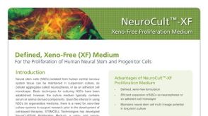 产品手册NeuroCult™-XF: Xeno-Free Culture Medium for the Proliferation of Human Neural Stem Cells
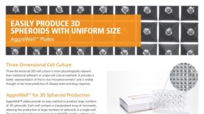 产品手册AggreWell™ for Spheroids
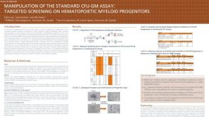 科学海报Manipulation of the Standard CFU-GM Assay Targeted Screening of Hematopoietic Myeloid Progenitors


 沪公网安备31010102008431号