Jiang P et al. (OCT 2014)
British Journal of Cancer 111 8 1562--1571
In vitro and in vivo anticancer effects of mevalonate pathway modulation on human cancer cells
BACKGROUND The increasing usage of statins (the 3-hydroxy-3-methylglutaryl-coenzyme A reductase inhibitors) has revealed a number of unexpected beneficial effects,including a reduction in cancer risk. METHODS We investigated the direct anticancer effects of different statins approved for clinical use on human breast and brain cancer cells. We also explored the effects of statins on cancer cells using in silico simulations. RESULTS In vitro studies showed that cerivastatin,pitavastatin,and fluvastatin were the most potent anti-proliferative,autophagy inducing agents in human cancer cells including stem cell-like primary glioblastoma cell lines. Consistently,pitavastatin was more effective than fluvastatin in inhibiting U87 tumour growth in vivo. Intraperitoneal injection was much better than oral administration in delaying glioblastoma growth. Following statin treatment,tumour cells were rescued by adding mevalonate and geranylgeranyl pyrophosphate. Knockdown of geranylgeranyl pyrophosphate synthetase-1 also induced strong cell autophagy and cell death in vitro and reduced U87 tumour growth in vivo. These data demonstrate that statins main effect is via targeting the mevalonate synthesis pathway in tumour cells. CONCLUSIONS Our study demonstrates the potent anticancer effects of statins. These safe and well-tolerated drugs need to be further investigated as cancer chemotherapeutics in comprehensive clinical studies.
View Publication
产品类型:
产品号#:
05700
05702
产品名:
NeuroCult™ 基础培养基(小鼠&大鼠)
NeuroCult™ 扩增试剂盒 (小鼠&大鼠)
Su H et al. (JUL 2013)
Stem Cell Research 11 1 529--539
Transplanted motoneurons derived from human induced pluripotent stem cells form functional connections with target muscle
Induced pluripotent stem cells (iPSCs) hold promise for the treatment of motoneuron diseases because of their distinct features including pluripotency,self-derivation and potential ability to differentiate into motoneurons. However,it is still unknown whether human iPSC-derived motoneurons can functionally innervate target muscles in vivo,which is the definitive sign of successful cell therapy for motoneuron diseases. In the present study,we demonstrated that human iPSCs derived from mesenchymal cells of the umbilical cord possessed a high yield in neural differentiation. Using a chemically-defined in vitro system,human iPSCs efficiently differentiated into motoneurons which displayed typical morphology,expressed specific molecules,and generated repetitive trains of action potentials. When transplanted into the injured musculocutaneous nerve of rats,they survived robustly,extended axons along the nerve,and formed functional connections with the target muscle (biceps brachii),thereby protecting the muscle from atrophy. Our study provides evidence for the first time that human iPSC-derived motoneurons are truly functional not only in vitro but also in vivo,and they have potential for stem cell-based therapies for motoneuron diseases. textcopyright 2013 Elsevier B.V.
View Publication
Francis KR et al. (APR 2016)
Nature medicine 22 4 388--396
Modeling Smith-Lemli-Opitz syndrome with induced pluripotent stem cells reveals a causal role for Wnt/$$-catenin defects in neuronal cholesterol synthesis phenotypes.
Smith-Lemli-Opitz syndrome (SLOS) is a malformation disorder caused by mutations in DHCR7,which impair the reduction of 7-dehydrocholesterol (7DHC) to cholesterol. SLOS results in cognitive impairment,behavioral abnormalities and nervous system defects,though neither affected cell types nor impaired signaling pathways are fully understood. Whether 7DHC accumulation or cholesterol loss is primarily responsible for disease pathogenesis is also unclear. Using induced pluripotent stem cells (iPSCs) from subjects with SLOS,we identified cellular defects that lead to precocious neuronal specification within SLOS derived neural progenitors. We also demonstrated that 7DHC accumulation,not cholesterol deficiency,is critical for SLOS-associated defects. We further identified downregulation of Wnt/$$-catenin signaling as a key initiator of aberrant SLOS iPSC differentiation through the direct inhibitory effects of 7DHC on the formation of an active Wnt receptor complex. Activation of canonical Wnt signaling prevented the neural phenotypes observed in SLOS iPSCs,suggesting that Wnt signaling may be a promising therapeutic target for SLOS.
View Publication
产品类型:
产品号#:
07923
85850
85857
产品名:
Dispase (1 U/mL)
mTeSR™1
mTeSR™1
Nakamura H et al. (OCT 2013)
Herpesviridae 4 1 2
Human cytomegalovirus induces apoptosis in neural stem/progenitor cells derived from induced pluripotent stem cells by generating mitochondrial dysfunction and endoplasmic reticulum stress
BACKGROUND Congenital human cytomegalovirus (HCMV) infection,a leading cause of birth defects,is most often manifested as neurological disorders. The pathogenesis of HCMV-induced neurological disorders is,however,largely unresolved,primarily because of limited availability of model systems to analyze the effects of HCMV infection on neural cells. METHODS An induced pluripotent stem cell (iPSC) line was established from the human fibroblast line MRC5 by introducing the Yamanaka's four factors and then induced to differentiate into neural stem/progenitor cells (NSPCs) by dual inhibition of the SMAD signaling pathway using Noggin and SB-431542. RESULTS iPSC-derived NSPCs (NSPC/iPSCs) were susceptible to HCMV infection and allowed the expression of both early and late viral gene products. HCMV-infected NSPC/iPSCs underwent apoptosis with the activation of caspase-3 and -9 as well as positive staining by the terminal deoxynucleotidyl transferase-mediated dUTP nick-end labeling (TUNEL). Cytochrome c release from mitochondria to cytosol was observed in these cells,indicating the involvement of mitochondrial dysfunction in their apoptosis. In addition,phosphorylation of proteins involved in the unfolded protein response (UPR),such as PKR-like eukaryotic initiation factor 2a kinase (PERK),c-Jun NH2-terminal kinase (JNK),inositol-requiring enzyme 1 (IRE1),and the alpha subunit of eukaryotic initiation factor 2 (eIF2$$) was observed in HCMV-infected NSPC/iPSCs. These results,coupled with the finding of increased expression of mRNA encoding the C/EBP-homologous protein (CHOP) and the detection of a spliced form of X-box binding protein 1 (XBP1) mRNA,suggest that endoplasmic reticulum (ER) stress is also involved in HCMV-induced apoptosis of these cells. CONCLUSIONS iPSC-derived NSPCs are thought to be a useful model to study HCMV neuropathogenesis and to analyze the mechanisms of HCMV-induced apoptosis in neural cells.
View Publication
产品类型:
产品号#:
85850
85857
产品名:
mTeSR™1
mTeSR™1
B. S. Souza et al. (dec 2016)
Scientific Reports 6 1 39775
Zika virus infection induces mitosis abnormalities and apoptotic cell death of human neural progenitor cells
Zika virus (ZIKV) infection has been associated with severe complications both in the developing and adult nervous system. To investigate the deleterious effects of ZIKV infection,we used human neural progenitor cells (NPC),derived from induced pluripotent stem cells (iPSC). We found that NPC are highly susceptible to ZIKV and the infection results in cell death. ZIKV infection led to a marked reduction in cell proliferation,ultrastructural alterations and induction of autophagy. Induction of apoptosis of Sox2 + cells was demonstrated by activation of caspases 3/7,8 and 9,and by ultrastructural and flow cytometry analyses. ZIKV-induced death of Sox2 + cells was prevented by incubation with the pan-caspase inhibitor,Z-VAD-FMK. By confocal microscopy analysis we found an increased number of cells with supernumerary centrosomes. Live imaging showed a significant increase in mitosis abnormalities,including multipolar spindle,chromosome laggards,micronuclei and death of progeny after cell division. FISH analysis for chromosomes 12 and 17 showed increased frequency of aneuploidy,such as monosomy,trisomy and polyploidy. Our study reinforces the link between ZIKV and abnormalities in the developing human brain,including microcephaly.
View Publication
产品类型:
产品号#:
05832
05833
19851
19851RF
19852
19852RF
19854
19854RF
05835
05839
产品名:
STEMdiff™ 神经花环选择试剂
STEMdiff™神经前体细胞培养基
EasySep™小鼠T细胞分选试剂盒
RoboSep™ 小鼠T细胞分选试剂盒
EasySep™小鼠CD4+ T细胞分选试剂盒
RoboSep™ 小鼠CD4+ T细胞分选试剂盒
EasySep™小鼠B细胞分选试剂盒
RoboSep™ 小鼠B细胞分选试剂盒
STEMdiff™ 神经诱导培养基
STEMdiff™ 神经诱导培养基
Wattanapanitch M et al. (SEP 2014)
PloS one 9 9 e106952
Dual small-molecule targeting of SMAD signaling stimulates human induced pluripotent stem cells toward neural lineages.
Incurable neurological disorders such as Parkinson's disease (PD),Huntington's disease (HD),and Alzheimer's disease (AD) are very common and can be life-threatening because of their progressive disease symptoms with limited treatment options. To provide an alternative renewable cell source for cell-based transplantation and as study models for neurological diseases,we generated induced pluripotent stem cells (iPSCs) from human dermal fibroblasts (HDFs) and then differentiated them into neural progenitor cells (NPCs) and mature neurons by dual SMAD signaling inhibitors. Reprogramming efficiency was improved by supplementing the histone deacethylase inhibitor,valproic acid (VPA),and inhibitor of p160-Rho associated coiled-coil kinase (ROCK),Y-27632,after retroviral transduction. We obtained a number of iPS colonies that shared similar characteristics with human embryonic stem cells in terms of their morphology,cell surface antigens,pluripotency-associated gene and protein expressions as well as their in vitro and in vivo differentiation potentials. After treatment with Noggin and SB431542,inhibitors of the SMAD signaling pathway,HDF-iPSCs demonstrated rapid and efficient differentiation into neural lineages. Six days after neural induction,neuroepithelial cells (NEPCs) were observed in the adherent monolayer culture,which had the ability to differentiate further into NPCs and neurons,as characterized by their morphology and the expression of neuron-specific transcripts and proteins. We propose that our study may be applied to generate neurological disease patient-specific iPSCs allowing better understanding of disease pathogenesis and drug sensitivity assays.
View Publication
产品类型:
产品号#:
07923
85850
85857
产品名:
Dispase (1 U/mL)
mTeSR™1
mTeSR™1
Belzile J-P et al. (APR 2014)
Journal of virology 88 8 4021--4039
Human cytomegalovirus infection of human embryonic stem cell-derived primitive neural stem cells is restricted at several steps but leads to the persistence of viral DNA.
UNLABELLED Congenital human cytomegalovirus (HCMV) infection is a major cause of central nervous system structural anomalies and sensory impairments. It is likely that the stage of fetal development,as well as the state of differentiation of susceptible cells at the time of infection,affects the severity of the disease. We used human embryonic stem (ES) cell-derived primitive prerosette neural stem cells (pNSCs) and neural progenitor cells (NPCs) maintained in chemically defined conditions to study HCMV replication in cells at the early stages of neural development. In contrast to what was observed previously using fetus-derived NPCs,infection of ES cell-derived pNSCs with HCMV was nonprogressive. At a low multiplicity of infection,we observed only a small percentage of cells expressing immediate-early genes (IE) and early genes. IE expression was found to be restricted to cells negative for the anterior marker FORSE-1,and treatment of pNSCs with retinoic acid restored IE expression. Differentiation of pNSCs into NPCs restored IE expression but not the transactivation of early genes. Virions produced in NPCs and pNSCs were exclusively cell associated and were mostly non-neural tropic. Finally,we found that viral genomes could persist in pNSC cultures for up to a month after infection despite the absence of detectable IE expression by immunofluorescence,and infectious virus could be produced upon differentiation of pNSCs to neurons. In conclusion,our results highlight the complex array of hurdles that HCMV must overcome in order to infect primitive neural stem cells and suggest that these cells might act as a reservoir for the virus. IMPORTANCE Human cytomegalovirus (HCMV) is a betaherpesvirus that is highly prevalent in the population. HCMV infection is usually asymptomatic but can lead to severe consequences in immunosuppressed individuals. HCMV is also the most important infectious cause of congenital developmental birth defects. Manifestations of fetal HCMV disease range from deafness and learning disabilities to more severe symptoms such as microcephaly. In this study,we have used embryonic stem cells to generate primitive neural stem cells and have used these to model HCMV infection of the fetal central nervous system (CNS) in vitro. Our results reveal that these cells,which are similar to those present in the developing neural tube,do not support viral replication but instead likely constitute a viral reservoir. Future work will define the effect of viral persistence on cellular functions as well as the exogenous signals leading to the reactivation of viral replication in the CNS.
View Publication
Yamamizu K et al. (DEC 2013)
Stem Cell Reports 1 6 545--559
Identification of Transcription Factors for Lineage-Specific ESC Differentiation
A network of transcription factors (TFs) determines cell identity,but identity can be altered by overexpressing a combination of TFs. However,choosing and verifying combinations of TFs for specific cell differentiation have been daunting due to the large number of possible combinations of 2,000 TFs. Here,we report the identification of individual TFs for lineage-specific cell differentiation based on the correlation matrix of global gene expression profiles. The overexpression of identified TFs-Myod1,Mef2c,Esx1,Foxa1,Hnf4a,Gata2,Gata3,Myc,Elf5,Irf2,Elf1,Sfpi1,Ets1,Smad7,Nr2f1,Sox11,Dmrt1,Sox9,Foxg1,Sox2,or Ascl1-can direct efficient,specific,and rapid differentiation into myocytes,hepatocytes,blood cells,and neurons. Furthermore,transfection of synthetic mRNAs of TFs generates their appropriate target cells. These results demonstrate both the utility of this approach to identify potent TFs for cell differentiation,and the unanticipated capacity of single TFs directly guides differentiation to specific lineage fates.
View Publication

 EasySep™小鼠TIL(CD45)正选试剂盒


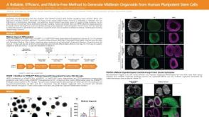 科学海报A Reliable, Efficient, and Matrix-Free Method to Generate Midbrain Organoids from Human Pluripotent Stem Cells
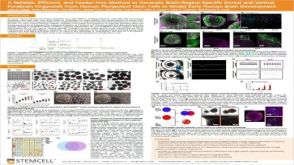 科学海报A Reliable, Efficient, and Feeder-Free Method to Generate Brain-Region-Specific Dorsal and Ventral Forebrain Organoids From Human Pluripotent Stem Cells to Model Early Human Brain Development
 科学海报A Human Pluripotent Stem Cell-Derived Organoid Model for Recapitulation of Central Nervous System (CNS) Barrier and Fluid Secretion Functions of the Choroid Plexus
 实验方案How to Tri-Culture Astrocytes, Microglia, and NGN2 mRNA-LNP-Induced Forebrain Neurons Derived from Human Pluripotent Stem Cells
 沪公网安备31010102008431号