Xia G et al. (JUN 2015)
Stem cells (Dayton,Ohio) 33 6 1829--38
Genome modification leads to phenotype reversal in human myotonic dystrophy type 1 induced pluripotent stem cell-derived neural stem cells.
Myotonic dystrophy type 1 (DM1) is caused by expanded CTG repeats in the 3'-untranslated region (3' UTR) of the DMPK gene. Correcting the mutation in DM1 stem cells would be an important step toward autologous stem cell therapy. The objective of this study is to demonstrate in vitro genome editing to prevent production of toxic mutant transcripts and reverse phenotypes in DM1 stem cells. Genome editing was performed in DM1 neural stem cells (NSCs) derived from human DM1 induced pluripotent stem (iPS) cells. An editing cassette containing SV40/bGH polyA signals was integrated upstream of the CTG repeats by TALEN-mediated homologous recombination (HR). The expression of mutant CUG repeats transcript was monitored by nuclear RNA foci,the molecular hallmarks of DM1,using RNA fluorescence in situ hybridization. Alternative splicing of microtubule-associated protein tau (MAPT) and muscleblind-like (MBNL) proteins were analyzed to further monitor the phenotype reversal after genome modification. The cassette was successfully inserted into DMPK intron 9 and this genomic modification led to complete disappearance of nuclear RNA foci. MAPT and MBNL 1,2 aberrant splicing in DM1 NSCs were reversed to normal pattern in genome-modified NSCs. Genome modification by integration of exogenous polyA signals upstream of the DMPK CTG repeat expansion prevents the production of toxic RNA and leads to phenotype reversal in human DM1 iPS-cells derived stem cells. Our data provide proof-of-principle evidence that genome modification may be used to generate genetically modified progenitor cells as a first step toward autologous cell transfer therapy for DM1.
View Publication
产品类型:
产品号#:
05833
05835
05839
产品名:
STEMdiff™神经前体细胞培养基
STEMdiff™ 神经诱导培养基
STEMdiff™ 神经诱导培养基
Vazin T et al. (FEB 2014)
Neurobiology of Disease 62 62--72
Efficient derivation of cortical glutamatergic neurons from human pluripotent stem cells: a model system to study neurotoxicity in Alzheimer's disease.
Alzheimer's disease (AD) is among the most prevalent forms of dementia affecting the aging population,and pharmacological therapies to date have not been successful in preventing disease progression. Future therapeutic efforts may benefit from the development of models that enable basic investigation of early disease pathology. In particular,disease-relevant models based on human pluripotent stem cells (hPSCs) may be promising approaches to assess the impact of neurotoxic agents in AD on specific neuronal populations and thereby facilitate the development of novel interventions to avert early disease mechanisms. We implemented an efficient paradigm to convert hPSCs into enriched populations of cortical glutamatergic neurons emerging from dorsal forebrain neural progenitors,aided by modulating Sonic hedgehog (Shh) signaling. Since AD is generally known to be toxic to glutamatergic circuits,we exposed glutamatergic neurons derived from hESCs to an oligomeric pre-fibrillar forms of Aβ known as globulomers"�
View Publication
产品类型:
产品号#:
05850
05857
05870
05875
85850
85857
85870
85875
产品名:
mTeSR™1
mTeSR™1
Drury-Stewart D et al. (AUG 2013)
Stem cell research & therapy 4 4 93
Highly efficient differentiation of neural precursors from human embryonic stem cells and benefits of transplantation after ischemic stroke in mice.
INTRODUCTION: Ischemic stroke is a leading cause of death and disability,but treatment options are severely limited. Cell therapy offers an attractive strategy for regenerating lost tissues and enhancing the endogenous healing process. In this study,we investigated the use of human embryonic stem cell-derived neural precursors as a cell therapy in a murine stroke model.backslashnbackslashnMETHODS: Neural precursors were derived from human embryonic stem cells by using a fully adherent SMAD inhibition protocol employing small molecules. The efficiency of neural induction and the ability of these cells to further differentiate into neurons were assessed by using immunocytochemistry. Whole-cell patch-clamp recording was used to demonstrate the electrophysiological activity of human embryonic stem cell-derived neurons. Neural precursors were transplanted into the core and penumbra regions of a focal ischemic stroke in the barrel cortex of mice. Animals received injections of bromodeoxyuridine to track regeneration. Neural differentiation of the transplanted cells and regenerative markers were measured by using immunohistochemistry. The adhesive removal test was used to determine functional improvement after stroke and intervention.backslashnbackslashnRESULTS: After 11 days of neural induction by using the small-molecule protocol,over 95% of human embryonic stem-derived cells expressed at least one neural marker. Further in vitro differentiation yielded cells that stained for mature neuronal markers and exhibited high-amplitude,repetitive action potentials in response to depolarization. Neuronal differentiation also occurred after transplantation into the ischemic cortex. A greater level of bromodeoxyuridine co-localization with neurons was observed in the penumbra region of animals receiving cell transplantation. Transplantation also improved sensory recovery in transplant animals over that in control animals.backslashnbackslashnCONCLUSIONS: Human embryonic stem cell-derived neural precursors derived by using a highly efficient small-molecule SMAD inhibition protocol can differentiate into electrophysiologically functional neurons in vitro. These cells also differentiate into neurons in vivo,enhance regenerative activities,and improve sensory recovery after ischemic stroke.
View Publication
Androgenetic embryonic stem cells form neural progenitor cells in vivo and in vitro.
Uniparental zygotes with two paternal (androgenetic [AG]) or two maternal (gynogenetic [GG]; parthenogenetic [PG]) genomes are not able to develop into viable offspring but can form blastocysts from which embryonic stem cells (ESCs) can be derived. Although some aspects of the in vitro and in vivo differentiation potential of PG and GG ESCs of several species have been studied,the developmental capacity of AG ESCs is much less clear. Here,we investigate the potential of murine AG ESCs to undergo neural differentiation. We observed that AG ESCs differentiate in vitro into pan-neural progenitor cells (pnPCs) that further give rise to cells that express neuronal- and astroglial-specific markers. Neural progeny of in vitro-differentiated AG ESCs exhibited fidelity of expression of six imprinted genes analyzed,with the exception of Ube3a. Bisulfite sequencing for two imprinting control regions suggested that pnPCs predominantly maintained their methylation pattern. Following blastocyst injection of AG and biparental (normal fertilized [N]) ESCs,we found widespread and evenly distributed contribution of ESC-derived cells in both AG and N chimeric early fetal brains. AG and N ESC-derived cells isolated from chimeric fetal brains by fluorescence-activated cell sorting exhibited similar neurosphere-initiating cell frequencies and neural multilineage differentiation potential. Our results indicate that AG ESC-derived neural progenitor/stem cells do not differ from N neural progenitor/stem cells in their self-renewal and neural multilineage differentiation potential. Disclosure of potential conflicts of interest is found at the end of this article.
View Publication
产品类型:
产品号#:
05703
产品名:
NeuroCult™ 分化添加物(小鼠和大鼠)
M. S. Fernandopulle et al. (JUN 2018)
Current protocols in cell biology 79 1 e51
Transcription Factor-Mediated Differentiation of Human iPSCs into Neurons.
Accurate modeling of human neuronal cell biology has been a long-standing challenge. However,methods to differentiate human induced pluripotent stem cells (iPSCs) to neurons have recently provided experimentally tractable cell models. Numerous methods that use small molecules to direct iPSCs into neuronal lineages have arisen in recent years. Unfortunately,these methods entail numerous challenges,including poor efficiency,variable cell type heterogeneity,and lengthy,expensive differentiation procedures. We recently developed a new method to generate stable transgenic lines of human iPSCs with doxycycline-inducible transcription factors at safe-harbor loci. Using a simple two-step protocol,these lines can be inducibly differentiated into either cortical (i3 Neurons) or lower motor neurons (i3 LMN) in a rapid,efficient,and scalable manner (Wang et al.,2017). In this manuscript,we describe a set of protocols to assist investigators in the culture and genetic engineering of iPSC lines to enable transcription factor-mediated differentiation of iPSCs into i3 Neurons or i3 LMNs,and we present neuronal culture conditions for various experimental applications. {\textcopyright} 2018 by John Wiley & Sons,Inc.
View Publication
产品类型:
产品号#:
07920
07922
05790
05792
05793
05794
05795
产品名:
ACCUTASE™
ACCUTASE™
BrainPhys™神经元培养基
BrainPhys™神经元培养基和SM1试剂盒
BrainPhys™ 神经元培养基N2-A和SM1试剂盒
BrainPhys™原代神经元试剂盒
BrainPhys™ hPSC 神经元试剂盒
Matthews TA et al. (JAN 2014)
Brain Research 1543 28--37
Expression of the CHOP-inducible carbonic anhydrase CAVI-b is required for BDNF-mediated protection from hypoxia
Carbonic anhydrases (CAs) comprise a family of zinc-containing enzymes that catalyze the reversible hydration of carbon dioxide. CAs contribute to a myriad of physiological processes,including pH regulation,anion transport and water balance. To date,16 known members of the mammalian alpha-CA family have been identified. Given that the catalytic family members share identical reaction chemistry,their physiologic roles are influenced greatly by their tissue and sub-cellular locations. CAVI is the lone secreted CA and exists in both saliva and the gastrointestinal mucosa. An alternative,stress-inducible isoform of CAVI (CAVI-b) has been shown to be expressed from a cryptic promoter that is activated by the CCAAT/Enhancer-Binding Protein Homologous Protein (CHOP). The CAVI-b isoform is not secreted and is currently of unknown physiological function. Here we use neuronal models,including a model derived using Car6 and CHOP gene ablations,to delineate a role for CAVI-b in ischemic protection. Our results demonstrate that CAVI-b expression,which is increased through CHOP-signaling in response to unfolded protein stress,is also increased by oxygen-glucose deprivation (OGD). While enforced expression of CAVI-b is not sufficient to protect against ischemia,CHOP regulation of CAVI-b is necessary for adaptive changes mediated by BDNF that reduce subsequent ischemic damage. These results suggest that CAVI-b comprises a necessary component of a larger adaptive signaling pathway downstream of CHOP.
View Publication
产品类型:
产品号#:
05700
05701
05702
05703
05704
产品名:
NeuroCult™ 基础培养基(小鼠和大鼠)
NeuroCult™ 扩增添加物(小鼠和大鼠)
NeuroCult™扩增试剂盒(小鼠和大鼠)
NeuroCult™ 分化添加物(小鼠和大鼠)
NeuroCult™ 分化试剂盒(小鼠和大鼠)
Wang L et al. (NOV 2008)
PLoS Biology 6 11 e289
Gamma-Secretase Represents a Therapeutic Target for the Treatment of Invasive Glioma Mediated by the p75 Neurotrophin Receptor
The multifunctional signaling protein p75 neurotrophin receptor (p75(NTR)) is a central regulator and major contributor to the highly invasive nature of malignant gliomas. Here,we show that neurotrophin-dependent regulated intramembrane proteolysis (RIP) of p75(NTR) is required for p75(NTR)-mediated glioma invasion,and identify a previously unnamed process for targeted glioma therapy. Expression of cleavage-resistant chimeras of p75(NTR) or treatment of animals bearing p75(NTR)-positive intracranial tumors with clinically applicable gamma-secretase inhibitors resulted in dramatically decreased glioma invasion and prolonged survival. Importantly,proteolytic processing of p75(NTR) was observed in p75(NTR)-positive patient tumor specimens and brain tumor initiating cells. This work highlights the importance of p75(NTR) as a therapeutic target,suggesting that gamma-secretase inhibitors may have direct clinical application for the treatment of malignant glioma.
View Publication
产品类型:
产品号#:
05750
05751
产品名:
NeuroCult™ NS-A 基础培养基(人)
NeuroCult™ NS-A 扩增试剂盒(人)
Abeysinghe HCS et al. (SEP 2015)
Stem cell research & therapy 6 1 186
Pre-differentiation of human neural stem cells into GABAergic neurons prior to transplant results in greater repopulation of the damaged brain and accelerates functional recovery after transient ischemic stroke.
INTRODUCTION Despite attempts to prevent brain injury during the hyperacute phase of stroke,most sufferers end up with significant neuronal loss and functional deficits. The use of cell-based therapies to recover the injured brain offers new hope. In the current study,we employed human neural stem cells (hNSCs) isolated from subventricular zone (SVZ),and directed their differentiation into GABAergic neurons followed by transplantation to ischemic brain. METHODS Pre-differentiated GABAergic neurons,undifferentiated SVZ-hNSCs or media alone were stereotaxically transplanted into the rat brain (n=7/group) 7 days after endothelin-1 induced stroke. Neurological outcome was assessed by neurological deficit scores and the cylinder test. Transplanted cell survival,cellular phenotype and maturation were assessed using immunohistochemistry and confocal microscopy. RESULTS Behavioral assessments revealed accelerated improvements in motor function 7 days post-transplant in rats treated with pre-differentiated GABAergic cells in comparison to media alone and undifferentiated hNSC treated groups. Histopathology 28 days-post transplant indicated that pre-differentiated cells maintained their GABAergic neuronal phenotype,showed evidence of synaptogenesis and up-regulated expression of both GABA and calcium signaling proteins associated with neurotransmission. Rats treated with pre-differentiated cells also showed increased neurogenic activity within the SVZ at 28 days,suggesting an additional trophic role of these GABAergic cells. In contrast,undifferentiated SVZ-hNSCs predominantly differentiated into GFAP-positive astrocytes and appeared to be incorporated into the glial scar. CONCLUSION Our study is the first to show enhanced exogenous repopulation of a neuronal phenotype after stroke using techniques aimed at GABAergic cell induction prior to delivery that resulted in accelerated and improved functional recovery.
View Publication
Azari H et al. (JAN 2011)
Journal of visualized experiments : JoVE 49
Neural-colony forming cell assay: an assay to discriminate bona fide neural stem cells from neural progenitor cells.
The neurosphere assay (NSA) is one of the most frequently used methods to isolate,expand and also calculate the frequency of neural stem cells (NSCs). Furthermore,this serum-free culture system has also been employed to expand stem cells and determine their frequency from a variety of tumors and normal tissues. It has been shown recently that a one-to-one relationship does not exist between neurosphere formation and NSCs. This suggests that the NSA as currently applied,overestimates the frequency of NSCs in a mixed population of neural precursor cells isolated from both the embryonic and adult mammalian brain. This video practically demonstrates a novel collagen based semi- solid assay,the neural-colony forming cell assay (N-CFCA),which has the ability to discriminate stem from progenitor cells based on their long-term proliferative potential,and thus provides a method to enumerate NSC frequency. In the N-CFCA,colonies ≥2 mm in diameter are derived from cells that meet all the functional criteria of a NSC,while colonies textless 2mm are derived from progenitors. The N-CFCA procedure can be used for cells prepared from different sources including primary and cultured adult or embryonic mouse CNS cells. Here we use cells prepared from passage one neurospheres generated from embryonic day 14 mice brain to perform N-CFCA. The cultures are replenished with proliferation medium every seven days for three weeks to allow the plated cells to exhibit their full proliferative potential and then the frequency of neural progenitor and bona fide neural stem cells is calculated respectively by counting the number of colonies that are textless 2mm and the ones that are ≥2mm in reference to the number of cells that were initially plated.
View Publication

 EasySep™小鼠TIL(CD45)正选试剂盒


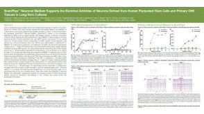 科学海报BrainPhys™ Neuronal Medium Supports the Electrical Activities of Neurons Derived from Human Pluripotent Stem Cells and Primary CNS Tissues in Long-Term Cultures
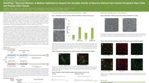 科学海报BrainPhys™ Neuronal Medium: A Medium Optimized to Support the Synaptic Activity of Neurons Derived from Human Pluripotent Stem Cells and Primary CNS Tissues
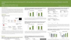 科学海报Complete Serum-Free Culture Kit and Protocols for Culturing High Yields of Functional Mature Neurons from Primary Embryonic Mouse CNS Tissues
 沪公网安备31010102008431号