Su CTE et al. (FEB 2015)
Journal of visualized experiments : JoVE 96 1--9
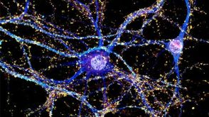
An Optogenetic Approach for Assessing Formation of Neuronal Connections in a Co-culture System.
Here we describe a protocol to generate a co-culture consisting of 2 different neuronal populations. Induced pluripotent stem cells (iPSCs) are reprogrammed from human fibroblasts using episomal vectors. Colonies of iPSCs can be observed 30 days after initiation of fibroblast reprogramming. Pluripotent colonies are manually picked and grown in neural induction medium to permit differentiation into neural progenitor cells (NPCs). iPSCs rapidly convert into neuroepithelial cells within 1 week and retain the capability to self-renew when maintained at a high culture density. Primary mouse NPCs are differentiated into astrocytes by exposure to a serum-containing medium for 7 days and form a monolayer upon which embryonic day 18 (E18) rat cortical neurons (transfected with channelrhodopsin-2 (ChR2)) are added. Human NPCs tagged with the fluorescent protein,tandem dimer Tomato (tdTomato),are then seeded onto the astrocyte/cortical neuron culture the following day and allowed to differentiate for 28 to 35 days. We demonstrate that this system forms synaptic connections between iPSC-derived neurons and cortical neurons,evident from an increase in the frequency of synaptic currents upon photostimulation of the cortical neurons. This co-culture system provides a novel platform for evaluating the ability of iPSC-derived neurons to create synaptic connections with other neuronal populations.
View Publication
产品类型:
产品号#:
05850
05857
05870
05875
85850
85857
85870
85875
产品名:
mTeSR™1
mTeSR™1
Rahman M et al. (MAR 2015)
Anatomy & cell biology 48 1 25--35
Neurosphere and adherent culture conditions are equivalent for malignant glioma stem cell lines.
Certain limitations of the neurosphere assay (NSA) have resulted in a search for alternative culture techniques for brain tumor-initiating cells (TICs). Recently,reports have described growing glioblastoma (GBM) TICs as a monolayer using laminin. We performed a side-by-side analysis of the NSA and laminin (adherent) culture conditions to compare the growth and expansion of GBM TICs. GBM cells were grown using the NSA and adherent culture conditions. Comparisons were made using growth in culture,apoptosis assays,protein expression,limiting dilution clonal frequency assay,genetic affymetrix analysis,and tumorigenicity in vivo. In vitro expansion curves for the NSA and adherent culture conditions were virtually identical (P=0.24) and the clonogenic frequencies (5.2% for NSA vs. 5.0% for laminin,P=0.9) were similar as well. Likewise,markers of differentiation (glial fibrillary acidic protein and beta tubulin III) and proliferation (Ki67 and MCM2) revealed no statistical difference between the sphere and attachment methods. Several different methods were used to determine the numbers of dead or dying cells (trypan blue,DiIC,caspase-3,and annexin V) with none of the assays noting a meaningful variance between the two methods. In addition,genetic expression analysis with microarrays revealed no significant differences between the two groups. Finally,glioma cells derived from both methods of expansion formed large invasive tumors exhibiting GBM features when implanted in immune-compromised animals. A detailed functional,protein and genetic characterization of human GBM cells cultured in serum-free defined conditions demonstrated no statistically meaningful differences when grown using sphere (NSA) or adherent conditions. Hence,both methods are functionally equivalent and remain suitable options for expanding primary high-grade gliomas in tissue culture.
View Publication
产品类型:
产品号#:
05750
05751
产品名:
NeuroCult™ NS-A 基础培养基(人)
NeuroCult™ NS-A 扩增试剂盒(人)
Katori S et al. (JUL 2009)
The Journal of neuroscience : the official journal of the Society for Neuroscience 29 29 9137--47
Protocadherin-alpha family is required for serotonergic projections to appropriately innervate target brain areas.
Serotonergic axons from the raphe nuclei in the brainstem project to every region of the brain,where they make connections through their extensive terminal arborizations. This serotonergic innervation contributes to various normal behaviors and psychiatric disorders. The protocadherin-alpha (Pcdha) family of clustered protocadherins consists of 14 cadherin-related molecules generated from a single gene cluster. We found that the Pcdhas were strongly expressed in the serotonergic neurons. To elucidate their roles,we examined serotonergic fibers in a mouse mutant (Pcdha(Delta CR/Delta CR)) lacking the Pcdha cytoplasmic region-encoding exons,which are common to the gene cluster. In the first week after birth,the distribution pattern of serotonergic fibers in Pcdha(Delta CR/Delta CR) mice was similar to wild-type,but by 3 weeks of age,when the serotonergic axonal termini complete their arborizations,the distribution of the projections was abnormal. In some target regions,notably the globus pallidus and substantia nigra,the normally even distribution of serotonin axonal terminals was,in the mutants,dense at the periphery of each region,but sparse in the center. In the stratum lacunosum-molecular of the hippocampus,the mutants showed denser serotonergic innervation than in wild-type,and in the dentate gyrus of the hippocampus and the caudate-putamen,the innervation was sparser. Together,the abnormalities suggested that Pcdha proteins are important in the late-stage maturation of serotonergic projections. Further examination of alternatively spliced exons encoding the cytoplasmic tail showed that the A-type (but not the B-type) cytoplasmic tail was essential for the normal development of serotonergic projections.
View Publication
产品类型:
产品号#:
03800
03801
03802
03803
03804
03805
03806
产品名:
ClonaCell™-HY杂交瘤试剂盒
ClonaCell™-HY培养基A
ClonaCell™-HY 培养基 B
ClonaCell™-HY 培养基 C
ClonaCell™-HY 培养基 D
ClonaCell™-HY 培养基 E
ClonaCell™-HY PEG
Bravard A et al. (JAN 2015)
Nucleic acids research 43 2 904--16
The prion protein is critical for DNA repair and cell survival after genotoxic stress.
The prion protein (PrP) is highly conserved and ubiquitously expressed,suggesting that it plays an important physiological function. However,despite decades of investigation,this role remains elusive. Here,by using animal and cellular models,we unveil a key role of PrP in the DNA damage response. Exposure of neurons to a genotoxic stress activates PRNP transcription leading to an increased amount of PrP in the nucleus where it interacts with APE1,the major mammalian endonuclease essential for base excision repair,and stimulates its activity. Preventing the induction of PRNP results in accumulation of abasic sites in DNA and impairs cell survival after genotoxic treatment. Brains from Prnp(-/-) mice display a reduced APE1 activity and a defect in the repair of induced DNA damage in vivo. Thus,PrP is required to maintain genomic stability in response to genotoxic stresses.
View Publication
产品类型:
产品号#:
05700
05701
05702
产品名:
NeuroCult™ 基础培养基(小鼠和大鼠)
NeuroCult™ 扩增添加物(小鼠和大鼠)
NeuroCult™扩增试剂盒(小鼠和大鼠)
Mao J et al. (OCT 2015)
Aging Cell 14 5 784--796
A herbal medicine for Alzheimer's disease and its active constituents promote neural progenitor proliferation
Aberrant neural progenitor cell (NPC) proliferation and self-renewal have been linked to age-related neurodegeneration and neurodegenerative disorders including Alzheimer's disease (AD). Rhizoma Acori tatarinowii is a traditional Chinese herbal medicine against cognitive decline. In this study,we found that the extract of Rhizoma Acori tatarinowii (AT) and its active constituents,asarones,promote NPC proliferation. Oral administration of AT enhanced NPC proliferation and neurogenesis in the hippocampi of adult and aged mice as well as that of transgenic AD model mice. AT and its fractions also enhanced the proliferation of NPCs cultured in vitro. Further analysis identified α-asarone and β-asarone as the two active constituents of AT in promoting neurogenesis. Our mechanistic study revealed that AT and asarones activated extracellular signal-regulated kinase (ERK) but not Akt,two critical kinase cascades for neurogenesis. Consistently,the inhibition of ERK activities effectively blocked the enhancement of NPC proliferation by AT or asarones. Our findings suggest that AT and asarones,which can be orally administrated,could serve as preventive and regenerative therapeutic agents to promote neurogenesis against age-related neurodegeneration and neurodegenerative disorders.
View Publication
Hartfield EM et al. (FEB 2014)
PLoS ONE 9 2 e87388
Physiological characterisation of human iPS-derived dopaminergic neurons
Human induced pluripotent stem cells (hiPSCs) offer the potential to study otherwise inaccessible cell types. Critical to this is the directed differentiation of hiPSCs into functional cell lineages. This is of particular relevance to research into neurological disease,such as Parkinson's disease (PD),in which midbrain dopaminergic neurons degenerate during disease progression but are unobtainable until post-mortem. Here we report a detailed study into the physiological maturation over time of human dopaminergic neurons in vitro. We first generated and differentiated hiPSC lines into midbrain dopaminergic neurons and performed a comprehensive characterisation to confirm dopaminergic functionality by demonstrating dopamine synthesis,release,and re-uptake. The neuronal cultures include cells positive for both tyrosine hydroxylase (TH) and G protein-activated inward rectifier potassium channel 2 (Kir3.2,henceforth referred to as GIRK2),representative of the A9 population of substantia nigra pars compacta (SNc) neurons vulnerable in PD. We observed for the first time the maturation of the slow autonomous pace-making (textless10 Hz) and spontaneous synaptic activity typical of mature SNc dopaminergic neurons using a combination of calcium imaging and electrophysiology. hiPSC-derived neurons exhibited inositol tri-phosphate (IP3) receptor-dependent release of intracellular calcium from the endoplasmic reticulum in neuronal processes as calcium waves propagating from apical and distal dendrites,and in the soma. Finally,neurons were susceptible to the dopamine neuron-specific toxin 1-methyl-4-phenylpyridinium (MPP+) which reduced mitochondrial membrane potential and altered mitochondrial morphology. Mature hiPSC-derived dopaminergic neurons provide a neurophysiologically-defined model of previously inaccessible vulnerable SNc dopaminergic neurons to bridge the gap between clinical PD and animal models.
View Publication
On-demand optogenetic activation of human stem-cell-derived neurons
The widespread application of human stem-cell-derived neurons for functional studies is impeded by complicated differentiation protocols,immaturity,and deficient optogene expression as stem cells frequently lose transgene expression over time. Here we report a simple but precise Cre-loxP-based strategy for generating conditional,and thereby stable,optogenetic human stem-cell lines. These cells can be easily and efficiently differentiated into functional neurons,and optogene expression can be triggered by administering Cre protein to the cultures. This conditional expression system may be applied to stem-cell-derived neurons whenever timed transgene expression could help to overcome silencing at the stem-cell level.
View Publication
产品类型:
产品号#:
05711
05790
05792
05793
05794
05795
100-1281
产品名:
NeuroCult™ SM1 神经添加物
BrainPhys™神经元培养基
BrainPhys™神经元培养基和SM1试剂盒
BrainPhys™ 神经元培养基N2-A和SM1试剂盒
BrainPhys™原代神经元试剂盒
BrainPhys™ hPSC 神经元试剂盒
NeuroCult™ SM1 神经添加物
Poloni A et al. (JAN 2015)
Journal of Molecular Neuroscience 55 1 91--98
Glial-Like Differentiation Potential of Human Mature Adipocytes
The potential ability to differentiate dedifferentiated adipocytes into a neural lineage is attracting strong interest as an emerging method of producing model cells for the treatment of a variety of neurological diseases. Here,we describe the efficient conversion of dedifferentiated adipocytes into a neural-like cell population. These cells grew in neurosphere-like structures and expressed a high level of the early neuroectodermal marker Nestin. These neurospheres could proliferate and express stemness genes,suggesting that these cells could be committed to the neural lineage. After neural induction,NeuroD1,Sox1,Double Cortin,and Eno2 were not expressed. Patch clamp data did not reveal different electrophysiological properties,indicating the inability of these cells to differentiate into mature neurons. In contrast,the differentiated cells expressed a high level of CLDN11,as demonstrated using molecular method,and stained positively for the glial cell markers CLDN11 and GFAP,as demonstrated using immunocytochemistry. These data were confirmed by quantitative results for glial cell line-derived neurotrophic factor production,which showed a higher secretion level in neurospheres and the differentiated cells compared with the untreated cells. In conclusion,our data demonstrate morphological,molecular,and immunocytochemical evidence of initial neural differentiation of mature adipocytes,committing to a glial lineage.
View Publication

 EasySep™小鼠TIL(CD45)正选试剂盒


 实验方案How to Culture Primary Rodent Neurons for MEA Analysis Using the Maestro MEA™ System


 沪公网安备31010102008431号