Gallia GL et al. (FEB 2009)
Molecular cancer therapeutics 8 2 386--93
Inhibition of Akt inhibits growth of glioblastoma and glioblastoma stem-like cells.
A commonly activated signaling cascade in many human malignancies,including glioblastoma multiforme,is the Akt pathway. This pathway can be activated via numerous upstream alterations including genomic amplification of epidermal growth factor receptor,PTEN deletion,or PIK3CA mutations. In this study,we screened phosphatidylinositol 3-kinase/Akt small-molecule inhibitors in an isogenic cell culture system with an activated Akt pathway secondary to a PIK3CA mutation. One small molecule,A-443654,showed the greatest selective inhibition of cells with the mutant phenotype. Based on these findings,this inhibitor was screened in vitro against a panel of glioblastoma multiforme cell lines. All cell lines tested were sensitive to A-443654 with a mean IC(50) of approximately 150 nmol/L. An analogue of A-443654,methylated at a region that blocks Akt binding,was on average 36-fold less active. Caspase assays and dual flow cytometric analysis showed an apoptotic mechanism of cell death. A-443654 was further tested in a rat intracranial model of glioblastoma multiforme. Animals treated intracranially with polymers containing A-443654 had significantly extended survival compared with control animals; animals survived 79% and 43% longer than controls when A-443654-containing polymers were implanted simultaneously or in a delayed fashion,respectively. This small molecule also inhibited glioblastoma multiforme stem-like cells with similar efficacy compared with traditionally cultured glioblastoma multiforme cell lines. These results suggest that local delivery of an Akt small-molecule inhibitor is effective against experimental intracranial glioma,with no observed resistance to glioblastoma multiforme cells grown in stem cell conditions.
View Publication
产品类型:
产品号#:
05751
产品名:
NeuroCult™ NS-A 扩增试剂盒(人)
Badizadegan K et al. (NOV 2014)
AJP: Gastrointestinal and Liver Physiology 307 10 G1002--G1012
Presence of intramucosal neuroglial cells in normal and aganglionic human colon
The enteric nervous system (ENS) is composed of neural crest-derived neurons (also known as ganglion cells) the cell bodies of which are located in the submucosal and myenteric plexuses of the intestinal wall. Intramucosal ganglion cells are known to exist but are rare and often considered ectopic. Also derived from the neural crest are enteric glial cells that populate the ganglia and the associated nerves,as well as the lamina propria of the intestinal mucosa. In Hirschsprung disease (HSCR),ganglion cells are absent from the distal gut because of a failure of neural crest-derived progenitor cells to complete their rostrocaudal migration during embryogenesis. The fate of intramucosal glial cells in human HSCR is essentially unknown. We demonstrate a network of intramucosal cells that exhibit dendritic morphology typical of neurons and glial cells. These dendritic cells are present throughout the human gut and express Tuj1,S100,glial fibrillary acidic protein,CD56,synaptophysin,and calretinin,consistent with mixed or overlapping neuroglial differentiation. The cells are present in aganglionic colon from patients with HSCR,but with an altered immunophenotype. Coexpression of Tuj1 and HNK1 in this cell population supports a neural crest origin. These findings extend and challenge the current understanding of ENS microanatomy and suggest the existence of an intramucosal population of neural crest-derived cells,present in HSCR,with overlapping immunophenotype of neurons and glia. Intramucosal neuroglial cells have not been previously recognized,and their presence in HSCR poses new questions about ENS development and the pathobiology of HSCR that merit further investigation.
View Publication
CXCR7 Mediates Neural Progenitor Cells Migration to CXCL12 Independent of CXCR4
Neural progenitor cell (NPC) migration is an essential process for brain development,adult neurogenesis,and neuroregeneration after brain injury. Stromal cell-derived factor-1 (SDF-1,CXCL12) and its traditional receptor CXCR4 are well known to regulate NPC migration. However,the discovery of CXCR7,a newly identified CXCL12 receptor,adds to the dynamics of the existing CXCL12/CXCR4 pair. Antagonists for either CXCR4 or CXCR7 blocked CXCL12-mediated NPC migration in a transwell chemotaxis assay,suggesting that both receptors are required for CXCL12 action. We derived NPC cultures from Cxcr4 knockout (KO) mice and used transwell and stripe assays to determine the cell migration. NPCs derived from Cxcr4 KO mice polarized and migrated in response to CXCL12 gradient,suggesting that CXCR7 could serve as an independent migration receptor. Furthermore,Cxcr4 KO NPCs transplanted into the adult mouse striatum migrated in response to the adjacent injection of CXCL12,an effect that was blocked by a CXCR7 antagonist,suggesting that CXCR7 also mediates NPC migration in vivo. Molecular mechanism studies revealed that CXCR7 interact with Rac1 in the leading edge of the polarized NPCs in the absence of CXCR4. Both CXCR7 and Rac1 are required for extracellular signal-regulated kinases (ERK) 1/2 activation and subsequent NPC migration,indicating that CXCR7 could serve as a functional receptor in CXCL12-mediated NPC migration independent of CXCR4. Together these results reveal an essential role of CXCR7 for CXCL12-mediated NPC migration that will be important to understand neurogenesis during development and in adulthood.
View Publication
Although human induced pluripotent stem cells (hiPSCs) hold great potential for the study of human diseases affecting disparate cell types,they have been underutilized in seeking mechanistic insights into the pathogenesis of congenital craniofacial disorders. Craniofrontonasal syndrome (CFNS) is a rare X-linked disorder caused by mutations in EFNB1 and characterized by craniofacial,skeletal,and neurological anomalies. Heterozygous females are more severely affected than hemizygous males,a phenomenon termed cellular interference that involves mosaicism for EPHRIN-B1 function. Although the mechanistic basis for cellular interference in CFNS has been hypothesized to involve Eph/ephrin-mediated cell segregation,no direct evidence for this has been demonstrated. Here,by generating hiPSCs from CFNS patients,we demonstrate that mosaicism for EPHRIN-B1 expression induced by random X inactivation in heterozygous females results in robust cell segregation in human neuroepithelial cells,thus supplying experimental evidence that Eph/ephrin-mediated cell segregation is relevant to pathogenesis in human CFNS patients.
View Publication
产品类型:
产品号#:
05835
05839
08581
08582
产品名:
STEMdiff™ 神经诱导培养基
STEMdiff™ 神经诱导培养基
STEMdiff™SMADi神经诱导试剂盒
STEMdiff™SMADi神经诱导试剂盒,2套
Kim MY et al. (MAR 2017)
Oncology letters 13 3 1767--1774
Accumulation of low-dose BIX01294 promotes metastatic potential of U251 glioblastoma cells.
BIX01294 (Bix) is known to be a euchromatic histone-lysine N-methyltransferase 2 inhibitor and treatment with Bix suppresses cancer cell survival and proliferation. In the present study,it was observed that sequential treatment with low-dose Bix notably increases glioblastoma cell migration and metastasis. It was demonstrated that U251 cells sequentially treated with low-dose Bix exhibited induced characteristic changes in critical epithelial-mesenchymal transition (EMT) markers,including E-cadherin,N-cadherin,β-catenin and zinc finger protein SNAI2. Notably,sequential treatment with Bix also increased the expression of cancer stem cell-associated markers,including sex determining region Y-box 2,octamer-binding transcription factor 4 and cluster of differentiation 133. Neurosphere formation was significantly enhanced in cells sequentially treated with Bix,compared with control cells (control: P=0.011; single treatment of Bix,P=0.045). The results of the present study suggest that accumulation of low-dose Bix enhanced the migration and metastatic potential of glioblastoma cells by regulating EMT-associated gene expression,which may be the cause of the altered properties of glioblastoma stem cells.
View Publication
产品类型:
产品号#:
05750
产品名:
NeuroCult™ NS-A 基础培养基(人)
Stipcevic T et al. (DEC 2013)
Acta Neurologica Belgica 113 4 501--506
Stimulation of adult neural stem cells with a novel glycolipid biosurfactant
Glycolipids are amphipathic molecules which are highly expressed on cell membranes in skin and brain where they mediate several key cellular processes. Neural stem cells are defined as undifferentiated,proliferative,multipotential cells with extensive self-renewal and are responsive to brain injury. Di-rhamnolipid: α-L-rhamnopyranosyl-(1-2)α-L-rhamnopyranosyl-3-hydroxydecanoyl-3-hydroxydecanoic acid,also referred to as di-rhamnolipid BAC-3,is a glycolipid isolated from the bacteria Pseudomonas aeruginosa. In the previous studies,di-rhamnolipid enhanced dermal tissue healing and regeneration. The present study provides the first assessment of di-rhamnolipid,and glycolipid biosurfactants in general,on the nervous system. Treatment of neural stem cells isolated from the lateral ventricle of adult mice and cultured in defined media containing growth factors at 0.5 and 1 μg/ml of di-rhamnolipid increased the number of neurospheres (2.7- and 2.8-fold,respectively) compared to controls and this effect remained even after passaging in the absence of di-rhamnolipid. In addition,neural stem cells treated with di-rhamnolipid at 50 and 100 μg/ml in defined media supplemented with fetal calf serum and without growth factors exhibited increased cell viability,indicating an interaction between di-rhamnolipid and serum components in the regulation of neural stem cells and neuroprogenitors. Intracerebroventricular administration of di-rhamnolipid at 300 and 120 ng/day increased the number of neurospheres (1.3- and 1.63-fold,respectively) that could be derived from the anterior lateral ventricles of adult mice. These results indicate that di-rhamnolipid stimulates proliferation of neural stem cells and increases their endogenous pools which may have therapeutic potential in managing neurodegenerative or neuropsychiatric disorders and promoting nervous tissue regeneration following injury.
View Publication
产品类型:
产品号#:
05700
产品名:
NeuroCult™ 基础培养基(小鼠&大鼠)
Xu G et al. (MAY 2013)
Neuroscience 238 195--208
Functional analysis of platelet-derived growth factor receptor-β in neural stem/progenitor cells
Activation of neural stem/progenitor cells (NSPCs) is a potential therapeutic strategy of neurological disorders. In this study,NSPCs of subventricular zone were isolated and cultured from platelet-derived growth factor-β-receptor-knockout (PDGFR-β(-/-)) mice of postnatal day 1 (P1) and P28,and the roles of PDGFR-β were examined in these cells. In PDGFR-β-preserving control NSPCs,stem cell activities,such as numbers and diameters of secondary neurospheres,cell proliferation and survival rates,were significantly higher in P1 NSPCs than those in P28 NSPCs. In PDGFR-β(-/-) NSPCs,most of these parameters were decreased as compared with age-matched controls. Among them,the decrease of secondary neurosphere formation was most striking in P1 and P28 PDGFR-β(-/-) NSPCs and in P28 control NSPCs as compared with P1 control NSPCs. PCR-array and following quantitative real-time PCR (qRT-PCR) analyses demonstrated that expressions of fibroblast growth factor-2 (FGF2) and exons IV-IX of brain-derived neurotrophic factor (BDNF) were decreased,and noggin was increased in P1 PDGFR-β(-/-) as compared with P1 controls. Addition of BDNF rescued the number and diameter of secondary neurospheres in P1 PDGFR-β(-/-) NSPCs to similar levels as controls. The expressions of PDGFs and PDGFRs in control NSPCs were increased along with the differentiation-induction,where phosphorylated PDGFR-β was co-localized with neuronal and astrocyte differentiation markers. In controls,the neuronal differentiation was decreased,and the glial differentiation was increased from P1 to P28 NSPCs. Compared with P1 controls,neuronal differentiation was reduced in P1 PDGFR-β(-/-) NSPCs,whereas glial differentiation was comparable between the two genotypes. These results suggest that PDGFR-β signaling is important for the self-renewal and multipotency of NSPCs,particularly in neonatal NSPCs. BDNF,FGF2,and noggin may be involved in the effects of PDGFR-β signaling in these cells. Accordingly,the activation of PDGFR-β in NSPCs may be a novel therapeutic strategy of neurological diseases.
View Publication
K. B. Langer et al. (APR 2018)
Stem cell reports 10 4 1282--1293
Retinal Ganglion Cell Diversity and Subtype Specification from Human Pluripotent Stem Cells.
Retinal ganglion cells (RGCs) are the projection neurons of the retina and transmit visual information to postsynaptic targets in the brain. While this function is shared among nearly all RGCs,this class of cell is remarkably diverse,comprised of multiple subtypes. Previous efforts have identified numerous RGC subtypes in animal models,but less attention has been paid to human RGCs. Thus,efforts of this study examined the diversity of RGCs differentiated from human pluripotent stem cells (hPSCs) and characterized defined subtypes through the expression of subtype-specific markers. Further investigation of these subtypes was achieved using single-cell transcriptomics,confirming the combinatorial expression of molecular markers associated with these subtypes,and also provided insight into more subtype-specific markers. Thus,the results of this study describe the derivation of RGC subtypes from hPSCs and will support the future exploration of phenotypic and functional diversity within human RGCs.
View Publication
产品类型:
产品号#:
05790
05792
05793
05794
05795
85850
85857
产品名:
BrainPhys™神经元培养基
BrainPhys™神经元培养基和SM1试剂盒
BrainPhys™ 神经元培养基N2-A和SM1试剂盒
BrainPhys™原代神经元试剂盒
BrainPhys™ hPSC 神经元试剂盒
mTeSR™1
mTeSR™1
I. Canals et al. (SEP 2018)
Nature methods 15 9 693--696
Rapid and efficient induction of functional astrocytes from human pluripotent stem cells.
The derivation of astrocytes from human pluripotent stem cells is currently slow and inefficient. We demonstrate that overexpression of the transcription factors SOX9 and NFIB in human pluripotent stem cells rapidly and efficiently yields homogeneous populations of induced astrocytes. In our study these cells exhibited molecular and functional properties resembling those of adult human astrocytes and were deemed suitable for disease modeling. Our method provides new possibilities for the study of human astrocytes in health and disease.
View Publication

 EasySep™小鼠TIL(CD45)正选试剂盒


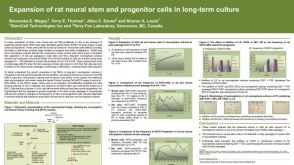 科学海报Expansion of Rat Neural Stem and Progenitor Cells in Long-Term Culture
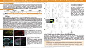 科学海报Generation of Functional 3D Spinal Cord Organoids from Human Pluripotent Stem Cells

 沪公网安备31010102008431号