Okabe S et al. (SEP 1996)
Mechanisms of development 59 1 89--102
Development of neuronal precursor cells and functional postmitotic neurons from embryonic stem cells in vitro.
To understand the mechanism of the sequential restriction of multipotency of stem cells during development,we have established culture conditions that allow the differentiation of neuroepithelial precursor cells from embryonic stem (ES) cells. A highly enriched population of neuroepithelial precursor cells derived from ES cells proliferates in the presence of basic fibroblast growth factor (bFGF). These cells differentiate into both neurons and glia following withdrawal of bFGF. By further differentiating the cells in serum-containing medium,the neurons express a wide variety of neuron-specific genes and generate both excitatory and inhibitory synaptic connections. The expression pattern of position-specific neural markers suggests the presence of a variety of central nervous system (CNS) neuronal cell types. These findings indicate that neuronal precursor cells can be isolated from ES cells and that these cells can efficiently differentiate into functional post-mitotic neurons of diverse CNS structures.
View Publication
Beckerman SR et al. (SEP 2015)
ASSAY and Drug Development Technologies 13 7 377--388
Phenotypic Assays to Identify Agents That Induce Reactive Gliosis: A Counter-Screen to Prioritize Compounds for Preclinical Animal Studies
Astrocyte phenotypes change in a process called reactive gliosis after traumatic central nervous system (CNS) injury. Astrogliosis is characterized by expansion of the glial fibrillary acidic protein (GFAP) cytoskeleton,adoption of stellate morphologies,and differential expression of some extracellular matrix molecules. The astrocytic response immediately after injury is beneficial,but in the chronic injury phase,reactive astrocytes produce inhibitory factors (i.e.,chondroitin sulfate proteoglycans [CSPGs]) that limit the regrowth of injured axons. There are no drugs that promote axon regeneration or functional recovery after CNS trauma in humans. To develop novel therapeutics for the injured CNS,we screened various libraries in a phenotypic assay to identify compounds that promote neurite outgrowth. However,the effects these compounds have on astrocytes are unknown. Specifically,we were interested in whether compounds could alter astrocytes in a manner that mimics the glial reaction to injury. To test this hypothesis,we developed cell-based phenotypic bioassays to measure changes in (1) GFAP morphology/localization and (2) CSPG expression/immunoreactivity from primary astrocyte cultures. These assays were optimized for six-point dose-response experiments in 96-well plates. The GFAP morphology assay is suitable for counter-screening with a Z-factor of 0.44±0.03 (mean±standard error of the mean; N=3 biological replicates). The CSPG assay is reproducible and informative,but does not satisfy common metrics for a screenable" assay. As proof of principle we tested a small set of hit compounds from our neurite outgrowth bioassay and identified one that can enhance axon growth without exacerbating the deleterious characteristics of reactive gliosis.
View Publication
产品类型:
产品号#:
05711
100-1281
产品名:
NeuroCult™ SM1 神经添加物
NeuroCult™ SM1 神经添加物
Chaumeil MM et al. ( 2016)
NeuroImage. Clinical 12 180--9
Hyperpolarized (13)C MR imaging detects no lactate production in mutant IDH1 gliomas: Implications for diagnosis and response monitoring.
Metabolic imaging of brain tumors using (13)C Magnetic Resonance Spectroscopy (MRS) of hyperpolarized [1-(13)C] pyruvate is a promising neuroimaging strategy which,after a decade of preclinical success in glioblastoma (GBM) models,is now entering clinical trials in multiple centers. Typically,the presence of GBM has been associated with elevated hyperpolarized [1-(13)C] lactate produced from [1-(13)C] pyruvate,and response to therapy has been associated with a drop in hyperpolarized [1-(13)C] lactate. However,to date,lower grade gliomas had not been investigated using this approach. The most prevalent mutation in lower grade gliomas is the isocitrate dehydrogenase 1 (IDH1) mutation,which,in addition to initiating tumor development,also induces metabolic reprogramming. In particular,mutant IDH1 gliomas are associated with low levels of lactate dehydrogenase A (LDHA) and monocarboxylate transporters 1 and 4 (MCT1,MCT4),three proteins involved in pyruvate metabolism to lactate. We therefore investigated the potential of (13)C MRS of hyperpolarized [1-(13)C] pyruvate for detection of mutant IDH1 gliomas and for monitoring of their therapeutic response. We studied patient-derived mutant IDH1 glioma cells that underexpress LDHA,MCT1 and MCT4,and wild-type IDH1 GBM cells that express high levels of these proteins. Mutant IDH1 cells and tumors produced significantly less hyperpolarized [1-(13)C] lactate compared to GBM,consistent with their metabolic reprogramming. Furthermore,hyperpolarized [1-(13)C] lactate production was not affected by chemotherapeutic treatment with temozolomide (TMZ) in mutant IDH1 tumors,in contrast to previous reports in GBM. Our results demonstrate the unusual metabolic imaging profile of mutant IDH1 gliomas,which,when combined with other clinically available imaging methods,could be used to detect the presence of the IDH1 mutation in vivo.
View Publication
产品类型:
产品号#:
05700
05750
05751
产品名:
NeuroCult™ 基础培养基(小鼠和大鼠)
NeuroCult™ NS-A 基础培养基(人)
NeuroCult™ NS-A 扩增试剂盒(人)
Choi SA et al. (NOV 2012)
Cancer Letters 324 2 221--230
A distinct subpopulation within CD133 positive brain tumor cells shares characteristics with endothelial progenitor cells
The cell surface marker CD133 has been proposed as a brain tumor stem cell marker. However,there have been substantial controversies regarding the necessity and role of CD133 in tumorigenesis. This study aimed to characterize CD133(+) cells in brain tumors. Human brain tumor specimens and whole blood were collected from the same patients (N=12). We carried out dual FACS staining for CD133/CD34 and functional tumorigenesis and angiogenesis analyses of CD133(+) cells from different origins. We also investigated the in vivo tumorigenic potential and histological characteristics of four distinct groups on the basis of expression of CD133/CD34 markers (CD133(+),CD133(+)/CD34(+),CD133(+)/CD34(-),and CD133(-)). CD133(+) brain tumor cells coexpressed significantly higher positivity for CD34 (70.7±5.2% in CD133(+) vs. 12.3±4.2% in CD133(-) cells,P<0.001). CD133(+) brain tumor cells formed neurosphere-like spheroids and differentiated into multiple nervous system lineages unlike CD133(+) blood cells. They showed biological characteristics of endothelial cells,including vWF expression,LDL uptake and tube formation in vitro,unlike CD133(-) brain tumors cells. Pathologic analysis of brains implanted with CD133(+) cells showed large,markedly hypervascular tumors with well-demarcated boundary. CD133(+)/CD34(-) cells produced smaller but highly infiltrative tumors. Notably,pure angiogenic cell fractions (CD133(+)/CD34(+)) and CD133(-) tumor cells did not generate tumors in vivo. Our data suggest the presence of a distinct subpopulation of CD133(+) cells isolated from human brain tumors,with characteristics of endothelial progenitor cells (EPCs).
View Publication
产品类型:
产品号#:
05750
05752
产品名:
NeuroCult™ NS-A 基础培养基(人)
NeuroCult™ NS-A 分化试剂盒(人)
Xia N et al. (FEB 2016)
Scientific Reports 6 20270
Transcriptional comparison of human induced and primary midbrain dopaminergic neurons
Generation of induced dopaminergic (iDA) neurons may provide a significant step forward towards cell replacement therapy for Parkinson's disease (PD). To study and compare transcriptional programs of induced cells versus primary DA neurons is a preliminary step towards characterizing human iDA neurons. We have optimized a protocol to efficiently generate iDA neurons from human pluripotent stem cells (hPSCs). We then sequenced the transcriptomes of iDA neurons derived from 6 different hPSC lines and compared them to that of primary midbrain (mDA) neurons. We identified a small subset of genes with altered expression in derived iDA neurons from patients with Parkinson's Disease (PD). We also observed that iDA neurons differ significantly from primary mDA neurons in global gene expression,especially in genes related to neuron maturation level. Results suggest iDA neurons from patient iPSCs could be useful for basic and translational studies,including in vitro modeling of PD. However,further refinement of methods of induction and maturation of neurons may better recapitulate full development of mDA neurons from hPSCs.
View Publication
产品类型:
产品号#:
05850
05857
05870
05875
85850
85857
85870
85875
产品名:
mTeSR™1
mTeSR™1
Pulvirenti T et al. (DEC 2011)
Cancer research 71 23 7280--90
Dishevelled 2 signaling promotes self-renewal and tumorigenicity in human gliomas.
Glioblastoma multiforme is the most common glioma variant in adults and is highly malignant. Tumors are thought to harbor a subpopulation of stem-like cancer cells,with the bulk resembling neural progenitor-like cells that are unable to fully differentiate. Although multiple pathways are known to be involved in glioma tumorigenesis,the role of Wnt signaling has been poorly described. Here,we show that Dishevelled 2 (Dvl2),a key component of the Wnt signaling pathway,is overexpressed in human gliomas. RNA interference-mediated depletion of Dvl2 blocked proliferation and promoted the differentiation of cultured human glioma cell lines and primary,patient-derived glioma cells. In addition,Dvl2 depletion inhibited tumor formation after intracranial injection of glioblastoma cells in immunodeficient mice. Inhibition of canonical Wnt/β-catenin signaling also blocked proliferation,but unlike Dvl2 depletion,did not induce differentiation. Finally,Wnt5a,a noncanonical Wnt ligand,was also required for glioma cell proliferation. The data therefore suggest that both canonical and noncanonical Wnt signaling pathways downstream of Dvl2 cooperate to maintain the proliferative capacity of human glioblastomas.
View Publication
产品类型:
产品号#:
05751
产品名:
NeuroCult™ NS-A 扩增试剂盒(人)
Keller GM (DEC 1995)
Current opinion in cell biology 7 6 862--9
In vitro differentiation of embryonic stem cells.
Under appropriate conditions in culture,embryonic stem cells will differentiate and form embryoid bodies that have been shown to contain cells of the hematopoietic,endothelial,muscle and neuronal lineages. Many aspects of the lineage-specific differentiation programs observed within the embryoid bodies reflect those found in the embryo,indicating that this model system provides access to early cell populations that develop in a normal fashion. Recent studies involving the differentiation of genetically altered embryonic stem cells highlight the potential of this in vitro differentiation system for defining the function of genes in early development.
View Publication
产品类型:
产品号#:
06902
06952
00321
00322
00323
00324
00325
产品名:
Amenduni M et al. (DEC 2011)
European Journal of Human Genetics 19131 10 1246--1255
ARTICLE iPS cells to model CDKL5-related disorders
Rett syndrome (RTT) is a progressive neurologic disorder representing one of the most common causes of mental retardation in females. To date mutations in three genes have been associated with this condition. Classic RTT is caused by mutations in the MECP2 gene,whereas variants can be due to mutations in either MECP2 or FOXG1 or CDKL5. Mutations in CDKL5 have been identified both in females with the early onset seizure variant of RTT and in males with X-linked epileptic encephalopathy. CDKL5 is a kinase protein highly expressed in neurons,but its exact function inside the cell is unknown. To address this issue we established a human cellular model for CDKL5-related disease using the recently developed technology of induced pluripotent stem cells (iPSCs). iPSCs can be expanded indefinitely and differentiated in vitro into many different cell types,including neurons. These features make them the ideal tool to study disease mechanisms directly on the primarily affected neuronal cells. We derived iPSCs from fibroblasts of one female with p.Q347X and one male with p.T288I mutation,affected by early onset seizure variant and X-linked epileptic encephalopathy,respectively. We demonstrated that female CDKL5-mutated iPSCs maintain X-chromosome inactivation and clones express either the mutant CDKL5 allele or the wild-type allele that serve as an ideal experimental control. Array CGH indicates normal isogenic molecular karyotypes without detection of de novo CNVs in the CDKL5-mutated iPSCs. Furthermore,the iPS cells can be differentiated into neurons and are thus suitable to model disease pathogenesis in vitro.
View Publication
Harris MA et al. (DEC 2008)
Cancer research 68 24 10051--9
Cancer stem cells are enriched in the side population cells in a mouse model of glioma.
The recent identification of cancer stem cells (CSCs) in multiple human cancers provides a new inroad to understanding tumorigenesis at the cellular level. CSCs are defined by their characteristics of self-renewal,multipotentiality,and tumor initiation upon transplantation. By testing for these defining characteristics,we provide evidence for the existence of CSCs in a transgenic mouse model of glioma,S100beta-verbB;Trp53. In this glioma model,CSCs are enriched in the side population (SP) cells. These SP cells have enhanced tumor-initiating capacity,self-renewal,and multipotentiality compared with non-SP cells from the same tumors. Furthermore,gene expression analysis comparing fluorescence-activated cell sorting-sorted cancer SP cells to non-SP cancer cells and normal neural SP cells identified 45 candidate genes that are differentially expressed in glioma stem cells. We validated the expression of two genes from this list (S100a4 and S100a6) in primary mouse gliomas and human glioma samples. Analyses of xenografted human glioblastoma multiforme cell lines and primary human glioma tissues show that S100A4 and S100A6 are expressed in a small subset of cancer cells and that their abundance is positively correlated to tumor grade. In conclusion,this study shows that CSCs exist in a mouse glioma model,suggesting that this model can be used to study the molecular and cellular characteristics of CSCs in vivo and to further test the CSC hypothesis.
View Publication

 EasySep™小鼠TIL(CD45)正选试剂盒


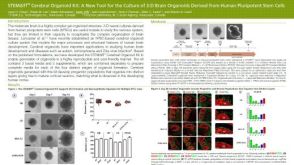 科学海报STEMdiff™ Cerebral Organoid Kit: A New Tool for the Culture of 3D Brain Organoids Derived from hPSCs


 沪公网安备31010102008431号