Micropatterning Facilitates the Long-Term Growth and Analysis of iPSC-Derived Individual Human Neurons and Neuronal Networks
The discovery of induced pluripotent stem cells (iPSCs) and their application to patient-specific disease models offers new opportunities for studying the pathophysiology of neurological disorders. However,current methods for culturing iPSC-derived neuronal cells result in clustering of neurons,which precludes the analysis of individual neurons and defined neuronal networks. To address this challenge,cultures of human neurons on micropatterned surfaces are developed that promote neuronal survival over extended periods of time. This approach facilitates studies of neuronal development,cellular trafficking,and related mechanisms that require assessment of individual neurons and specific network connections. Importantly,micropatterns support the long-term stability of cultured neurons,which enables time-dependent analysis of cellular processes in living neurons. The approach described in this paper allows mechanistic studies of human neurons,both in terms of normal neuronal development and function,as well as time-dependent pathological processes,and provides a platform for testing of new therapeutics in neuropsychiatric disorders.
View Publication
BACKGROUND Type 2 diabetes (T2D) is a recognized risk factor for the development of cognitive impairment (CI) and/or dementia,although the exact nature of the molecular pathology of T2D-associated CI remains obscure. One link between T2D and CI might involve decreased insulin signaling in brain and/or neurons in either animal or postmortem human brains as has been reported as a feature of Alzheimer's disease (AD). Here we asked if neuronal insulin resistance is a cell autonomous phenomenon in a familial form of AD. METHODS We have applied a newly developed protocol for deriving human basal forebrain cholinergic neurons (BFCN) from skin fibroblasts via induced pluripotent stem cell (iPSC) technology. We generated wildtype and familial AD mutant PSEN2 N141I (presenilin 2) BFCNs and assessed if insulin signaling,insulin regulation of the major AD proteins Abeta$ and/or tau,and/or calcium fluxes is altered by the PSEN2 N141I mutation. RESULTS We report herein that wildtype,PSEN2 N141I and CRISPR/Cas9-corrected iPSC-derived BFCNs (and their precursors) show indistinguishable insulin signaling profiles as determined by the phosphorylation of canonical insulin signaling pathway molecules. Chronic insulin treatment of BFCNs of all genotypes led to a reduction in the Abeta$42/40 ratio. Unexpectedly,we found a CRISPR/Cas9-correctable effect of PSEN2 N141I on calcium flux,which could be prevented by chronic exposure of BFCNs to insulin. CONCLUSIONS Our studies indicate that the familial AD mutation PSEN2 N141I does not induce neuronal insulin resistance in a cell autonomous fashion. The ability of insulin to correct calcium fluxes and to lower Abeta$42/40 ratio suggests that insulin acts to oppose an AD-pathophysiology. Hence,our results are consistent with a potential physiological role for insulin as a mediator of resilience by counteracting specific metabolic and molecular features of AD.
View Publication
产品类型:
产品号#:
05790
05791
05792
05793
05794
05795
07920
07922
85850
85857
产品名:
BrainPhys™神经元培养基
BrainPhys™ 无酚红
BrainPhys™神经元培养基和SM1试剂盒
BrainPhys™ 神经元培养基N2-A和SM1试剂盒
BrainPhys™原代神经元试剂盒
BrainPhys™ hPSC 神经元试剂盒
ACCUTASE™
ACCUTASE™
mTeSR™1
mTeSR™1
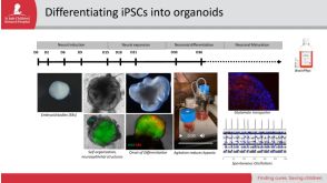
E. Gabriel et al. (JAN 2016)
Stem cell reports 7 4 678--692
Development and Dynamic Regulation of Mitochondrial Network in Human Midbrain Dopaminergic Neurons Differentiated from iPSCs.
Mitochondria are critical to neurogenesis,but the mechanisms of mitochondria in neurogenesis have not been well explored. We fully characterized mitochondrial alterations and function in relation to the development of human induced pluripotent stem cell (hiPSC)-derived dopaminergic (DA) neurons. Following directed differentiation of hiPSCs to DA neurons,mitochondria in these neurons exhibit pronounced changes during differentiation,including mature neurophysiology characterization and functional synaptic network formation. Inhibition of mitochondrial respiratory chains via application of complex IV inhibitor KCN (potassium cyanide) or complex I inhibitor rotenone restricted neurogenesis of DA neurons. These results demonstrated the direct importance of mitochondrial development and bioenergetics in DA neuronal differentiation. Our study also provides a neurophysiologic model of mitochondrial involvement in neurogenesis,which will enhance our understanding of the role of mitochondrial dysfunctions in neurodegenerative diseases.
View Publication
产品类型:
产品号#:
05832
05835
05839
08581
08582
05833
05790
05792
05794
05795
05793
产品名:
STEMdiff™ 神经花环选择试剂
STEMdiff™ 神经诱导培养基
STEMdiff™ 神经诱导培养基
STEMdiff™SMADi神经诱导试剂盒
STEMdiff™SMADi神经诱导试剂盒,2套
STEMdiff™神经前体细胞培养基
BrainPhys™神经元培养基
BrainPhys™神经元培养基和SM1试剂盒
BrainPhys™原代神经元试剂盒
BrainPhys™ hPSC 神经元试剂盒
BrainPhys™ 神经元培养基N2-A和SM1试剂盒
Belle K et al. (JAN 2017)
Neuroscience letters 637 201--206
Generation of disease-specific autopsy-confirmed iPSCs lines from postmortem isolated Peripheral Blood Mononuclear Cells
Understanding the molecular mechanisms that underlie neurodegenerative disorders has been hampered by a lack of readily available model systems that replicate the complexity of the human disease. Recent advances in stem cell technology have facilitated the derivation of patient-specific stem cells from a variety of differentiated cell types. These induced pluripotent stem cells (iPSCs) are attractive disease models since they can be grown and differentiated to produce large numbers of disease-relevant cell types. However,most iPSC lines are derived in advance of,and without the benefit of,neuropathological confirmation of the donor - the gold standard for many disease classifications and measurement of disease severity. While others have reported the generation of autopsy-confirmed iPSC lines from patient explants,these methods require outgrowth of cadaver tissue,which require additional time and is often only successul 50% of the time. Here we report the rapid generation of autopsy-confirmed iPSC lines from peripheral blood mononuclear cells (PBMCs) drawn postmortem. Since this approach doesn't require the propagation of previously frozen cadaver tissue,iPSC can be rapidly and efficiently produced from patients with autopsy-confirmed pathology. These matched iPSC-derived patient-specific neurons and postmortem brain tissue will support studies of specific mechanisms that drive the pathogenesis of neurodegenerative diseases.
View Publication
产品类型:
产品号#:
05833
05835
05839
07811
07861
18060
18061
85450
85460
85850
85857
86450
86460
产品名:
STEMdiff™神经前体细胞培养基
STEMdiff™ 神经诱导培养基
STEMdiff™ 神经诱导培养基
Lymphoprep™
Lymphoprep™
Lymphoprep™
Lymphoprep™
SepMate™-50 (IVD)
SepMate™-50 (IVD)
mTeSR™1
mTeSR™1
SepMate™-50 (RUO)
SepMate™-50 (RUO)
Tomov ML et al. (DEC 2016)
Scientific Reports 6 1 37637
Distinct and Shared Determinants of Cardiomyocyte Contractility in Multi-Lineage Competent Ethnically Diverse Human iPSCs
The realization of personalized medicine through human induced pluripotent stem cell (iPSC) technology can be advanced by transcriptomics,epigenomics,and bioinformatics that inform on genetic pathways directing tissue development and function. When possible,population diversity should be included in new studies as resources become available. Previously we derived replicate iPSC lines of African American,Hispanic-Latino and Asian self-designated ethnically diverse (ED) origins with normal karyotype,verified teratoma formation,pluripotency biomarkers,and tri-lineage in vitro commitment. Here we perform bioinformatics of RNA-Seq and ChIP-seq pluripotency data sets for two replicate Asian and Hispanic-Latino ED-iPSC lines that reveal differences in generation of contractile cardiomyocytes but similar and robust differentiation to multiple neural,pancreatic,and smooth muscle cell types. We identify shared and distinct genes and contributing pathways in the replicate ED-iPSC lines to enhance our ability to understand how reprogramming to iPSC impacts genes and pathways contributing to cardiomyocyte contractility potential.
View Publication
Although human induced pluripotent stem cells (hiPSCs) hold great potential for the study of human diseases affecting disparate cell types,they have been underutilized in seeking mechanistic insights into the pathogenesis of congenital craniofacial disorders. Craniofrontonasal syndrome (CFNS) is a rare X-linked disorder caused by mutations in EFNB1 and characterized by craniofacial,skeletal,and neurological anomalies. Heterozygous females are more severely affected than hemizygous males,a phenomenon termed cellular interference that involves mosaicism for EPHRIN-B1 function. Although the mechanistic basis for cellular interference in CFNS has been hypothesized to involve Eph/ephrin-mediated cell segregation,no direct evidence for this has been demonstrated. Here,by generating hiPSCs from CFNS patients,we demonstrate that mosaicism for EPHRIN-B1 expression induced by random X inactivation in heterozygous females results in robust cell segregation in human neuroepithelial cells,thus supplying experimental evidence that Eph/ephrin-mediated cell segregation is relevant to pathogenesis in human CFNS patients.
View Publication
A. M. Tukker et al. (JUL 2018)
Neurotoxicology 67 215--225
Human iPSC-derived neuronal models for in vitro neurotoxicity assessment.
Neurotoxicity testing still relies on ethically debated,expensive and time consuming in vivo experiments,which are unsuitable for high-throughput toxicity screening. There is thus a clear need for a rapid in vitro screening strategy that is preferably based on human-derived neurons to circumvent interspecies translation. Recent availability of commercially obtainable human induced pluripotent stem cell (hiPSC)-derived neurons and astrocytes holds great promise in assisting the transition from the current standard of rat primary cortical cultures to an animal-free alternative. We therefore composed several hiPSC-derived neuronal models with different ratios of excitatory and inhibitory neurons in the presence or absence of astrocytes. Using immunofluorescent stainings and multi-well micro-electrode array (mwMEA) recordings we demonstrate that these models form functional neuronal networks that become spontaneously active. The differences in development of spontaneous neuronal activity and bursting behavior as well as spiking patterns between our models confirm the importance of the presence of astrocytes. Preliminary neurotoxicity assessment demonstrates that these cultures can be modulated with known seizurogenic compounds,such as picrotoxin (PTX) and endosulfan,and the neurotoxicant methylmercury (MeHg). However,the chemical-induced effects on different parameters for neuronal activity,such as mean spike rate (MSR) and mean burst rate (MBR),may depend on the ratio of inhibitory and excitatory neurons. Our results thus indicate that hiPSC-derived neuronal models must be carefully designed and characterized prior to large-scale use in neurotoxicity screening.
View Publication
产品类型:
产品号#:
05790
05792
05793
05794
05795
产品名:
BrainPhys™神经元培养基
BrainPhys™神经元培养基和SM1试剂盒
BrainPhys™ 神经元培养基N2-A和SM1试剂盒
BrainPhys™原代神经元试剂盒
BrainPhys™ hPSC 神经元试剂盒
Patriarchi T et al. (JUN 2016)
European journal of human genetics : EJHG 24 6 871--880
Imbalance of excitatory/inhibitory synaptic protein expression in iPSC-derived neurons from FOXG1(+/-) patients and in foxg1(+/-) mice.
Rett syndrome (RTT) is a severe neurodevelopmental disorder associated with mutations in either MECP2,CDKL5 or FOXG1. The precise molecular mechanisms that lead to the pathogenesis of RTT have yet to be elucidated. We recently reported that expression of GluD1 (orphan glutamate receptor $\$-1 subunit) is increased in iPSC-derived neurons obtained from patients with mutations in either MECP2 or CDKL5. GluD1 controls synaptic differentiation and shifts the balance between excitatory and inhibitory synapses toward the latter. Thus,an increase in GluD1 might be a critical factor in the etiology of RTT by affecting the excitatory/inhibitory balance in the developing brain. To test this hypothesis,we generated iPSC-derived neurons from FOXG1(+/-) patients. We analyzed mRNA and protein levels of GluD1 together with key markers of excitatory and inhibitory synapses in these iPSC-derived neurons and in Foxg1(+/-) mouse fetal (E11.5) and adult (P70) brains. We found strong correlation between iPSC-derived neurons and fetal mouse brains,where GluD1 and inhibitory synaptic markers (GAD67 and GABA AR-$\$1) were increased,whereas the levels of a number of excitatory synaptic markers (VGLUT1,GluA1,GluN1 and PSD-95) were decreased. In adult mice,GluD1 was decreased along with all GABAergic and glutamatergic markers. Our findings further the understanding of the etiology of RTT by introducing a new pathological event occurring in the brain of FOXG1(+/-) patients during embryonic development and its time-dependent shift toward a general decrease in brain synapses.
View Publication

 EasySep™小鼠TIL(CD45)正选试剂盒


 30:53
线上讲座Highly Characterized Human iPSCs and NPCs for Downstream Differentiation Applications发布日期: 07/19/2023
30:53
线上讲座Highly Characterized Human iPSCs and NPCs for Downstream Differentiation Applications发布日期: 07/19/2023

 沪公网安备31010102008431号