Prolactin stimulates precursor cells in the adult mouse hippocampus.
In the search for ways to combat degenerative neurological disorders,neurogenesis-stimulating factors are proving to be a promising area of research. In this study,we show that the hormonal factor prolactin (PRL) can activate a pool of latent precursor cells in the adult mouse hippocampus. Using an in vitro neurosphere assay,we found that the addition of exogenous PRL to primary adult hippocampal cells resulted in an approximate 50% increase in neurosphere number. In addition,direct infusion of PRL into the adult dentate gyrus also resulted in a significant increase in neurosphere number. Together these data indicate that exogenous PRL can increase hippocampal precursor numbers both in vitro and in vivo. Conversely,PRL null mice showed a significant reduction (approximately 80%) in the number of hippocampal-derived neurospheres. Interestingly,no deficit in precursor proliferation was observed in vivo,indicating that in this situation other niche factors can compensate for a loss in PRL. The PRL loss resulted in learning and memory deficits in the PRL null mice,as indicated by significant deficits in the standard behavioral tests requiring input from the hippocampus. This behavioral deficit was rescued by direct infusion of recombinant PRL into the hippocampus,indicating that a lack of PRL in the adult mouse hippocampus can be correlated with impaired learning and memory.
View Publication
产品类型:
产品号#:
05700
05701
05702
产品名:
NeuroCult™ 基础培养基(小鼠&大鼠)
NeuroCult™ 扩增添加物 (小鼠&大鼠)
NeuroCult™ 扩增试剂盒 (小鼠&大鼠)
Zhang L et al. (APR 2016)
Human Reproduction 31 4 832--843
Protein kinase A inhibitor, H89, enhances survival and clonogenicity of dissociated human embryonic stem cells through Rho-associated coiled-coil containing protein kinase (ROCK) inhibition
H89 inhibits the dissociation-induced phosphorylation of PKA and two substrates of Rho-associated coiled-coil containing protein kinase (ROCK),myosin light chain (MLC2) and myosin phosphatase target subunit 1 (MYPT1),significantly increases cell survival and colony formation,and strongly depresses dissociation-induced cell death and cell blebbing without affecting the pluripotency of hESCs and their differentiation in vitro.
View Publication
产品类型:
产品号#:
05835
05839
产品名:
STEMdiff™ 神经诱导培养基
STEMdiff™ 神经诱导培养基
Lee S-HH et al. (JUN 2000)
Nature biotechnology 18 6 675--9
Efficient generation of midbrain and hindbrain neurons from mouse embryonic stem cells.
Embryonic stem (ES) cells are clonal cell lines derived from the inner cell mass of the developing blastocyst that can proliferate extensively in vitro and are capable of adopting all the cell fates in a developing embryo. Clinical interest in the use of ES cells has been stimulated by studies showing that isolated human cells with ES properties from the inner cell mass or developing germ cells can provide a source of somatic precursors. Previous studies have defined in vitro conditions for promoting the development of specific somatic fates,specifically,hematopoietic,mesodermal,and neurectodermal. In this study,we present a method for obtaining dopaminergic (DA) and serotonergic neurons in high yield from mouse ES cells in vitro. Furthermore,we demonstrate that the ES cells can be obtained in unlimited numbers and that these neuron types are generated efficiently. We generated CNS progenitor populations from ES cells,expanded these cells and promoted their differentiation into dopaminergic and serotonergic neurons in the presence of mitogen and specific signaling molecules. The differentiation and maturation of neuronal cells was completed after mitogen withdrawal from the growth medium. This experimental system provides a powerful tool for analyzing the molecular mechanisms controlling the functions of these neurons in vitro and in vivo,and potentially for understanding and treating neurodegenerative and psychiatric diseases.
View Publication
产品类型:
产品号#:
06902
06952
07152
07157
00321
00322
00323
00324
00325
产品名:
N2 添加物-A
Daynac M et al. (DEC 2014)
STEM CELLS 32 12 3257--3265
TGFβ Lengthens the G1 Phase of Stem Cells in Aged Mouse Brain
Neurogenesis decreases during aging causing a progressive cognitive decline but it is still controversial whether proliferation defects in neurogenic niches result from a loss of neural stem cells or from an impairment of their progression through the cell cycle. Using an accurate fluorescence-activated cell sorting technique,we show that the pool of neural stem cells is maintained in the subventricular zone of middle-aged mice while they have a reduced proliferative potential eventually leading to the subsequent decrease of their progeny. In addition,we demonstrate that the G1 phase is lengthened during aging specifically in activated stem cells,but not in transit-amplifying cells,and directly impacts on neurogenesis. Finally,we report that inhibition of TGFβ signaling restores cell cycle progression defects in stem cells. Our data highlight the significance of cell cycle dysregulation in stem cells in the aged brain and provide an attractive foundation for the development of anti-TGFβ regenerative therapies based on stimulating endogenous neural stem cells.
View Publication
Heterotopically transplanted CVO neural stem cells generate neurons and migrate with SVZ cells in the adult mouse brain.
Production of new neurons throughout adulthood has been well characterized in two brain regions,the subventricular zone (SVZ) of the anterolateral ventricle and the subgranular zone (SGZ) of the hippocampus. The neurons produced from these regions arise from neural stem cells (NSCs) found in highly regulated stem cell niches. We recently showed that midline structures called circumventricular organs (CVOs) also contain NSCs capable of neurogenesis and/or astrogliogenesis in vitro and in situ (Bennett et al.). The present study demonstrates that NSCs derived from two astrogliogenic CVOs,the median eminence and organum vasculosum of the lamina terminalis of the nestin-GFP mouse,possess the potential to integrate into the SVZ and differentiate into cells with a neuronal phenotype. These NSCs,following expansion and BrdU-labeling in culture and heterotopic transplantation into a region proximal to the SVZ in adult mice,migrate caudally to the SVZ and express early neuronal markers (TUC-4,PSA-NCAM) as they migrate along the rostral migratory stream. CVO-derived BrdU(+) cells ultimately reach the olfactory bulb where they express early (PSA-NCAM) and mature (NeuN) neuronal markers. Collectively,these data suggest that although NSCs derived from the ME and OVLT CVOs are astrogliogenic in situ,they produce cells phenotypic of neurons in vivo when placed in a neurogenic environment. These findings may have implications for neural repair in the adult brain.
View Publication
产品类型:
产品号#:
05700
05701
05702
产品名:
NeuroCult™ 基础培养基(小鼠&大鼠)
NeuroCult™ 扩增添加物 (小鼠&大鼠)
NeuroCult™ 扩增试剂盒 (小鼠&大鼠)
Harris MA et al. (DEC 2008)
Cancer research 68 24 10051--9
Cancer stem cells are enriched in the side population cells in a mouse model of glioma.
The recent identification of cancer stem cells (CSCs) in multiple human cancers provides a new inroad to understanding tumorigenesis at the cellular level. CSCs are defined by their characteristics of self-renewal,multipotentiality,and tumor initiation upon transplantation. By testing for these defining characteristics,we provide evidence for the existence of CSCs in a transgenic mouse model of glioma,S100beta-verbB;Trp53. In this glioma model,CSCs are enriched in the side population (SP) cells. These SP cells have enhanced tumor-initiating capacity,self-renewal,and multipotentiality compared with non-SP cells from the same tumors. Furthermore,gene expression analysis comparing fluorescence-activated cell sorting-sorted cancer SP cells to non-SP cancer cells and normal neural SP cells identified 45 candidate genes that are differentially expressed in glioma stem cells. We validated the expression of two genes from this list (S100a4 and S100a6) in primary mouse gliomas and human glioma samples. Analyses of xenografted human glioblastoma multiforme cell lines and primary human glioma tissues show that S100A4 and S100A6 are expressed in a small subset of cancer cells and that their abundance is positively correlated to tumor grade. In conclusion,this study shows that CSCs exist in a mouse glioma model,suggesting that this model can be used to study the molecular and cellular characteristics of CSCs in vivo and to further test the CSC hypothesis.
View Publication
Nardosinone Improves the Proliferation, Migration and Selective Differentiation of Mouse Embryonic Neural Stem Cells
In this study,we investigated the impact of Nardosinone,a bioactive component in Nardostachys root,on the proliferation and differentiation of neural stem cells. The neural stem cells were isolated from cerebrums of embryonic day 14 CD1 mice. The proliferation of cells was monitored using the cell counting kit-8 assay,bromodeoxyuridine incorporation and cell cycle analysis. Cell migration and differentiation were investigated with the neurosphere assay and cell specific markers,respectively. The results showed that Nardosinone promotes cells proliferation and increases cells migration distance in a dose-dependent manner. Nardosinone also induces the selective differentiation of neural stem cells to neurons and oligodendrocytes,as indicated by the expression of microtubule-associated protein-2 and myelin basic protein,respectively. Nardosinone also increases the expression of phospho-extracellular signal-regulated kinase and phospho-cAMP response element binding protein during proliferation and differentiation. In conclusion,this study reveals the regulatory effects of Nardosinone on neural stem cells,which may have significant implications for the treatment of brain injury and neurodegenerative diseases.
View Publication
产品类型:
产品号#:
05700
05702
05704
产品名:
NeuroCult™ 基础培养基(小鼠&大鼠)
NeuroCult™ 扩增试剂盒 (小鼠&大鼠)
NeuroCult™ 分化试剂盒 (小鼠&大鼠)
Rosa AI et al. (DEC 2016)
Frontiers in cellular neuroscience 10 284
Heterocellular Contacts with Mouse Brain Endothelial Cells Via Laminin and α6β1 Integrin Sustain Subventricular Zone (SVZ) Stem/Progenitor Cells Properties.
Neurogenesis in the subventricular zone (SVZ) is regulated by diffusible factors and cell-cell contacts. In vivo,SVZ stem cells are associated with the abluminal surface of blood vessels and such interactions are thought to regulate their neurogenic capacity. SVZ neural stem cells (NSCs) have been described to contact endothelial-derived laminin via α6β1 integrin. To elucidate whether heterocellular contacts with brain endothelial cells (BEC) regulate SVZ cells neurogenic capacities,cocultures of SVZ neurospheres and primary BEC,both obtained from C57BL/6 mice,were performed. The involvement of laminin-integrin interactions in SVZ homeostasis was tested in three ways. Firstly,SVZ cells were analyzed following incubation of BEC with the protein synthesis inhibitor cycloheximide (CHX) prior to coculture,a treatment expected to decrease membrane proteins. Secondly,SVZ cells were cocultured with BEC in the presence of an anti-α6 integrin neutralizing antibody. Thirdly,BEC were cultured with β1-/- SVZ cells. We showed that contact with BEC supports,at least in part,proliferation and stemness of SVZ cells,as evaluated by the number of BrdU positive (+) and Sox2+ cells in contact with BEC. These effects are dependent on BEC-derived laminin binding to α6β1 integrin and are decreased in cocultures incubated with anti-α6 integrin neutralizing antibody and in cocultures with SVZ β1-/- cells. Moreover,BEC-derived laminin sustains stemness in SVZ cell cultures via activation of the Notch and mTOR signaling pathways. Our results show that BEC/SVZ interactions involving α6β1 integrin binding to laminin,contribute to SVZ cell proliferation and stemness.
View Publication
Mujtaba T et al. (OCT 1999)
Developmental biology 214 1 113--27
Lineage-restricted neural precursors can be isolated from both the mouse neural tube and cultured ES cells.
We have previously identified multipotent neuroepithelial (NEP) stem cells and lineage-restricted,self-renewing precursor cells termed NRPs (neuron-restricted precursors) and GRPs (glial-restricted precursors) present in the developing rat spinal cord (A. Kalyani,K. Hobson,and M. S. Rao,1997,Dev. Biol. 186,202-223; M. S. Rao and M. Mayer-Proschel,1997,Dev. Biol. 188,48-63; M. Mayer-Proschel,A. J. Kalyani,T. Mujtaba,and M. S. Rao,1997,Neuron 19,773-785). We now show that cells identical to rat NEPs,NRPs,and GRPs are present in mouse neural tubes and that immunoselection against cell surface markers E-NCAM and A2B5 can be used to isolate NRPs and GRPs,respectively. Restricted precursors similar to NRPs and GRPs can also be isolated from mouse embryonic stem cells (ES cells). ES cell-derived NRPs are E-NCAM immunoreactive,undergo self-renewal in defined medium,and differentiate into multiple neuronal phenotypes in mass culture. ES cells also generate A2B5-immunoreactive cells that are similar to E9 NEP-cell-derived GRPs and can differentiate into oligodendrocytes and astrocytes. Thus,lineage restricted precursors can be generated in vitro from cultured ES cells and these restricted precursors resemble those derived from mouse neural tubes. These results demonstrate the utility of using ES cells as a source of late embryonic precursor cells.
View Publication

 EasySep™小鼠TIL(CD45)正选试剂盒


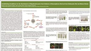 科学海报Establishing Conditions for the Enrichment of Oligodendrocytes from Cultures of Neurospheres Derived from Embryonic Rat and Mouse Brains
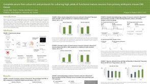 科学海报Complete Serum-Free Culture Kit and Protocols for Culturing High Yields of Functional Mature Neurons from Primary Embryonic Mouse CNS Tissues
 科学海报Comparative Performance of Neural-Specific Media in Differentiating and Maturing Human Neural Progenitor Cell-Derived Forebrain Neurons
 沪公网安备31010102008431号