Bain G et al. (APR 1995)
Developmental biology 168 2 342--57
Embryonic stem cells express neuronal properties in vitro.
Mouse embryonic stem (ES) cells cultured as aggregates and exposed to retinoic acid are induced to express multiple phenotypes normally associated with neurons. A large percentage of treated aggregates produce a rich neuritic outgrowth. Dissociating the induced aggregates with trypsin and plating the cells as a monolayer results in cultures in which a sizable percentage of the cells have a neuronal appearance. These neuron-like cells express class III beta-tubulin and the neurofilament M subunit. Induced cultures express transcripts for neural-associated genes including the neurofilament L subunit,glutamate receptor subunits,the transcription factor Brn-3,and GFAP. Levels of neurofilament L and GAD67 and GAD65 transcripts rise dramatically upon induction. Physiological studies show that the neuron-like cells generate action potentials and express TTX-sensitive sodium channels,as well as voltage-gated potassium channels and calcium channels. We conclude that a complex system of neuronal gene expression can be activated in cultured ES cells. This system should be favorable for investigating some of the mechanisms that regulate neuronal differentiation.
View Publication
产品类型:
产品号#:
06902
06952
00321
00322
00323
00324
00325
产品名:
Badr CE et al. (MAY 2013)
JNCI: Journal of the National Cancer Institute 105 9 643--653
Targeting Cancer Cells With the Natural Compound Obtusaquinone
BACKGROUND Tumor cells present high levels of oxidative stress. Cancer therapeutics exploiting such biochemical changes by increasing reactive oxygen species (ROS) production or decreasing intracellular ROS scavengers could provide a powerful treatment strategy. METHODS To test the effect of our compound,obtusaquinone (OBT),we used several cell viability assays on seven different glioblastoma (GBM) cell lines and primary cells and on 12 different cell lines representing various cancer types in culture as well as on subcutaneous (n = 7 mice per group) and two intracranial GBM (n = 6-8 mice per group) and breast cancer (n = 6 mice per group) tumor models in vivo. Immunoblotting,immunostaining,flow cytometry,and biochemical assays were used to investigate the OBT mechanism of action. Histopathological analysis (n = 2 mice per group) and blood chemistry (n = 2 mice per group) were used to test for any compound-related toxicity. Statistical tests were two-sided. RESULTS OBT induced rapid increase in intracellular ROS levels,downregulation of cellular glutathione levels and increase in its oxidized form,and activation of cellular stress pathways and DNA damage,subsequently leading to apoptosis. Oxidative stress is believed to be the main mechanism through which this compounds targets cancer cells. OBT was well tolerated in mice,slowed tumor growth,and statistically prolonged survival in GBM tumor models. The ratio of median survival in U251 intracranial model in OBT vs control was 1.367 (95% confidence interval [CI] of ratio = 1.031 to 1.367,P = .008). Tumor growth inhibition was also observed in a mouse breast cancer model (average tumor volume per mouse,OBT vs control: 36.3 vs 200.4mm(3),difference = 164.1mm(3),95% CI =72.6 to 255.6mm(3),P = .005). CONCLUSIONS Given its properties and efficacy in cancer killing,our results suggest that OBT is a promising cancer therapeutic.
View Publication
产品类型:
产品号#:
05750
05751
产品名:
NeuroCult™ NS-A 基础培养基(人)
NeuroCult™ NS-A 扩增试剂盒(人)
Donangelo I et al. (JAN 2014)
Endocrine Related Cancer 21 2 203--216
Sca1+ murine pituitary adenoma cells show tumor-growth advantage
The role of tumor stem cells in benign tumors such as pituitary adenomas remains unclear. In this study,we investigated whether the cells within pituitary adenomas that spontaneously develop in Rb+/- mice are hierarchically distributed with a subset being responsible for tumor growth. Cells derived directly from such tumors grew as spheres in serum-free culture medium supplemented with epidermal growth factor and basic fibroblast growth factor. Some cells within growing pituitary tumor spheres (PTS) expressed common stem cell markers (Sca1,Sox2,Nestin,and CD133),but were devoid of hormone-positive differentiated cells. Under subsequent differentiating conditions (matrigel-coated growth surface),PTS expressed all six pituitary hormones. We next searched for specific markers of the stem cell population and isolated a Sca1(+) cell population that showed increased sphere formation potential,lower mRNA hormone expression,higher expression of stem cell markers (Notch1,Sox2,and Nestin),and increased proliferation rates. When transplanted into non-obese diabetic-severe combined immunodeficiency gamma mice brains,Sca1(+) pituitary tumor cells exhibited higher rates of tumor formation (brain tumors observed in 11/11 (100%) vs 7/12 (54%) of mice transplanted with Sca1(+) and Sca1(-) cells respectively). Magnetic resonance imaging and histological analysis of brain tumors showed that tumors derived from Sca1(+) pituitary tumor cells were also larger and plurihormonal. Our findings show that Sca1(+) cells derived from benign pituitary tumors exhibit an undifferentiated expression profile and tumor-proliferative advantages,and we propose that they could represent putative pituitary tumor stem/progenitor cells.
View Publication
Induced pluripotent stem cells with a mitochondrial dna deletion
In congenital mitochondrial DNA (mtDNA) disorders,a mixture of normal and mutated mtDNA (termed heteroplasmy) exists at varying levels in different tissues,which determines the severity and phenotypic expression of disease. Pearson marrow pancreas syndrome (PS) is a congenital bone marrow failure disorder caused by heteroplasmic deletions in mtDNA. The cause of the hematopoietic failure in PS is unknown,and adequate cellular and animal models are lacking. Induced pluripotent stem (iPS) cells are particularly amenable for studying mtDNA disorders,as cytoplasmic genetic material is retained during direct reprogramming. Here,we derive and characterize iPS cells from a patient with PS. Taking advantage of the tendency for heteroplasmy to change with cell passage,we isolated isogenic PS-iPS cells without detectable levels of deleted mtDNA. We found that PS-iPS cells carrying a high burden of deleted mtDNA displayed differences in growth,mitochondrial function,and hematopoietic phenotype when differentiated in vitro,compared to isogenic iPS cells without deleted mtDNA. Our results demonstrate that reprogramming somatic cells from patients with mtDNA disorders can yield pluripotent stem cells with varying burdens of heteroplasmy that might be useful in the study and treatment of mitochondrial diseases. STEM CELLS2013;31:1287–1297
View Publication
Stapelberg M et al. (FEB 2014)
Free Radical Biology and Medicine 67 41--50
Indoleamine-2,3-dioxygenase elevated in tumor-initiating cells is suppressed by mitocans
Tumor-initiating cells (TICs) often survive therapy and give rise to second-line tumors. We tested the plausibility of sphere cultures as models of TICs. Microarray data and microRNA data analysis confirmed the validity of spheres as models of TICs for breast and prostate cancer as well as mesothelioma cell lines. Microarray data analysis revealed the Trp pathway as the only pathway upregulated significantly in all types of studied TICs,with increased levels of indoleamine-2,3-dioxygenase-1 (IDO1),the rate-limiting enzyme of Trp metabolism along the kynurenine pathway. All types of TICs also expressed higher levels of the Trp uptake system consisting of CD98 and LAT1 with functional consequences. IDO1 expression was regulated via both transcriptional and posttranscriptional mechanisms,depending on the cancer type. Serial transplantation of TICs in mice resulted in gradually increased IDO1. Mitocans,represented by α-tocopheryl succinate and mitochondrially targeted vitamin E succinate (MitoVES),suppressed IDO1 in TICs. MitoVES suppressed IDO1 in TICs with functional mitochondrial complex II,involving transcriptional and posttranscriptional mechanisms. IDO1 increase and its suppression by VE analogues were replicated in TICs from primary human glioblastomas. Our work indicates that IDO1 is increased in TICs and that mitocans suppress the protein.
View Publication
产品类型:
产品号#:
05750
05751
产品名:
NeuroCult™ NS-A 基础培养基(人)
NeuroCult™ NS-A 扩增试剂盒(人)
Wee S et al. (DEC 2014)
PloS one 9 12 e115698
Selective calcium sensitivity in immature glioma cancer stem cells.
Tumor-initiating cells are a subpopulation in aggressive cancers that exhibit traits shared with stem cells,including the ability to self-renew and differentiate,commonly referred to as stemness. In addition,such cells are resistant to chemo- and radiation therapy posing a therapeutic challenge. To uncover stemness-associated functions in glioma-initiating cells (GICs),transcriptome profiles were compared to neural stem cells (NSCs) and gene ontology analysis identified an enrichment of Ca2+ signaling genes in NSCs and the more stem-like (NSC-proximal) GICs. Functional analysis in a set of different GIC lines regarding sensitivity to disturbed homeostasis using A23187 and Thapsigargin,revealed that NSC-proximal GICs were more sensitive,corroborating the transcriptome data. Furthermore,Ca2+ drug sensitivity was reduced in GICs after differentiation,with most potent effect in the NSC-proximal GIC,supporting a stemness-associated Ca2+ sensitivity. NSCs and the NSC-proximal GIC line expressed a larger number of ion channels permeable to potassium,sodium and Ca2+. Conversely,a higher number of and higher expression levels of Ca2+ binding genes that may buffer Ca2+,were expressed in NSC-distal GICs. In particular,expression of the AMPA glutamate receptor subunit GRIA1,was found to associate with Ca2+ sensitive NSC-proximal GICs,and decreased as GICs differentiated along with reduced Ca2+ drug sensitivity. The correlation between high expression of Ca2+ channels (such as GRIA1) and sensitivity to Ca2+ drugs was confirmed in an additional nine novel GIC lines. Calcium drug sensitivity also correlated with expression of the NSC markers nestin (NES) and FABP7 (BLBP,brain lipid-binding protein) in this extended analysis. In summary,NSC-associated NES+/FABP7+/GRIA1+ GICs were selectively sensitive to disturbances in Ca2+ homeostasis,providing a potential target mechanism for eradication of an immature population of malignant cells.
View Publication
产品类型:
产品号#:
05750
05751
产品名:
NeuroCult™ NS-A 基础培养基(人)
NeuroCult™ NS-A 扩增试剂盒(人)
Birbrair A et al. (JAN 2013)
Experimental cell research 319 1 45--63
Skeletal muscle neural progenitor cells exhibit properties of NG2-glia.
Reversing brain degeneration and trauma lesions will depend on cell therapy. Our previous work identified neural precursor cells derived from the skeletal muscle of Nestin-GFP transgenic mice,but their identity,origin,and potential survival in the brain are only vaguely understood. In this work,we show that Nestin-GFP+ progenitor cells share morphological and molecular markers with NG2-glia,including NG2,PDGFRα,O4,NGF receptor (p75),glutamate receptor-1(AMPA),and A2B5 expression. Although these cells exhibit NG2,they do not express other pericyte markers,such as α-SMA or connexin-43,and do not differentiate into the muscle lineage. Patch-clamp studies displayed outward potassium currents,probably carried through Kir6.1 channels. Given their potential therapeutic application,we compared their abundance in tissues and concluded that skeletal muscle is the richest source of predifferentiated neural precursor cells. We found that these cells migrate toward the neurogenic subventricular zone displaying their typical morphology and nestin-GFP expression two weeks after brain injection. For translational purposes,we sought to identify these neural progenitor cells in wild-type species by developing a DsRed expression vector under Nestin-Intron II control. This approach revealed them in nonhuman primates and aging rodents throughout the lifespan.
View Publication
产品类型:
产品号#:
05700
05701
05702
05703
05704
05715
产品名:
NeuroCult™ 基础培养基(小鼠和大鼠)
NeuroCult™ 扩增添加物(小鼠和大鼠)
NeuroCult™扩增试剂盒(小鼠和大鼠)
NeuroCult™ 分化添加物(小鼠和大鼠)
NeuroCult™ 分化试剂盒(小鼠和大鼠)
NeuroCult™成年中枢神经系统(CNS)组织酶解试剂盒(小鼠和大鼠)
Yuki N et al. (AUG 2004)
Proceedings of the National Academy of Sciences 101 31 11404--09
Carbohydrate mimicry between human ganglioside GM1 and Campylobacter jejuni lipooligosaccharide causes Guillain-Barre syndrome
Molecular mimicry between microbial and self-components is postulated as the mechanism that accounts for the antigen and tissue specificity of immune responses in postinfectious autoimmune diseases. Little direct evidence exists,and research in this area has focused principally on T cell-mediated,antipeptide responses,rather than on humoral responses to carbohydrate structures. Guillain-Barré syndrome,the most frequent cause of acute neuromuscular paralysis,occurs 1-2 wk after various infections,in particular,Campylobacter jejuni enteritis. Carbohydrate mimicry [Galbeta1-3GalNAcbeta1-4(NeuAcalpha2-3)Galbeta1-] between the bacterial lipooligosaccharide and human GM1 ganglioside is seen as having relevance to the pathogenesis of Guillain-Barré syndrome,and conclusive evidence is reported here. On sensitization with C. jejuni lipooligosaccharide,rabbits developed anti-GM1 IgG antibody and flaccid limb weakness. Paralyzed rabbits had pathological changes in their peripheral nerves identical with those present in Guillain-Barré syndrome. Immunization of mice with the lipooligosaccharide generated a mAb that reacted with GM1 and bound to human peripheral nerves. The mAb and anti-GM1 IgG from patients with Guillain-Barré syndrome did not induce paralysis but blocked muscle action potentials in a muscle-spinal cord coculture,indicating that anti-GM1 antibody can cause muscle weakness. These findings show that carbohydrate mimicry is an important cause of autoimmune neuropathy.
View Publication
Kerosuo L et al. (DEC 2008)
Journal of cell science 121 Pt 23 3941--50
Myc increases self-renewal in neural progenitor cells through Miz-1.
The mechanisms underlying the decision of a stem or progenitor cell to either self-renew or differentiate are incompletely understood. To address the role of Myc in this process,we expressed different forms of the proto-oncogene Myc in multipotent neural progenitor cells (NPCs) using retroviral transduction. Expression of Myc in neurospheres increased the proportion of self-renewing cells fivefold,and 1% of the Myc-overexpressing cells,but none of the control cells,retained self-renewal capacity even under differentiation-inducing conditions. A Myc mutant (MycV394D) deficient in binding to Miz-1,did not increase the percentage of self-renewing cells but was able to stimulate proliferation of NPCs as efficiently as wild-type Myc,indicating that these two cellular phenomena are regulated by at least partially different pathways. Our results suggest that Myc,through Miz-1,enhances self-renewal of NPCs and influences the way progenitor cells react to the environmental cues that normally dictate the cellular identity of tissues containing self-renewing cells.
View Publication
产品类型:
产品号#:
05707
产品名:
NeuroCult™化学解离试剂盒(小鼠)
Flanagan LA et al. (MAR 2008)
Stem cells (Dayton,Ohio) 26 3 656--65
Unique dielectric properties distinguish stem cells and their differentiated progeny.
The relatively new field of stem cell biology is hampered by a lack of sufficient means to accurately determine the phenotype of cells. Cell-type-specific markers,such as cell surface proteins used for flow cytometry or fluorescence-activated cell sorting,are limited and often recognize multiple members of a stem cell lineage. We sought to develop a complementary approach that would be less dependent on the identification of particular markers for the subpopulations of cells and would instead measure their overall character. We tested whether a microfluidic system using dielectrophoresis (DEP),which induces a frequency-dependent dipole in cells,would be useful for characterizing stem cells and their differentiated progeny. We found that populations of mouse neural stem/precursor cells (NSPCs),differentiated neurons,and differentiated astrocytes had different dielectric properties revealed by DEP. By isolating NSPCs from developmental ages at which they are more likely to generate neurons,or astrocytes,we were able to show that a shift in dielectric property reflecting their fate bias precedes detectable marker expression in these cells and identifies specific progenitor populations. In addition,experimental data and mathematical modeling suggest that DEP curve parameters can indicate cell heterogeneity in mixed cultures. These findings provide evidence for a whole cell property that reflects stem cell fate bias and establish DEP as a tool with unique capabilities for interrogating,characterizing,and sorting stem cells.
View Publication

 EasySep™小鼠TIL(CD45)正选试剂盒


 技术公告Identification of Viable Stem and Progenitor Cells with ALDEFLUOR™
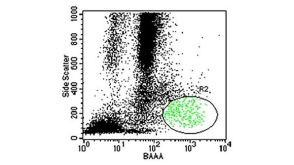

 沪公网安备31010102008431号