Xia G et al. (JUN 2015)
Stem cells (Dayton,Ohio) 33 6 1829--38
Genome modification leads to phenotype reversal in human myotonic dystrophy type 1 induced pluripotent stem cell-derived neural stem cells.
Myotonic dystrophy type 1 (DM1) is caused by expanded CTG repeats in the 3'-untranslated region (3' UTR) of the DMPK gene. Correcting the mutation in DM1 stem cells would be an important step toward autologous stem cell therapy. The objective of this study is to demonstrate in vitro genome editing to prevent production of toxic mutant transcripts and reverse phenotypes in DM1 stem cells. Genome editing was performed in DM1 neural stem cells (NSCs) derived from human DM1 induced pluripotent stem (iPS) cells. An editing cassette containing SV40/bGH polyA signals was integrated upstream of the CTG repeats by TALEN-mediated homologous recombination (HR). The expression of mutant CUG repeats transcript was monitored by nuclear RNA foci,the molecular hallmarks of DM1,using RNA fluorescence in situ hybridization. Alternative splicing of microtubule-associated protein tau (MAPT) and muscleblind-like (MBNL) proteins were analyzed to further monitor the phenotype reversal after genome modification. The cassette was successfully inserted into DMPK intron 9 and this genomic modification led to complete disappearance of nuclear RNA foci. MAPT and MBNL 1,2 aberrant splicing in DM1 NSCs were reversed to normal pattern in genome-modified NSCs. Genome modification by integration of exogenous polyA signals upstream of the DMPK CTG repeat expansion prevents the production of toxic RNA and leads to phenotype reversal in human DM1 iPS-cells derived stem cells. Our data provide proof-of-principle evidence that genome modification may be used to generate genetically modified progenitor cells as a first step toward autologous cell transfer therapy for DM1.
View Publication
Daynac M et al. (FEB 2016)
Scientific reports 6 21505
Age-related neurogenesis decline in the subventricular zone is associated with specific cell cycle regulation changes in activated neural stem cells.
Although neural stem cells (NSCs) sustain continuous neurogenesis throughout the adult lifespan of mammals,they progressively exhibit proliferation defects that contribute to a sharp reduction in subventricular neurogenesis during aging. However,little is known regarding the early age-related events in neurogenic niches. Using a fluorescence-activated cell sorting technique that allows for the prospective purification of the main neurogenic populations from the subventricular zone (SVZ),we demonstrated an early decline in adult neurogenesis with a dramatic loss of progenitor cells in 4 month-old young adult mice. Whereas the activated and quiescent NSC pools remained stable up to 12 months,the proliferative status of activated NSCs was already altered by 6 months,with an overall extension of the cell cycle resulting from a specific lengthening of G1. Whole genome analysis of activated NSCs from 2- and 6-month-old mice further revealed distinct transcriptomic and molecular signatures,as well as a modulation of the TGFβ signalling pathway. Our microarray study constitutes a cogent identification of new molecular players and signalling pathways regulating adult neurogenesis and its early modifications.
View Publication
产品类型:
产品号#:
05700
05701
05702
产品名:
NeuroCult™ 基础培养基(小鼠&大鼠)
NeuroCult™ 扩增添加物 (小鼠&大鼠)
NeuroCult™ 扩增试剂盒 (小鼠&大鼠)
Vazin T et al. (FEB 2014)
Neurobiology of Disease 62 62--72
Efficient derivation of cortical glutamatergic neurons from human pluripotent stem cells: a model system to study neurotoxicity in Alzheimer's disease.
Alzheimer's disease (AD) is among the most prevalent forms of dementia affecting the aging population,and pharmacological therapies to date have not been successful in preventing disease progression. Future therapeutic efforts may benefit from the development of models that enable basic investigation of early disease pathology. In particular,disease-relevant models based on human pluripotent stem cells (hPSCs) may be promising approaches to assess the impact of neurotoxic agents in AD on specific neuronal populations and thereby facilitate the development of novel interventions to avert early disease mechanisms. We implemented an efficient paradigm to convert hPSCs into enriched populations of cortical glutamatergic neurons emerging from dorsal forebrain neural progenitors,aided by modulating Sonic hedgehog (Shh) signaling. Since AD is generally known to be toxic to glutamatergic circuits,we exposed glutamatergic neurons derived from hESCs to an oligomeric pre-fibrillar forms of Aβ known as globulomers"�
View Publication
产品类型:
产品号#:
85850
85857
产品名:
mTeSR™1
mTeSR™1
Namba T et al. (MAY 2010)
Neuroscience 167 2 372--83
Pigment epithelium-derived factor up-regulation induced by memantine, an N-methyl-D-aspartate receptor antagonist, is involved in increased proliferation of hippocampal progenitor cells.
Memantine is classified as an NMDA receptor antagonist. We recently reported that memantine promoted the proliferation of neural progenitor cells and the production of mature granule neurons in the adult hippocampus. However,the molecular mechanism responsible for the memantine-induced promotion of cellular proliferation remains unknown. In this study we searched for a factor that mediates memantine-induced cellular proliferation,and found that pigment epithelium-derived factor (PEDF),a broad-acting neurotrophic factor,is up-regulated in the dentate gyrus of adult mice after the injection of memantine. PEDF mRNA expression increased significantly by 3.5-fold at 1 day after the injection of memantine. In addition,the expression level of PEDF protein also increased by 1.8-fold at 2 days after the injection of memantine. Immunohistochemical study using anti-PEDF antibody showed that the majority of the PEDF-expressing cells were protoplasmic and perivascular astrocytes. Using a neurosphere assay,we confirmed that PEDF enhanced cellular proliferation under the presence of fibroblast growth factor-2 (FGF-2) and epidermal growth factor (EGF) but was not involved in the multilineage potency of hippocampal progenitor cells. Over expression of PEDF by adeno-associated virus,however,did not stimulate cellular proliferation,suggesting PEDF per se does not promote cellular proliferation in vivo. These findings suggest that the memantine induced PEDF up-regulation is involved in increased proliferation of hippocampal progenitor cells.
View Publication
产品类型:
产品号#:
05700
05701
05702
产品名:
NeuroCult™ 基础培养基(小鼠&大鼠)
NeuroCult™ 扩增添加物 (小鼠&大鼠)
NeuroCult™ 扩增试剂盒 (小鼠&大鼠)
Young KM et al. (AUG 2007)
The Journal of neuroscience : the official journal of the Society for Neuroscience 27 31 8286--96
Subventricular zone stem cells are heterogeneous with respect to their embryonic origins and neurogenic fates in the adult olfactory bulb.
We determined the embryonic origins of adult forebrain subventricular zone (SVZ) stem cells by Cre-lox fate mapping in transgenic mice. We found that all parts of the telencephalic neuroepithelium,including the medial ganglionic eminence and lateral ganglionic eminence (LGE) and the cerebral cortex,contribute multipotent,self-renewing stem cells to the adult SVZ. Descendants of the embryonic LGE and cortex settle in ventral and dorsal aspects of the dorsolateral SVZ,respectively. Both populations contribute new (5-bromo-2'-deoxyuridine-labeled) tyrosine hydroxylase- and calretinin-positive interneurons to the adult olfactory bulb. However,calbindin-positive interneurons in the olfactory glomeruli were generated exclusively by LGE-derived stem cells. Thus,different SVZ stem cells have different embryonic origins,colonize different parts of the SVZ,and generate different neuronal progeny,suggesting that some aspects of embryonic patterning are preserved in the adult SVZ. This could have important implications for the design of endogenous stem cell-based therapies in the future.
View Publication
产品类型:
产品号#:
05700
05701
05702
产品名:
NeuroCult™ 基础培养基(小鼠&大鼠)
NeuroCult™ 扩增添加物 (小鼠&大鼠)
NeuroCult™ 扩增试剂盒 (小鼠&大鼠)
Drury-Stewart D et al. (AUG 2013)
Stem cell research & therapy 4 4 93
Highly efficient differentiation of neural precursors from human embryonic stem cells and benefits of transplantation after ischemic stroke in mice.
INTRODUCTION: Ischemic stroke is a leading cause of death and disability,but treatment options are severely limited. Cell therapy offers an attractive strategy for regenerating lost tissues and enhancing the endogenous healing process. In this study,we investigated the use of human embryonic stem cell-derived neural precursors as a cell therapy in a murine stroke model.backslashnbackslashnMETHODS: Neural precursors were derived from human embryonic stem cells by using a fully adherent SMAD inhibition protocol employing small molecules. The efficiency of neural induction and the ability of these cells to further differentiate into neurons were assessed by using immunocytochemistry. Whole-cell patch-clamp recording was used to demonstrate the electrophysiological activity of human embryonic stem cell-derived neurons. Neural precursors were transplanted into the core and penumbra regions of a focal ischemic stroke in the barrel cortex of mice. Animals received injections of bromodeoxyuridine to track regeneration. Neural differentiation of the transplanted cells and regenerative markers were measured by using immunohistochemistry. The adhesive removal test was used to determine functional improvement after stroke and intervention.backslashnbackslashnRESULTS: After 11 days of neural induction by using the small-molecule protocol,over 95% of human embryonic stem-derived cells expressed at least one neural marker. Further in vitro differentiation yielded cells that stained for mature neuronal markers and exhibited high-amplitude,repetitive action potentials in response to depolarization. Neuronal differentiation also occurred after transplantation into the ischemic cortex. A greater level of bromodeoxyuridine co-localization with neurons was observed in the penumbra region of animals receiving cell transplantation. Transplantation also improved sensory recovery in transplant animals over that in control animals.backslashnbackslashnCONCLUSIONS: Human embryonic stem cell-derived neural precursors derived by using a highly efficient small-molecule SMAD inhibition protocol can differentiate into electrophysiologically functional neurons in vitro. These cells also differentiate into neurons in vivo,enhance regenerative activities,and improve sensory recovery after ischemic stroke.
View Publication
产品类型:
产品号#:
85850
85857
产品名:
mTeSR™1
mTeSR™1
Biasini E et al. (JAN 2012)
PloS one 7 3 e33472
The toxicity of a mutant prion protein is cell-autonomous, and can be suppressed by wild-type prion protein on adjacent cells.
Insight into the normal function of PrP(C),and how it can be subverted to produce neurotoxic effects,is provided by PrP molecules carrying deletions encompassing the conserved central region. The most neurotoxic of these mutants,Δ105-125 (called ΔCR),produces a spontaneous neurodegenerative illness when expressed in transgenic mice,and this phenotype can be dose-dependently suppressed by co-expression of wild-type PrP. Whether the toxic activity of ΔCR PrP and the protective activity or wild-type PrP are cell-autonomous,or can be exerted on neighboring cells,is unknown. To investigate this question,we have utilized co-cultures of differentiated neural stem cells derived from mice expressing ΔCR or wild-type PrP. Cells from the two kinds of mice,which are marked by the presence or absence of GFP,are differentiated together to yield neurons,astrocytes,and oligodendrocytes. As a surrogate read-out of ΔCR PrP toxicity,we assayed sensitivity of the cells to the cationic antibiotic,Zeocin. In a previous study,we reported that cells expressing ΔCR PrP are hypersensitive to the toxic effects of several cationic antibiotics,an effect that is suppressed by co-expression of wild type PrP,similar to the rescue of the neurodegenerative phenotype observed in transgenic mice. Using this system,we find that while ΔCR-dependent toxicity is cell-autonomous,the rescuing activity of wild-type PrP can be exerted in trans from nearby cells. These results provide important insights into how ΔCR PrP subverts a normal physiological function of PrP(C),and the cellular mechanisms underlying the rescuing process.
View Publication
产品类型:
产品号#:
05700
05701
05702
05703
05704
产品名:
NeuroCult™ 基础培养基(小鼠&大鼠)
NeuroCult™ 扩增添加物 (小鼠&大鼠)
NeuroCult™ 扩增试剂盒 (小鼠&大鼠)
NeuroCult™ 分化添加物 (小鼠&大鼠)
NeuroCult™ 分化试剂盒 (小鼠&大鼠)
Sun Y et al. (MAR )
PLOS ONE 3 e0118771
Properties of Neurons Derived from Induced Pluripotent Stem Cells of Gaucher Disease Type 2 Patient Fibroblasts: Potential Role in Neuropathology
Gaucher disease (GD) is caused by insufficient activity of acid $\$-glucosidase (GCase) resulting from mutations in GBA1. To understand the pathogenesis of the neuronopathic GD,induced pluripotent stem cells (iPSCs) were generated from fibroblasts isolated from three GD type 2 (GD2) and 2 unaffected (normal and GD carrier) individuals. The iPSCs were converted to neural precursor cells (NPCs) which were further differentiated into neurons. Parental GD2 fibroblasts as well as iPSCs,NPCs,and neurons had similar degrees of GCase deficiency. Lipid analyses showed increases of glucosylsphingosine and glucosylceramide in the GD2 cells. In addition,GD2 neurons showed increased $\$-synuclein protein compared to control neurons. Whole cell patch-clamping of the GD2 and control iPSCs-derived neurons demonstrated excitation characteristics of neurons,but intriguingly,those from GD2 exhibited consistently less negative resting membrane potentials with various degree of reduction in action potential amplitudes,sodium and potassium currents. Culture of control neurons in the presence of the GCase inhibitor (conduritol B epoxide) recapitulated these findings,providing a functional link between decreased GCase activity in GD and abnormal neuronal electrophysiological properties. To our knowledge,this study is first to report abnormal electrophysiological properties in GD2 iPSC-derived neurons that may underlie the neuropathic phenotype in Gaucher disease.
View Publication
产品类型:
产品号#:
05835
05839
05854
05855
34811
34815
34821
34825
34850
34860
85850
85857
产品名:
STEMdiff™ 神经诱导培养基
STEMdiff™ 神经诱导培养基
mFreSR™
mFreSR™
AggreWell™ 800 24孔板,1个
AggreWell™ 800 24孔板,5个
AggreWell™ 800 6孔板,1个
AggreWell™ 800 6孔板,5个
AggreWell™ 800 24孔板启动套装
AggreWell™ 800 6孔板启动套装
mTeSR™1
mTeSR™1
Kim S-J et al. (AUG 2010)
Neuroscience letters 479 3 292--6
Omega-3 and omega-6 fatty acids suppress ER- and oxidative stress in cultured neurons and neuronal progenitor cells from mice lacking PPT1.
Reactive oxygen species (ROS) damage brain lipids,carbohydrates,proteins,as well as DNA and may contribute to neurodegeneration. We previously reported that ER- and oxidative stress cause neuronal apoptosis in infantile neuronal ceroid lipofuscinosis (INCL),a lethal neurodegenerative storage disease,caused by palmitoyl-protein thioesterase-1 (PPT1) deficiency. Polyunsaturated fatty acids (PUFA) are essential components of cell membrane phospholipids in the brain and excessive ROS may cause oxidative damage of PUFA leading to neuronal death. Using cultured neurons and neuroprogenitor cells from mice lacking Ppt1,which mimic INCL,we demonstrate that Ppt1-deficient neurons and neuroprogenitor cells contain high levels of ROS,which may cause peroxidation of PUFA and render them incapable of providing protection against oxidative stress. We tested whether treatment of these cells with omega-3 or omega-6 PUFA protects the neurons and neuroprogenitor cells from oxidative stress and suppress apoptosis. We report here that both omega-3 and omega-6 fatty acids protect the Ppt1-deficient cells from ER- as well as oxidative stress and suppress apoptosis. Our results suggest that PUFA supplementation may have neuroprotective effects in INCL.
View Publication

 EasySep™小鼠TIL(CD45)正选试剂盒


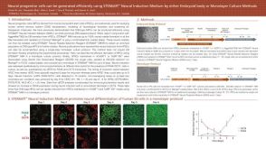 科学海报Neural Progenitor Cells can be Generated Efficiently Using STEMdiff™ Neural Induction Medium By Either Embryoid Body or Monolayer Culture Methods
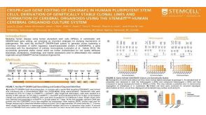 科学海报CRISPR-Cas9 Gene Editing Of CDK5RAP2 In Human Pluripotent Stem Cells, Derivation Of Genetically Stable Clonal Lines And Formation Of Cerebral Organoids
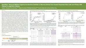 科学海报BrainPhys™ Neuronal Medium Supports the Electrical Activities of Neurons Derived from Human Pluripotent Stem Cells and Primary CNS Tissues in Long-Term Cultures
 沪公网安备31010102008431号