Kandasamy M et al. (MAR 2017)
Cell and Tissue Research 368 3 531--549
Glycoconjugates reveal diversity of human neural stem cells (hNSCs) derived from human induced pluripotent stem cells (hiPSCs)
Neural stem cells (NSCs) have the ability to self-renew and to differentiate into various cell types of the central nervous system. This potential can be recapitulated by human induced pluripotent stem cells (hiPSCs) in vitro. The differentiation capacity of hiPSCs is characterized by several stages with distinct morphologies and the expression of various marker molecules. We used the monoclonal antibodies (mAbs) 487(LeX),5750(LeX) and 473HD to analyze the expression pattern of particular carbohydrate motifs as potential markers at six differentiation stages of hiPSCs. Mouse ESCs were used as a comparison. At the pluripotent stage,487(LeX)-,5750(LeX)- and 473HD-related glycans were differently expressed. Later,cells of the three germ layers in embryoid bodies (hEBs) and,even after neuralization of hEBs,subpopulations of cells were labeled with these surface antibodies. At the human rosette-stage of NSCs (hR-NSC),LeX- and 473HD-related epitopes showed antibody-specific expression patterns. We also found evidence that these surface antibodies could be used to distinguish the hR-NSCs from the hSR-NSCs stages. Characterization of hNSCs(FGF-2/EGF) derived from hSR-NSCs revealed that both LeX antibodies and the 473HD antibody labeled subpopulations of hNSCs(FGF-2/EGF). Finally,we identified potential LeX carrier molecules that were spatiotemporally regulated in early and late stages of differentiation. Our study provides new insights into the regulation of glycoconjugates during early human stem cell development. The mAbs 487(LeX),5750(LeX) and 473HD are promising tools for identifying distinct stages during neural differentiation.
View Publication
Baarine M et al. (NOV 2015)
PLoS ONE 10 11 e0143238
Functional characterization of IPSC-derived brain cells as a model for X-linked adrenoleukodystrophy
X-ALD is an inherited neurodegenerative disorder where mutations in the ABCD1 gene result in clinically diverse phenotypes: the fatal disorder of cerebral childhood ALD (cALD) or a milder disorder of adrenomyeloneuropathy (AMN). The various models used to study the pathobiology of X-ALD disease lack the appropriate presentation for different phenotypes of cALD vs AMN. This study demonstrates that induced pluripotent stem cells (IPSC) derived brain cells astrocytes (Ast),neurons and oligodendrocytes (OLs) express morphological and functional activities of the respective brain cell types. The excessive accumulation of saturated VLCFA,a hallmark" of X-ALD�
View Publication
产品类型:
产品号#:
05850
05857
05870
05875
05940
85850
85857
85870
85875
27845
27945
27840
27865
27940
27965
05835
05839
08581
08582
产品名:
mTeSR™1
mTeSR™1
STEMdiff™ 神经诱导培养基
STEMdiff™ 神经诱导培养基
STEMdiff™SMADi神经诱导试剂盒
STEMdiff™SMADi神经诱导试剂盒,2套
Chestkov IV et al. (JAN 2014)
Acta Naturae 6 1 54--60
The genetic reprogramming technology allows one to generate pluripotent stem cells for individual patients. These cells,called induced pluripotent stem cells (iPSCs),can be an unlimited source of specialized cell types for the body. Thus,autologous somatic cell replacement therapy becomes possible,as well as the generation of in vitro cell models for studying the mechanisms of disease pathogenesis and drug discovery. Amyotrophic lateral sclerosis (ALS) is an incurable neurodegenerative disorder that leads to a loss of upper and lower motor neurons. About 10% of cases are genetically inherited,and the most common familial form of ALS is associated with mutations in the SOD1 gene. We used the reprogramming technology to generate induced pluripotent stem cells with patients with familial ALS. Patient-specific iPS cells were obtained by both integration and transgene-free delivery methods of reprogramming transcription factors. These iPS cells have the properties of pluripotent cells and are capable of direct differentiation into motor neurons.
View Publication
产品类型:
产品号#:
05850
05857
05870
05875
85850
85857
85870
85875
产品名:
mTeSR™1
mTeSR™1
Burkhardt MF et al. (SEP 2013)
Molecular and Cellular Neuroscience 56 355--364
A cellular model for sporadic ALS using patient-derived induced pluripotent stem cells
Development of therapeutics for genetically complex neurodegenerative diseases such as sporadic amyotrophic lateral sclerosis (ALS) has largely been hampered by lack of relevant disease models. Reprogramming of sporadic ALS patients' fibroblasts into induced pluripotent stem cells (iPSC) and differentiation into affected neurons that show a disease phenotype could provide a cellular model for disease mechanism studies and drug discovery. Here we report the reprogramming to pluripotency of fibroblasts from a large cohort of healthy controls and ALS patients and their differentiation into motor neurons. We demonstrate that motor neurons derived from three sALS patients show de novo TDP-43 aggregation and that the aggregates recapitulate pathology in postmortem tissue from one of the same patients from which the iPSC were derived. We configured a high-content chemical screen using the TDP-43 aggregate endpoint both in lower motor neurons and upper motor neuron like cells and identified FDA-approved small molecule modulators including Digoxin demonstrating the feasibility of patient-derived iPSC-based disease modeling for drug screening.
View Publication
产品类型:
产品号#:
05850
05857
05870
05875
85850
85857
85870
85875
产品名:
mTeSR™1
mTeSR™1
Busskamp V et al. (NOV 2014)
Molecular systems biology 10 11 760
Rapid neurogenesis through transcriptional activation in human stem cells.
Advances in cellular reprogramming and stem cell differentiation now enable ex vivo studies of human neuronal differentiation. However,it remains challenging to elucidate the underlying regulatory programs because differentiation protocols are laborious and often result in low neuron yields. Here,we overexpressed two Neurogenin transcription factors in human-induced pluripotent stem cells and obtained neurons with bipolar morphology in 4 days,at greater than 90% purity. The high purity enabled mRNA and microRNA expression profiling during neurogenesis,thus revealing the genetic programs involved in the rapid transition from stem cell to neuron. The resulting cells exhibited transcriptional,morphological and functional signatures of differentiated neurons,with greatest transcriptional similarity to prenatal human brain samples. Our analysis revealed a network of key transcription factors and microRNAs that promoted loss of pluripotency and rapid neurogenesis via progenitor states. Perturbations of key transcription factors affected homogeneity and phenotypic properties of the resulting neurons,suggesting that a systems-level view of the molecular biology of differentiation may guide subsequent manipulation of human stem cells to rapidly obtain diverse neuronal types.
View Publication
产品类型:
产品号#:
05854
05855
05850
05857
05870
05875
85850
85857
85870
85875
产品名:
mFreSR™
mFreSR™
mTeSR™1
mTeSR™1
Siney EJ et al. (JUL 2017)
Molecular neurobiology 54 5 3893--3905
Metalloproteinases ADAM10 and ADAM17 Mediate Migration and Differentiation in Glioblastoma Sphere-Forming Cells.
Glioblastoma is the most common form of primary malignant brain tumour. These tumours are highly proliferative and infiltrative resulting in a median patient survival of only 14 months from diagnosis. The current treatment regimens are ineffective against the small population of cancer stem cells residing in the tumourigenic niche; however,a new therapeutic approach could involve the removal of these cells from the microenvironment that maintains the cancer stem cell phenotype. We have isolated multipotent sphere-forming cells from human high grade glioma (glioma sphere-forming cells (GSCs)) to investigate the adhesive and migratory properties of these cells in vitro. We have focused on the role of two closely related metalloproteinases ADAM10 and ADAM17 due to their high expression in glioblastoma and GSCs and their ability to activate cytokines and growth factors. Here,we report that ADAM10 and ADAM17 inhibition selectively increases GSC,but not neural stem cell,migration and that the migrated GSCs exhibit a differentiated phenotype. We also observed a correlation between nestin,a stem/progenitor marker,and fibronectin,an extracellular matrix protein,expression in high grade glioma tissues. GSCs adherence on fibronectin is mediated by α5β1 integrin,where fibronectin further promotes GSC migration and is an effective candidate for in vivo cancer stem cell migration out of the tumourigenic niche. Our results suggest that therapies against ADAM10 and ADAM17 may promote cancer stem cell migration away from the tumourigenic niche resulting in a differentiated phenotype that is more susceptible to treatment.
View Publication

 EasySep™小鼠TIL(CD45)正选试剂盒


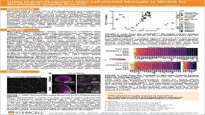 科学海报Using Human Pluripotent Stem Cell-Derived Microglia As Models For Neurological Disease Research
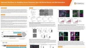 科学海报Optimized Workflows for Modelling Human Pluripotent Stem Cell-Derived Neuron and Glial Interactions
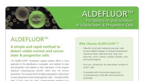 产品手册ALDEFLUOR™: For Identification and Isolation of Viable Stem & Progenitor Cells
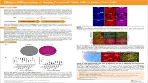 科学海报Efficient Generation of Human Pluripotent Stem Cells to Neural Crest Cells
 挂图Derivation and Applications of Human Pluripotent Stem Cells Overview of the derivation of human embryonic stem cells (hESCs) and induced pluripotent stem cells (iPSCs)发布日期: 11/26/2020

 沪公网安备31010102008431号