Gonzalez-Velasquez FJ and Moss MA (JAN 2008)
Journal of neurochemistry 104 2 500--13
Soluble aggregates of the amyloid-beta protein activate endothelial monolayers for adhesion and subsequent transmigration of monocyte cells.
Increasing evidence suggests that the deposition of amyloid plaques,composed primarily of the amyloid-beta protein (Abeta),within the cerebrovasculature is a frequent occurrence in Alzheimer's disease and may play a significant role in disease progression. Accordingly,the pathogenic mechanisms by which Abeta can alter vascular function may have therapeutic implications. Despite observations that Abeta elicits a number of physiological responses in endothelial cells,ranging from alteration of protein expression to cell death,the Abeta species accountable for these responses remains unexplored. In the current study,we show that isolated soluble Abeta aggregation intermediates activate human brain microvascular endothelial cells for both adhesion and subsequent transmigration of monocyte cells in the absence of endothelial cell death and monolayer disruption. In contrast,unaggregated Abeta monomer and mature Abeta fibril fail to induce any change in endothelial adhesion or transmigration. Correlations between average Abeta aggregate size and observed increases in adhesion illustrate that smaller soluble aggregates are more potent activators of endothelium. These results support previous studies demonstrating heightened neuronal activity of soluble Abeta aggregates,including Abeta-derived diffusible ligands,oligomers,and protofibrils,and further show that soluble aggregates also selectively exhibit activity in a vascular cell model.
View Publication
产品类型:
产品号#:
70034
200-0167
200-0166
产品名:
冻存的人外周血单核细胞
人外周血单核细胞,冷冻
人外周血单核细胞,冷冻
Robinson M et al. (AUG 2016)
Stem Cell Reviews and Reports 12 4 476--483
Functionalizing Ascl1 with Novel Intracellular Protein Delivery Technology for Promoting Neuronal Differentiation of Human Induced Pluripotent Stem Cells
Pluripotent stem cells can become any cell type found in the body. Accordingly,one of the major challenges when working with pluripotent stem cells is producing a highly homogenous population of differentiated cells,which can then be used for downstream applications such as cell therapies or drug screening. The transcription factor Ascl1 plays a key role in neural development and previous work has shown that Ascl1 overexpression using viral vectors can reprogram fibroblasts directly into neurons. Here we report on how a recombinant version of the Ascl1 protein functionalized with intracellular protein delivery technology (Ascl1-IPTD) can be used to rapidly differentiate human induced pluripotent stem cells (hiPSCs) into neurons. We first evaluated a range of Ascl1-IPTD concentrations to determine the most effective amount for generating neurons from hiPSCs cultured in serum free media. Next,we looked at the frequency of Ascl1-IPTD supplementation in the media on differentiation and found that one time supplementation is sufficient enough to trigger the neural differentiation process. Ascl1-IPTD was efficiently taken up by the hiPSCs and enabled rapid differentiation into TUJ1-positive and NeuN-positive populations with neuronal morphology after 8 days. After 12 days of culture,hiPSC-derived neurons produced by Ascl1-IPTD treatment exhibited greater neurite length and higher numbers of branch points compared to neurons derived using a standard neural progenitor differentiation protocol. This work validates Ascl1-IPTD as a powerful tool for engineering neural tissue from pluripotent stem cells.
View Publication
产品类型:
产品号#:
05832
05833
05838
05872
05873
05940
07174
07180
07183
07190
27147
07191
36254
07930
07931
07940
07955
07956
07959
07954
27845
27945
27840
27865
27940
27965
05835
05839
08581
08582
100-0483
100-0484
100-1061
07952
100-0763
100-0485
100-1077
产品名:
STEMdiff™ 神经花环选择试剂
STEMdiff™神经前体细胞培养基
STEMdiff™神经祖细胞冻存液
Vitronectin XF™
CellAdhere™ 稀释缓冲液
DMEM/F-12 with 15 mM HEPES
CryoStor® CS10
CryoStor® CS10
CryoStor® CS10
CryoStor® CS10
CryoStor® CS10
STEMdiff™ 神经诱导培养基
STEMdiff™ 神经诱导培养基
STEMdiff™SMADi神经诱导试剂盒
STEMdiff™SMADi神经诱导试剂盒,2套
Hausser Scientificᵀᴹ 明线血球计数板
ReLeSR™
CryoStor® CS10
CryoStor® CS10
Vitronectin XF™
温和细胞解离试剂
ReLeSR™
Kim J et al. (NOV 2013)
Stem Cell Research 11 3 978--989
Alginate microcapsule as a 3D platform for the efficient differentiation of human embryonic stem cells to dopamine neurons
Human embryonic stem cells (hESCs) are emerging as an attractive alternative source for cell replacement therapy since the cells can be expanded in culture indefinitely and differentiated into any cell types in the body. In order to optimize cell-to-cell interaction,cell proliferation and differentiation into specific lineages as well as tissue organization,it is important to provide a microenvironment for the hESCs which mimics the stem cell niche. One approach is to provide a three-dimensional (3D) environment such as encapsulation. We present an approach to culture and differentiate hESCs into midbrain dopamine (mdDA) neurons in a 3D microenvironment using alginate microcapsules for the first time. A detailed gene and protein expression analysis during neuronal differentiation showed an increased gene and protein expression of various specific DA neuronal markers,particularly tyrosine hydroxylase (TH) by textgreater100 folds after 2weeks and at least 50% higher expression after 4weeks respectively,compared to cells differentiated under conventional two-dimensional (2D) platform. The encapsulated TH+ cells co-expressed mdDA neuronal markers,forkhead box protein A-2 (FOXA2) and pituitary homeobox-3 (PITX3) after 4weeks and secreted approximately 60pg/ml/106 cells higher DA level when induced. We propose that the 3D platform facilitated an early onset of DA neuronal generation compared to that with conventional 2D system which also secretes more DA under potassium-induction. It is a very useful model to study the proliferation and directed differentiation of hESCs to various lineages,particularly to mdDA neurons. This 3D system also allows the separation of feeder cells from hESCs during the process of differentiation and also has potential for immune-isolation during transplantation studies. ?? 2013 Elsevier B.V.
View Publication
Rushkevich YN et al. (AUG 2015)
Bulletin of experimental biology and medicine 159 4 576--81
The Use of Autologous Mesenchymal Stem Cells for Cell Therapy of Patients with Amyotrophic Lateral Sclerosis in Belarus.
We studied a new method of treatment of amyotrophic lateral sclerosis with autologous mesenchymal stem cells. Autologous mesenchymal stem cells were injected intravenously (intact cells) or via lumbar puncture (cells committed to neuronal differentiation). Evaluation of the results of cell therapy after 12-month follow-up revealed slowing down of the disease progression in 10 patients in comparison with the control group consisting of 15 patients. The cell therapy was safe for the patients.
View Publication
产品类型:
产品号#:
05761
产品名:
用于小鼠和大鼠神经干细胞和祖细胞分化培养的试剂盒
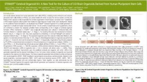
Chailangkarn T et al. (AUG 2016)
Nature 536 7616 338--343
A human neurodevelopmental model for Williams syndrome.
Williams syndrome is a genetic neurodevelopmental disorder characterized by an uncommon hypersociability and a mosaic of retained and compromised linguistic and cognitive abilities. Nearly all clinically diagnosed individuals with Williams syndrome lack precisely the same set of genes,with breakpoints in chromosome band 7q11.23 (refs 1-5). The contribution of specific genes to the neuroanatomical and functional alterations,leading to behavioural pathologies in humans,remains largely unexplored. Here we investigate neural progenitor cells and cortical neurons derived from Williams syndrome and typically developing induced pluripotent stem cells. Neural progenitor cells in Williams syndrome have an increased doubling time and apoptosis compared with typically developing neural progenitor cells. Using an individual with atypical Williams syndrome,we narrowed this cellular phenotype to a single gene candidate,frizzled 9 (FZD9). At the neuronal stage,layer V/VI cortical neurons derived from Williams syndrome were characterized by longer total dendrites,increased numbers of spines and synapses,aberrant calcium oscillation and altered network connectivity. Morphometric alterations observed in neurons from Williams syndrome were validated after Golgi staining of post-mortem layer V/VI cortical neurons. This model of human induced pluripotent stem cells fills the current knowledge gap in the cellular biology of Williams syndrome and could lead to further insights into the molecular mechanism underlying the disorder and the human social brain.
View Publication
An ID2-dependent mechanism for VHL inactivation in cancer.
Mechanisms that maintain cancer stem cells are crucial to tumour progression. The ID2 protein supports cancer hallmarks including the cancer stem cell state. HIFα transcription factors,most notably HIF2α (also known as EPAS1),are expressed in and required for maintenance of cancer stem cells (CSCs). However,the pathways that are engaged by ID2 or drive HIF2α accumulation in CSCs have remained unclear. Here we report that DYRK1A and DYRK1B kinases phosphorylate ID2 on threonine 27 (Thr27). Hypoxia downregulates this phosphorylation via inactivation of DYRK1A and DYRK1B. The activity of these kinases is stimulated in normoxia by the oxygen-sensing prolyl hydroxylase PHD1 (also known as EGLN2). ID2 binds to the VHL ubiquitin ligase complex,displaces VHL-associated Cullin 2,and impairs HIF2α ubiquitylation and degradation. Phosphorylation of Thr27 of ID2 by DYRK1 blocks ID2-VHL interaction and preserves HIF2α ubiquitylation. In glioblastoma,ID2 positively modulates HIF2α activity. Conversely,elevated expression of DYRK1 phosphorylates Thr27 of ID2,leading to HIF2α destabilization,loss of glioma stemness,inhibition of tumour growth,and a more favourable outcome for patients with glioblastoma.
View Publication
产品类型:
产品号#:
05700
05701
05702
产品名:
NeuroCult™ 基础培养基(小鼠&大鼠)
NeuroCult™ 扩增添加物 (小鼠&大鼠)
NeuroCult™ 扩增试剂盒 (小鼠&大鼠)
Rahman M et al. (SEP 2013)
Future Oncology 9 9 1389--1396
Controlling tumor invasion: bevacizumab and BMP4 for glioblastoma
AIM Bevacizumab has been reported to result in increased tumor invasion when used to treat malignant glioma. We hypothesized that BMP4 would prevent diffuse tumor infiltration induced by bevacizumab for malignant glioma in a xenograft model. METHODS Human glioblastoma (GBM) tumor cells were implanted in the striatum of immunocompromised mice. The animals were treated with bevacizumab and BMP4. Tumor growth and invasion were measured. RESULTS The bevacizumab-treated mice had increased survival compared with control animals (p = 0.02). BMP4 alone did not result in improved survival (p = 1.0). The bevacizumab (p = 0.006) and bevacizumab plus BMP4 (p = 0.006) groups demonstrated significantly decreased total tumor size compared with control. Tumor invasion was significantly decreased in the bevacizumab (p = 0.005),BMP4 (p = 0.04) alone and bevacizumab plus BMP4 (p = 0.002) groups compared with control. No synergistic effect between bevacizumab and BMP4 was observed. CONCLUSION Bevacizumab treatment did not result in diffuse infiltration of human GBM in a mouse xenograft model. BMP4 did have an independent favorable effect on GBM that was not synergistic with bevacizumab treatment.
View Publication
产品类型:
产品号#:
05750
05751
产品名:
NeuroCult™ NS-A 基础培养基(人)
NeuroCult™ NS-A 扩增试剂盒(人)
Francis KR et al. (APR 2016)
Nature medicine 22 4 388--396
Modeling Smith-Lemli-Opitz syndrome with induced pluripotent stem cells reveals a causal role for Wnt/$$-catenin defects in neuronal cholesterol synthesis phenotypes.
Smith-Lemli-Opitz syndrome (SLOS) is a malformation disorder caused by mutations in DHCR7,which impair the reduction of 7-dehydrocholesterol (7DHC) to cholesterol. SLOS results in cognitive impairment,behavioral abnormalities and nervous system defects,though neither affected cell types nor impaired signaling pathways are fully understood. Whether 7DHC accumulation or cholesterol loss is primarily responsible for disease pathogenesis is also unclear. Using induced pluripotent stem cells (iPSCs) from subjects with SLOS,we identified cellular defects that lead to precocious neuronal specification within SLOS derived neural progenitors. We also demonstrated that 7DHC accumulation,not cholesterol deficiency,is critical for SLOS-associated defects. We further identified downregulation of Wnt/$$-catenin signaling as a key initiator of aberrant SLOS iPSC differentiation through the direct inhibitory effects of 7DHC on the formation of an active Wnt receptor complex. Activation of canonical Wnt signaling prevented the neural phenotypes observed in SLOS iPSCs,suggesting that Wnt signaling may be a promising therapeutic target for SLOS.
View Publication