
 EasySep™小鼠TIL(CD45)正选试剂盒


搜索结果: 'methocult media formulations for mouse hematopoietic cells serum containing'
-

产品类型:
产品号#:
05700
05701
05702
产品名:
NeuroCult™ 基础培养基(小鼠&大鼠)
NeuroCult™ 扩增添加物 (小鼠&大鼠)
NeuroCult™ 扩增试剂盒 (小鼠&大鼠)
-
产品类型:
产品号#:
05700
05704
产品名:
NeuroCult™ 基础培养基(小鼠&大鼠)
NeuroCult™ 分化试剂盒 (小鼠&大鼠)
-
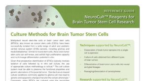 产品手册NeuroCult™: Reagents for Brain Tumor Stem Cell Research
产品类型:
品牌:
NeuroCult
产品号#:
01700
01705
01701
01702
05700
05702
05704
05707
05715
05740
05742
05751
05752
05761
05771
05772
07920
产品名:
ALDEFLUOR™ 试剂盒
ALDEFLUOR™ DEAB试剂
ALDEFLUOR™测定缓冲液
NeuroCult™ 基础培养基(小鼠&大鼠)
NeuroCult™ 扩增试剂盒 (小鼠&大鼠)
NeuroCult™ 分化试剂盒 (小鼠&大鼠)
NeuroCult™化学解离试剂盒(小鼠)
NeuroCult™成年中枢神经系统(CNS)组织酶解试剂盒(小鼠和大鼠)
NeuroCult™ NS-A 扩增试剂盒(人)
NeuroCult™ NS-A 分化试剂盒 (人)
用于小鼠和大鼠神经干细胞和祖细胞分化培养的试剂盒
ACCUTASE™
-
产品类型:
产品号#:
05790
05792
05793
05794
05795
R1061
R1034
R1116
产品名:
BrainPhys™神经元培养基
BrainPhys™神经元培养基和SM1试剂盒
BrainPhys™ 神经元培养基N2-A和SM1试剂盒
BrainPhys™原代神经元试剂盒
BrainPhys™ hPSC 神经元试剂盒
-
产品类型:
产品号#:
01801
产品名:
NeuroFluor™NeuO
-
 31:49
线上讲座Using Human Pluripotent Stem Cell-Derived Neural Organoids for Disease Modeling发布日期: 08/01/2024
31:49
线上讲座Using Human Pluripotent Stem Cell-Derived Neural Organoids for Disease Modeling发布日期: 08/01/2024 -
 30:53
线上讲座Highly Characterized Human iPSCs and NPCs for Downstream Differentiation Applications发布日期: 07/19/2023
30:53
线上讲座Highly Characterized Human iPSCs and NPCs for Downstream Differentiation Applications发布日期: 07/19/2023 -
产品类型:
产品号#:
05700
05701
05702
产品名:
NeuroCult™ 基础培养基(小鼠&大鼠)
NeuroCult™ 扩增添加物 (小鼠&大鼠)
NeuroCult™ 扩增试剂盒 (小鼠&大鼠)


 沪公网安备31010102008431号