Ma I and Allan AL (JUN 2011)
Stem cell reviews 7 2 292--306
The role of human aldehyde dehydrogenase in normal and cancer stem cells.
Normal stem cells and cancer stem cells (CSCs) share similar properties,in that both have the capacity to self-renew and differentiate into multiple cell types. In both the normal stem cell and cancer stem cell fields,there has been a great need for a universal marker that can effectively identify and isolate these rare populations of cells in order to characterize them and use this information for research and therapeutic purposes. Currently,it would appear that certain isoenzymes of the aldehyde dehydrogenase (ALDH) superfamily may be able to fulfill this role as a marker for both normal and cancer stem cells. ALDH has been identified as an important enzyme in the protection of normal hematopoietic stem cells,and is now also widely used as a marker to identify and isolate various types of normal stem cells and CSCs. In addition,emerging evidence suggests that ALDH1 is not only a marker for stem cells,but may also play important functional roles related to self-protection,differentiation,and expansion. This comprehensive review discusses the role that ALDH plays in normal stem cells and CSCs,with focus on ALDH1 and ALDH3A1. Discrepancies in the functional themes between cell types and future perspectives for therapeutic applications will also be discussed.
View Publication
产品类型:
产品号#:
01700
01705
01702
产品名:
ALDEFLUOR™ 试剂盒
ALDEFLUOR™ DEAB试剂, 1.5 mM, 1 mL
ALDEFLUOR™检测缓冲液
Gonzalez-Velasquez FJ and Moss MA (JAN 2008)
Journal of neurochemistry 104 2 500--13
Soluble aggregates of the amyloid-beta protein activate endothelial monolayers for adhesion and subsequent transmigration of monocyte cells.
Increasing evidence suggests that the deposition of amyloid plaques,composed primarily of the amyloid-beta protein (Abeta),within the cerebrovasculature is a frequent occurrence in Alzheimer's disease and may play a significant role in disease progression. Accordingly,the pathogenic mechanisms by which Abeta can alter vascular function may have therapeutic implications. Despite observations that Abeta elicits a number of physiological responses in endothelial cells,ranging from alteration of protein expression to cell death,the Abeta species accountable for these responses remains unexplored. In the current study,we show that isolated soluble Abeta aggregation intermediates activate human brain microvascular endothelial cells for both adhesion and subsequent transmigration of monocyte cells in the absence of endothelial cell death and monolayer disruption. In contrast,unaggregated Abeta monomer and mature Abeta fibril fail to induce any change in endothelial adhesion or transmigration. Correlations between average Abeta aggregate size and observed increases in adhesion illustrate that smaller soluble aggregates are more potent activators of endothelium. These results support previous studies demonstrating heightened neuronal activity of soluble Abeta aggregates,including Abeta-derived diffusible ligands,oligomers,and protofibrils,and further show that soluble aggregates also selectively exhibit activity in a vascular cell model.
View Publication
K. B. Langer et al. (APR 2018)
Stem cell reports 10 4 1282--1293
Retinal Ganglion Cell Diversity and Subtype Specification from Human Pluripotent Stem Cells.
Retinal ganglion cells (RGCs) are the projection neurons of the retina and transmit visual information to postsynaptic targets in the brain. While this function is shared among nearly all RGCs,this class of cell is remarkably diverse,comprised of multiple subtypes. Previous efforts have identified numerous RGC subtypes in animal models,but less attention has been paid to human RGCs. Thus,efforts of this study examined the diversity of RGCs differentiated from human pluripotent stem cells (hPSCs) and characterized defined subtypes through the expression of subtype-specific markers. Further investigation of these subtypes was achieved using single-cell transcriptomics,confirming the combinatorial expression of molecular markers associated with these subtypes,and also provided insight into more subtype-specific markers. Thus,the results of this study describe the derivation of RGC subtypes from hPSCs and will support the future exploration of phenotypic and functional diversity within human RGCs.
View Publication
产品类型:
产品号#:
05790
05792
05793
05794
05795
85850
85857
产品名:
BrainPhys™神经元培养基
BrainPhys™神经元培养基和SM1试剂盒
BrainPhys™ 神经元培养基N2-A和SM1试剂盒
BrainPhys™原代神经元试剂盒
BrainPhys™ hPSC 神经元试剂盒
mTeSR™1
mTeSR™1
I. Canals et al. (SEP 2018)
Nature methods 15 9 693--696
Rapid and efficient induction of functional astrocytes from human pluripotent stem cells.
The derivation of astrocytes from human pluripotent stem cells is currently slow and inefficient. We demonstrate that overexpression of the transcription factors SOX9 and NFIB in human pluripotent stem cells rapidly and efficiently yields homogeneous populations of induced astrocytes. In our study these cells exhibited molecular and functional properties resembling those of adult human astrocytes and were deemed suitable for disease modeling. Our method provides new possibilities for the study of human astrocytes in health and disease.
View Publication
Piccirillo SGM et al. (DEC 2006)
Nature 444 7120 761--5
Bone morphogenetic proteins inhibit the tumorigenic potential of human brain tumour-initiating cells.
Transformed,oncogenic precursors,possessing both defining neural-stem-cell properties and the ability to initiate intracerebral tumours,have been identified in human brain cancers. Here we report that bone morphogenetic proteins (BMPs),amongst which BMP4 elicits the strongest effect,trigger a significant reduction in the stem-like,tumour-initiating precursors of human glioblastomas (GBMs). Transient in vitro exposure to BMP4 abolishes the capacity of transplanted GBM cells to establish intracerebral GBMs. Most importantly,in vivo delivery of BMP4 effectively blocks the tumour growth and associated mortality that occur in 100% of mice after intracerebral grafting of human GBM cells. We demonstrate that BMPs activate their cognate receptors (BMPRs) and trigger the Smad signalling cascade in cells isolated from human glioblastomas (GBMs). This is followed by a reduction in proliferation,and increased expression of markers of neural differentiation,with no effect on cell viability. The concomitant reduction in clonogenic ability,in the size of the CD133+ population and in the growth kinetics of GBM cells indicates that BMP4 reduces the tumour-initiating cell pool of GBMs. These findings show that the BMP-BMPR signalling system--which controls the activity of normal brain stem cells--may also act as a key inhibitory regulator of tumour-initiating,stem-like cells from GBMs and the results also identify BMP4 as a novel,non-cytotoxic therapeutic effector,which may be used to prevent growth and recurrence of GBMs in humans.
View Publication
Harlow DE et al. (JAN 2014)
Journal of Neuroscience 34 4 1333--1343
Expression of Proteolipid Protein Gene in Spinal Cord Stem Cells and Early Oligodendrocyte Progenitor Cells Is Dispensable for Normal Cell Migration and Myelination
Plp1 gene expression occurs very early in development,well before the onset of myelination,creating a conundrum with regard to the function of myelin proteolipid protein (PLP),one of the major proteins in compact myelin. Using PLP-EGFP mice to investigate Plp1 promoter activity,we found that,at very early time points,PLP-EGFP was expressed in Sox2+ undifferentiated precursors in the spinal cord ventricular zone (VZ),as well as in the progenitors of both neuronal and glial lineages. As development progressed,most PLP-EGFP-expressing cells gave rise to oligodendrocyte progenitor cells (OPCs). The expression of PLP-EGFP in the spinal cord was quite dynamic during development. PLP-EGFP was highly expressed as cells delaminated from the VZ. Expression was downregulated as cells moved laterally through the cord,and then robustly upregulated as OPCs differentiated into mature myelinating oligodendrocytes. The presence of PLP-EGFP expression in OPCs raises the question of its role in this migratory population. We crossed PLP-EGFP reporter mice into a Plp1-null background to investigate the role of PLP in early OPC development. In the absence of PLP,normal numbers of OPCs were generated and their distribution throughout the spinal cord was unaffected. However,the orientation and length of OPC processes during migration was abnormal in Plp1-null mice,suggesting that PLP plays a role either in the structural integrity of OPC processes or in their response to extracellular cues that orient process outgrowth.
View Publication
产品类型:
产品号#:
05707
产品名:
NeuroCult™化学解离试剂盒(小鼠)
Pei Y et al. (MAY 2016)
Brain research 1638 Pt A 57--73
Comparative neurotoxicity screening in human iPSC-derived neural stem cells, neurons and astrocytes.
Induced pluripotent stem cells (iPSC) and their differentiated derivatives offer a unique source of human primary cells for toxicity screens. Here,we report on the comparative cytotoxicity of 80 compounds (neurotoxicants,developmental neurotoxicants,and environmental compounds) in iPSC as well as isogenic iPSC-derived neural stem cells (NSC),neurons,and astrocytes. All compounds were tested over a 24-h period at 10 and 100$\$,in duplicate,with cytotoxicity measured using the MTT assay. Of the 80 compounds tested,50 induced significant cytotoxicity in at least one cell type; per cell type,32,38,46,and 41 induced significant cytotoxicity in iPSC,NSC,neurons,and astrocytes,respectively. Four compounds (valinomycin,3,3',5,5'-tetrabromobisphenol,deltamethrin,and triphenyl phosphate) were cytotoxic in all four cell types. Retesting these compounds at 1,10,and 100$\$ using the same exposure protocol yielded consistent results as compared with the primary screen. Using rotenone,we extended the testing to seven additional iPSC lines of both genders; no substantial difference in the extent of cytotoxicity was detected among the cell lines. Finally,the cytotoxicity assay was simplified by measuring luciferase activity using lineage-specific luciferase reporter iPSC lines which were generated from the parental iPSC line. This article is part of a Special Issue entitled SI: PSC and the brain.
View Publication
产品类型:
产品号#:
05850
05857
05870
05875
85850
85857
85870
85875
产品名:
mTeSR™1
mTeSR™1
Li Y et al. (MAR 2015)
PLoS ONE 10 3 e0118266
A comprehensive library of familial human amyotrophic lateral sclerosis induced pluripotent stem cells
Amyotrophic lateral sclerosis is a progressive disease characterized by the loss of upper and lower motor neurons,leading to paralysis of voluntary muscles. About 10% of all ALS cases are familial (fALS),among which 15-20% are linked to Cu/Zn superoxide dismutase (SOD1) mutations,usually inherited in an autosomal dominant manner. To date only one FDA approved drug is available which increases survival moderately. Our understanding of ALS disease mechanisms is largely derived from rodent model studies,however due to the differences between rodents and humans,it is necessary to have humanized models for studies of disease pathogenesis as well as drug development. Therefore,we generated a comprehensive library of a total 22 of fALS patient-specific induced pluripotent stem cell (iPSC) lines. These cells were thoroughly characterized before being deposited into the library. The library of cells includes a variety of C9orf72 mutations,sod1 mutations,FUS,ANG and FIG4 mutations. Certain mutations are represented with more than one line,which allows for studies of variable genetic backgrounds. In addition,these iPSCs can be successfully differentiated to astroglia,a cell type known to play a critical role in ALS disease progression. This library represents a comprehensive resource that can be used for ALS disease modeling and the development of novel therapeutics.
View Publication

 EasySep™小鼠TIL(CD45)正选试剂盒


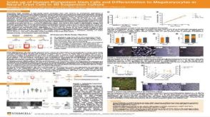 科学海报Scale-up of Human Pluripotent Stem Cells and Differentiation to Megakaryocytes or Neural Crest Cells in 3D Suspension Culture
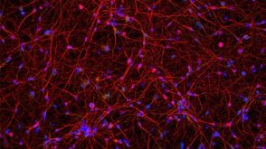 实验方案How to Culture Human Pluripotent Stem Cell (hPSC)-Derived Forebrain Neurons for MEA Analysis Using the Maestro MEA™ System
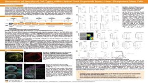 科学海报Generation of Functional 3D Spinal Cord Organoids from Human Pluripotent Stem Cells
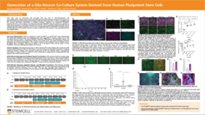 科学海报Generation of a Glia-Neuron Co-Culture System Derived From Human Pluripotent Stem Cells
 沪公网安备31010102008431号