Ware CB et al. (MAR 2014)
Proceedings of the National Academy of Sciences of the United States of America 111 12 4484--9
Derivation of naive human embryonic stem cells.
The naïve pluripotent state has been shown in mice to lead to broad and more robust developmental potential relative to primed mouse epiblast cells. The human naïve ES cell state has eluded derivation without the use of transgenes,and forced expression of OCT4,KLF4,and KLF2 allows maintenance of human cells in a naïve state [Hanna J,et al. (2010) Proc Natl Acad Sci USA 107(20):9222-9227]. We describe two routes to generate nontransgenic naïve human ES cells (hESCs). The first is by reverse toggling of preexisting primed hESC lines by preculture in the histone deacetylase inhibitors butyrate and suberoylanilide hydroxamic acid,followed by culture in MEK/ERK and GSK3 inhibitors (2i) with FGF2. The second route is by direct derivation from a human embryo in 2i with FGF2. We show that human naïve cells meet mouse criteria for the naïve state by growth characteristics,antibody labeling profile,gene expression,X-inactivation profile,mitochondrial morphology,microRNA profile and development in the context of teratomas. hESCs can exist in a naïve state without the need for transgenes. Direct derivation is an elusive,but attainable,process,leading to cells at the earliest stage of in vitro pluripotency described for humans. Reverse toggling of primed cells to naïve is efficient and reproducible.
View Publication
产品类型:
产品号#:
05860
05880
产品名:
Inagi R et al. (NOV 2007)
Nephrology,dialysis,transplantation : official publication of the European Dialysis and Transplant Association - European Renal Association 22 11 3311--7
Establishment of a sandwich ELISA for human megsin, a kidney-specific serine protease inhibitor.
BACKGROUND: We previously identified a novel serine protease inhibitor (serpin),megsin,which is predominantly expressed in the kidney. Megsin expression is up-regulated in human and experimental renal diseases associated with mesangial proliferation and expansion,suggesting that urinary megsin may be a novel diagnostic marker for some renal diseases. METHODS: We established a specific and sensitive sandwich enzyme-linked immunosorbent assay (ELISA) for megsin and measured urinary megsin of patients with various renal diseases. RESULTS: Megsin ELISA specifically detected megsin but not other serpins. The detection limit was 0.04 ng/ml,which allowed detection of urinary megsin in 3.6% of healthy individuals. The antigenic epitope in the urine detected by the ELISA was confirmed as megsin protein by time-of-flight mass spectrometry. Among patients with rapidly progressive glomerulonephritis (n = 18),55.6% were urinary megsin-positive,while 24.1% in IgA nephropathy (n = 112) and 15.1% in chronic non-IgA glomerulonephritis (n = 245) were urinary megsin-positive,respectively. Among patients with chronic renal failure due to unknown causes (n = 74),18.9% were positive for urinary megsin. In diabetic patients with or without nephropathy (n = 1073),12.3% were urinary megsin-positive,while positivity of urinary megsin in patients with non-renal diseases (n = 768) was equivalent (3.3%) to that of healthy individuals. Of note,when urinary megsin-positive patients with diabetic nephropathy (n = 71) were classified into four stages by their proteinuria and estimated glomerular filtration rate,urinary megsin excretion increased as the stage progressed up to stage 3A,suggesting correlation of that with mesangial expansion level. Urinary megsin decreased in the advanced stage,probably reflecting development of glomerulosclerosis. CONCLUSION: We established a high-sensitive megsin ELISA,which detects urinary megsin in some patients with renal diseases and in only a few healthy subjects. Megsin ELISA may be a novel diagnostic tool for renal diseases.
View Publication
产品类型:
产品号#:
03800
03801
03802
03803
03804
03805
03806
产品名:
ClonaCell™-HY杂交瘤试剂盒
ClonaCell™-HY培养基A
ClonaCell™-HY 培养基 B
ClonaCell™-HY 培养基 C
ClonaCell™-HY 培养基 D
ClonaCell™-HY 培养基 E
ClonaCell™-HY PEG
Pei Y et al. (MAR 2015)
Scientific reports 5 9205
A platform for rapid generation of single and multiplexed reporters in human iPSC lines.
Induced pluripotent stem cells (iPSC) are important tools for drug discovery assays and toxicology screens. In this manuscript,we design high efficiency TALEN and ZFN to target two safe harbor sites on chromosome 13 and 19 in a widely available and well-characterized integration-free iPSC line. We show that these sites can be targeted in multiple iPSC lines to generate reporter systems while retaining pluripotent characteristics. We extend this concept to making lineage reporters using a C-terminal targeting strategy to endogenous genes that express in a lineage-specific fashion. Furthermore,we demonstrate that we can develop a master cell line strategy and then use a Cre-recombinase induced cassette exchange strategy to rapidly exchange reporter cassettes to develop new reporter lines in the same isogenic background at high efficiency. Equally important we show that this recombination strategy allows targeting at progenitor cell stages,further increasing the utility of the platform system. The results in concert provide a novel platform for rapidly developing custom single or dual reporter systems for screening assays.
View Publication
产品类型:
产品号#:
05850
05857
05870
05875
85850
85857
85870
85875
产品名:
mTeSR™1
mTeSR™1
Cao N et al. ( 2015)
1212 113--125
Generation, expansion, and differentiation of cardiovascular progenitor cells from human pluripotent stem cells.
Cardiovascular progenitor cells (CVPCs) derived from human embryonic stem cells and human induced pluripotent stem cells represent an invaluable potential source for the study of early embryonic cardiovascular development and stem cell-based therapies for congenital and acquired heart diseases. To fully realize their values,it is essential to establish an efficient and stable differentiation system for the induction of these pluripotent stem cells (PSCs) into the CVPCs and robustly expand them in culture,and then further differentiate these CVPCs into multiple cardiovascular cell types. Here we describe the protocols for efficient derivation,expansion,and differentiation of CVPCs from hPSCs in a chemically defined medium under feeder- and serum-free culture conditions.
View Publication
产品类型:
产品号#:
05850
05857
05870
05875
07920
85850
85857
85870
85875
07922
产品名:
ACCUTASE™
mTeSR™1
mTeSR™1
ACCUTASE™
Su RJ et al. ( 2014)
1357 1341 57--69
Generation of iPS Cells from Human Peripheral Blood Mononuclear Cells Using Episomal Vectors
Peripheral blood is the easy-to-access,minimally invasive,and the most abundant cell source to use for cell reprogramming. The episomal vector is among the best approaches for generating integration-free induced pluripotent stem (iPS) cells due to its simplicity and affordability. Here we describe the detailed protocol for the efficient generation of integration-free iPS cells from peripheral blood mononuclear cells. With this optimized protocol,one can readily generate hundreds of iPS cell colonies from 1 ml of peripheral blood.
View Publication
Lancaster MA and Knoblich JA (OCT 2014)
Nature protocols 9 10 2329--2340
Generation of cerebral organoids from human pluripotent stem cells.
Human brain development exhibits several unique aspects,such as increased complexity and expansion of neuronal output,that have proven difficult to study in model organisms. As a result,in vitro approaches to model human brain development and disease are an intense area of research. Here we describe a recently established protocol for generating 3D brain tissue,so-called cerebral organoids,which closely mimics the endogenous developmental program. This method can easily be implemented in a standard tissue culture room and can give rise to developing cerebral cortex,ventral telencephalon,choroid plexus and retinal identities,among others,within 1-2 months. This straightforward protocol can be applied to developmental studies,as well as to the study of a variety of human brain diseases. Furthermore,as organoids can be maintained for more than 1 year in long-term culture,they also have the potential to model later events such as neuronal maturation and survival.
View Publication
产品类型:
产品号#:
05850
05857
05870
05875
85850
85857
85870
85875
产品名:
mTeSR™1
mTeSR™1
Park Y et al. (MAR 2014)
Journal of Biotechnology 174 1 39--48
Hepatic differentiation of human embryonic stem cells on microcarriers
Translation of stem cell research to industrial and clinical settings mostly requires large quantities of cells,especially those involving large organs such as the liver. A scalable reactor system is desirable to ensure a reliable supply of sufficient quantities of differentiated cells. To increase the culture efficiency in bioreactor system,high surface to volume ratio needs to be achieved. We employed a microcarrier culture system for the expansion of undifferentiated human embryonic stem cells (hESCs) as well as for directed differentiation of these cells to hepatocyte-like cells. Cells in single cell suspension were attached to the bead surface in even distribution and were expanded to 1??106cells/ml within 2 days of hESC culture with maintenance of the level of pluripotency markers. Directed differentiation into hepatocyte-like cells on microcarriers,both in static culture and stirred bioreactors,induced similar levels of hepatocyte-like cell differentiation as observed with cells cultured in conventional tissue culture plates. The cells expressed both immature and mature hepatocyte-lineage genes and proteins such as asialoglycoprotein receptor-1 (ASGPR-1) and albumin. Differentiated cells exhibited functional characteristics such as secretion of albumin and urea,and CYP3A4 activity could be detected. Microcarriers thus offer the potential for large-scale expansion and differentiation of hESCs induced hepatocyte-like cells in a more controllable bioreactor environment. ?? 2014.
View Publication
产品类型:
产品号#:
05850
05857
05870
05875
85850
85857
85870
85875
产品名:
mTeSR™1
mTeSR™1
Kuroki MM et al. ( 2005)
Anticancer Research 25 6A 3733--9
Preparation of human IgG and IgM monoclonal antibodies for MK-1/Ep-CAM by using human immunoglobulin gene-transferred mouse and gene cloning of their variable regions.
For antibody-based therapy of cancer,monoclonal antibodies (mAbs) of human origin are superior to mouse,mouse/human chimeric or humanized mAbs,because of their minimum immunogenicity to humans and their efficient collaboration with human effector cells. In the present study,human mAbs were prepared against a pancarcinoma antigen,MK-1 (Ep-CAM),using a genetically-engineered mouse (KM mouse) that contains the human immunoglobulin genes. Spleen cells from KM mice,immunized with recombinant MK-1,were fused with P3-U1 mouse myeloma cells. Of 44 anti-MK-1 clones analyzed,two were of IgG4 and the others of IgM clones. Although the two IgG4 clones were suggested to recognize the same antigenic determinant or two closely located determinants,their VK regions were encoded by different light-chain genes while their VH sequences were identical. The two IgG4 and one of the IgM clones tested revealed antibody-dependent cell-mediated cytotoxicity and complement-dependent cytotoxicity,respectively,against MK-1-expressing cells in vitro,suggesting that these fully human mAbs produced against MK-1 and their V-region genes,which are applicable for the preparation of engineered antibody fragments that may be useful for antibody-based therapy of cancer.
View Publication
产品类型:
产品号#:
03800
03801
03802
03803
03804
03805
03806
产品名:
ClonaCell™-HY杂交瘤试剂盒
ClonaCell™-HY培养基A
ClonaCell™-HY 培养基 B
ClonaCell™-HY 培养基 C
ClonaCell™-HY 培养基 D
ClonaCell™-HY 培养基 E
ClonaCell™-HY PEG
Zimmerman Z et al. (AUG 2005)
Biology of Blood and Marrow Transplantation 11 8 576--86
Effector cells derived from host CD8 memory T cells mediate rapid resistance against minor histocompatibility antigen-mismatched allogeneic marrow grafts without participation of perforin, Fas ligand, and the simultaneous inhibition of 3 tumor necrosis Fa
Reduced-intensity conditioning regimens for transplant recipients have heightened awareness of immunologic resistance to allogeneic bone marrow transplants (BMT). Although T cell-mediated cytotoxicity has been assumed to play a role in the resistance against donor allogeneic hematopoietic stem and progenitor cell grafts,several studies have reported relatively unimpaired resistance by recipients who lack perforin,Fas ligand (FasL),and other cytotoxic mediators. This study compared the early kinetics of T cell-mediated resistance in B6 (H2b) cytotoxically normal versus deficient recipients after transplantation with major histocompatibility complex-matched,minor histocompatibility antigen (MiHA)-mismatched allogeneic marrow grafts. Wild-type B6 or cytotoxic double-deficient perforin-/-/ gld+/+ (B6-cdd) mice were sensitized against major histocompatibility complex-matched BALB.B or C3H.SW (H2b) MiHA and transplanted with a high dose (1 ?? 107) of T cell-depleted bone marrow. CD8 T memory cells were shown to be present in recipients before BMT,and anti-CD8 monoclonal antibody infusion abolished resistance,thus demonstrating that CD8 T cells are the host effector population. Donor-committed and high proliferative potential progenitor numbers were markedly diminished by 48 hours after transplantation in both wild-type B6 and B6-cdd anti-donor MiHA-sensitized recipients. These observations indicate that the resistance pathway used in the cytotoxic deficient mice was both potent and rapidly induced - consistent with a CD8 memory T-cell response. To examine the role of Tumor necrosis factor-like weak inducer of apoptosis (TWEAK)- and TL1A-mediated cytotoxicity in this strong resistance,newly generated monoclonal antibodies specific for these ligands were administered to B6-cdd recipients sensitized to donor antigens. Recipients of syngeneic B6-gfp bone marrow exhibited significant donor colony-forming unit numbers after BMT. In contrast,low or absent colony-forming unit levels were detected in allogeneic recipients,including those that lacked perforin and FasL and that received anti-TWEAK,anti-tumor necrosis factor-related apoptosis-inducing ligand,and anti-TL1A monoclonal antibodies. These findings extend previous observations by demonstrating the existence of a rapidly effected resistance pathway mediated by memory CD8 effector T cells independent of the 2 major pathways of cytotoxicity. Together with previous findings,these results support the notion that effector cells derived from memory CD8 T-cell populations can mediate strong resistance against donor allogeneic MiHA-disparate hematopoietic engraftment by using a mechanism that is independent of the contribution of perforin,FasL,and the known death ligand receptor pathways. ?? 2005 American Society for Blood and Marrow Transplantation.
View Publication
产品类型:
产品号#:
03800
03801
03802
03803
03804
03805
03806
产品名:
ClonaCell™-HY杂交瘤试剂盒
ClonaCell™-HY培养基A
ClonaCell™-HY 培养基 B
ClonaCell™-HY 培养基 C
ClonaCell™-HY 培养基 D
ClonaCell™-HY 培养基 E
ClonaCell™-HY PEG
Richardson T et al. (DEC 2013)
Tissue Engineering: Part A 20 23-24 Epub ahead of print
Alginate encapsulation of human embryonic stem cells to enhance directed differentiation to pancreatic islet-like cells
The pluripotent property of hESCs makes them attractive for treatment of degenerative diseases such as diabetes. We have developed a stage-wise directed differentiation protocol to produce alginate-encapsulated islet-like cells derived from hESCs,which can be directly implanted for diabetes therapy. The advantage of alginate encapsulation lies in its capability to immunoisolate,along with the added possibility of scalable culture. We have evaluated the possibility of encapsulating hESCs at different stages of differentiation. Encapsulation of predifferentiated cells resulted in insufficient cellular yield and differentiation. On the other hand,encapsulation of undifferentiated hESCs followed by differentiation induction upon encapsulation,resulted in the highest viability and differentiation. More striking was that alginate encapsulation resulted in a much stronger differentiation compared to parallel 2D cultures,resulting in 20-fold increase in c-peptide protein synthesis. To elucidate the mechanism contributing to encapsulation-mediated enhancement in hESC maturation,investigation of the signaling pathways revealed interesting insight. While the phospho-protein levels of all the tested signaling molecules were lower under encapsulation,the ratio of pSMAD/pAKT was significantly higher,indicating a more efficient signal transduction under encapsulation. These results clearly demonstrate that alginate encapsulation of hESCs and differentiation to islet-cells types provides a potentially translatable treatment option for type1 diabetes.
View Publication

 EasySep™小鼠TIL(CD45)正选试剂盒


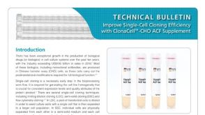 产品手册ClonaCell™-CHO ACF Supplement for Robust Growth of CHO Cells at Low Cell Density
 沪公网安备31010102008431号