Yano M and Pirofski L-a (JAN 2011)
Clinical and vaccine immunology : CVI 18 1 59--66
Characterization of gene use and efficacy of mouse monoclonal antibodies to Streptococcus pneumoniae serotype 8.
Streptococcus pneumoniae is the most common cause of community-acquired pneumonia in the United States and globally. Despite the availability of pneumococcal capsular polysaccharide (PPS) and protein conjugate-based vaccines,the prevalence of antibiotic-resistant pneumococcal strains,serotype (ST) replacement in nonconjugate vaccine strains,and uncertainty as to whether the PPS vaccine that is used in adults protects against pneumonia emphasize the need for continued efforts to understand the nature of protective PPS antibody responses. In this study,we generated mouse monoclonal antibodies (MAbs) to a conjugate consisting of the PPS of serotype 8 (PPS8) S. pneumoniae and tetanus toxoid. Thirteen MAbs,including four IgMs that bound to PPS8 and phosphorylcholine (PC) and five IgMs and four IgG1s that bound to PPS8 but not PC,were produced,and their nucleotide sequences,epitope and fine specificity,and efficacy against lethal challenge with ST8 S. pneumoniae were determined. MAbs that bound to PPS8 exhibited gene use that was distinct from that exhibited by MAbs that bound to PC. Only PPS8-binding MAbs that did not bind PC were protective in mice. All 13 MAbs used germ line variable-region heavy (V(H)) and light (V(L)) chain genes,with no evidence of somatic hypermutation. Our data reveal a relationship between PPS specificity and V(H) gene use and MAb efficacy in mice. These findings provide insight into the relationship between antibody molecular structure and function and hold promise for the development of novel surrogates for pneumococcal vaccine efficacy.
View Publication
产品类型:
产品号#:
03800
03801
03802
03803
03804
03805
03806
产品名:
ClonaCell™-HY杂交瘤试剂盒
ClonaCell™-HY培养基A
ClonaCell™-HY 培养基 B
ClonaCell™-HY 培养基 C
ClonaCell™-HY 培养基 D
ClonaCell™-HY 培养基 E
ClonaCell™-HY PEG
Mou H et al. (APR 2012)
Cell stem cell 10 4 385--397
Generation of multipotent lung and airway progenitors from mouse ESCs and patient-specific cystic fibrosis iPSCs
Deriving lung progenitors from patient-specific pluripotent cells is a key step in producing differentiated lung epithelium for disease modeling and transplantation. By mimicking the signaling events that occur during mouse lung development,we generated murine lung progenitors in a series of discrete steps. Definitive endoderm derived from mouse embryonic stem cells (ESCs) was converted into foregut endoderm,then into replicating Nkx2.1+ lung endoderm,and finally into multipotent embryonic lung progenitor and airway progenitor cells. We demonstrated that precisely-timed BMP,FGF,and WNT signaling are required for NKX2.1 induction. Mouse ESC-derived Nkx2.1+ progenitor cells formed respiratory epithelium (tracheospheres) when transplanted subcutaneously into mice. We then adapted this strategy to produce disease-specific lung progenitor cells from human Cystic Fibrosis induced pluripotent stem cells (iPSCs),creating a platform for dissecting human lung disease. These disease-specific human lung progenitors formed respiratory epithelium when subcutaneously engrafted into immunodeficient mice.
View Publication
产品类型:
产品号#:
05850
05857
05870
05875
85850
85857
85870
85875
产品名:
mTeSR™1
mTeSR™1
Dorrell C et al. (JUN 2011)
Molecular and Cellular Endocrinology 339 1-2 144--150
Isolation of mouse pancreatic alpha, beta, duct and acinar populations with cell surface markers
Tools permitting the isolation of live pancreatic cell subsets for culture and/or molecular analysis are limited. To address this,we developed a collection of monoclonal antibodies with selective surface labeling of endocrine and exocrine pancreatic cell types. Cell type labeling specificity and cell surface reactivity were validated on mouse pancreatic sections and by gene expression analysis of cells isolated using FACS. Five antibodies which marked populations of particular interest were used to isolate and study viable populations of purified pancreatic ducts,acinar cells,and subsets of acinar cells from whole pancreatic tissue or of alpha or beta cells from isolated mouse islets. Gene expression analysis showed the presence of known endocrine markers in alpha and beta cell populations and revealed that TTR and DPPIV are primarily expressed in alpha cells whereas DGKB and GPM6A have a beta cell specific expression profile.
View Publication
Smith Sa et al. (MAR 2012)
Journal of Virology 86 5 2665--75
Persistence of circulating memory B cell clones with potential for Dengue virus disease enhancement for decades following infection
Symptomatic dengue virus infection ranges in disease severity from an influenza-like illness to life-threatening shock. One model of the mechanism underlying severe disease proposes that weakly neutralizing,dengue serotype cross-reactive antibodies induced during a primary infection facilitate virus entry into Fc receptor-bearing cells during a subsequent secondary infection,increasing viral replication and the release of cytokines and vasoactive mediators,culminating in shock. This process has been termed antibody-dependent enhancement of infection and has significantly hindered vaccine development. Much of our understanding of this process has come from studies using mouse monoclonal antibodies (MAbs); however,antibody responses in mice typically exhibit less complexity than those in humans. A better understanding of the humoral immune response to natural dengue virus infection in humans is sorely needed. Using a high-efficiency human hybridoma technology,we isolated 37 hybridomas secreting human MAbs to dengue viruses from 12 subjects years or even decades following primary or secondary infection. The majority of the human antibodies recovered were broadly cross-reactive,directed against either envelope or premembrane proteins,and capable of enhancement of infection in vitro; few exhibited serotype-specific binding or potent neutralizing activity. Memory B cells encoding enhancing antibodies predominated in the circulation,even two or more decades following infection. Mapping the epitopes and activity of naturally occurring dengue antibodies should prove valuable in determining whether the enhancing and neutralizing activity of antibodies can be separated. Such principles could be used in the rational design of vaccines that enhance the induction of neutralizing antibodies,while lowering the risk of dengue shock syndrome.
View Publication

 EasySep™小鼠TIL(CD45)正选试剂盒


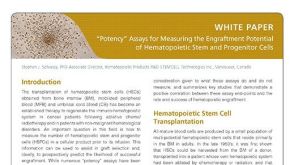 技术公告Potency Assays for Measuring the Engraftment Potential of HSPCs
 技术公告Protocol for Cloning CHO Cell Lines Using ClonaCell™-CHO Semi-Solid Medium
 31:49
线上讲座Using Human Pluripotent Stem Cell-Derived Neural Organoids for Disease Modeling发布日期: 08/01/2024
31:49
线上讲座Using Human Pluripotent Stem Cell-Derived Neural Organoids for Disease Modeling发布日期: 08/01/2024 技术公告Protocol for Producing Monoclonal Cell Lines Using ClonaCell™ FLEX Semi-Solid Medium


 沪公网安备31010102008431号