Fischbach NA et al. (FEB 2005)
Blood 105 4 1456--66
HOXB6 overexpression in murine bone marrow immortalizes a myelomonocytic precursor in vitro and causes hematopoietic stem cell expansion and acute myeloid leukemia in vivo.
The HOX family of homeobox genes plays an important role in normal and malignant hematopoiesis. Dysregulated HOX gene expression profoundly effects the proliferation and differentiation of hematopoietic stem cells (HSCs) and committed progenitors,and aberrant activation of HOX genes is a common event in human myeloid leukemia. HOXB6 is frequently overexpressed in human acute myeloid leukemia (AML). To gain further insight into the role of HOXB6 in hematopoiesis,we overexpressed HOXB6 in murine bone marrow using retrovirus-mediated gene transfer. We also explored structure-function relationships using mutant HOXB6 proteins unable to bind to DNA or a key HOX-binding partner,pre-B-cell leukemia transcription factor-1 (PBX1). Additionally,we investigated the potential cooperative interaction with myeloid ecotropic viral integration site 1 homolog (MEIS1). In vivo,HOXB6 expanded HSCs and myeloid precursors while inhibiting erythropoiesis and lymphopoiesis. Overexpression of HOXB6 resulted in AML with a median latency of 223 days. Coexpression of MEIS1 dramatically shortened the onset of AML. Cytogenetic analysis of a subset of HOXB6-induced AMLs revealed recurrent deletions of chromosome bands 2D-E4,a region frequently deleted in HOXA9-induced AMLs. In vitro,HOXB6 immortalized a factor-dependent myelomonocytic precursor capable of granulocytic and monocytic differentiation. These biologic effects of HOXB6 were largely dependent on DNA binding but independent of direct interaction with PBX1.
View Publication
产品类型:
产品号#:
03434
03444
28600
产品名:
MethoCult™GF M3434
MethoCult™GF M3434
L-Calc™有限稀释软件
Thirukkumaran CM et al. (JUL 2003)
Blood 102 1 377--87
Reovirus oncolysis as a novel purging strategy for autologous stem cell transplantation.
Hematologic stem cell rescue after high-dose cytotoxic therapy is extensively used for the treatment of many hematopoietic and solid cancers. Gene marking studies suggest that occult tumor cells within the autograft may contribute to clinical relapse. To date purging of autografts contaminated with cancer cells has been unsuccessful. The selective oncolytic property of reovirus against myriad malignant histologies in in vitro,in vivo,and ex vivo systems has been previously demonstrated. In the present study we have shown that reovirus can successfully purge cancer cells within autografts. Human monocytic and myeloma cell lines as well as enriched ex vivo lymphoma,myeloma,and Waldenström macroglobulinemia patient tumor specimens were used in an experimental purging model. Viability of the cell lines or purified ex vivo tumor cells of diffuse large B-cell lymphoma,chronic lymphocytic leukemia,Waldenström macroglobulinemia,and small lymphocytic lymphoma was significantly reduced after reovirus treatment. Further,[35S]-methionine labeling and sodium dodecyl sulfate-polyacrylamide gel electrophoresis (SDS-PAGE) of cellular proteins demonstrated reovirus protein synthesis and disruption of host cell protein synthesis as early as 24 hours. Admixtures of apheresis product with the abovementioned tumor cells and cell lines treated with reovirus showed complete purging of disease. In contrast,reovirus purging of enriched ex vivo multiple myeloma,Burkitt lymphoma,and follicular lymphoma was incomplete. The oncolytic action of reovirus did not affect CD34+ stem cells or their long-term colony-forming assays even after granulocyte colony-stimulating factor (G-CSF) stimulation. Our results indicate the ex vivo use of an unattenuated oncolytic virus as an attractive purging strategy for autologous stem cell transplantations.
View Publication
产品类型:
产品号#:
04434
04444
09600
09650
84434
84444
产品名:
MethoCult™H4434经典
MethoCult™H4434经典
StemSpan™ SFEM
StemSpan™ SFEM
Jiang J et al. (SEP 2010)
Cancer research 70 18 7242--52
Crucial roles for protein kinase C isoforms in tumor-specific killing by apoptin.
The chicken anemia virus-derived protein apoptin induces apoptosis in a variety of human malignant and transformed cells but not in normal cells. However,the mechanisms through which apoptin achieves its selective killing effects are not well understood. We developed a lentiviral vector encoding a green fluorescent protein-apoptin fusion gene (LV-GFP-AP) that can efficiently deliver apoptin into hematopoietic cells. Apoptin selectively killed the human multiple myeloma cell lines MM1.R and MM1.S,and the leukemia cell lines K562,HL60,U937,KG1,and NB4. In contrast,normal CD34(+) cells were not killed and maintained their differentiation potential in multilineage colony formation assays. In addition,dexamethasone-resistant MM1.R cells were found to be more susceptible to apoptin-induced cell death than the parental matched MM1.S cells. Death susceptibility correlated with increased phosphorylation and activation of the apoptin protein in MM1.R cells. Expression array profiling identified differential kinase profiles between MM1.R and MM1.S cells. Among these kinases,protein kinase Cβ (PKCβ) was found by immunoprecipitation and in vitro kinase studies to be a candidate kinase responsible for apoptin phosphorylation. Indeed,shRNA knockdown or drug-mediated inhibition of PKCβ significantly reduced apoptin phosphorylation. Furthermore,apoptin-mediated cell death proceeded through the upregulation of PKCβ,activation of caspase-9/3,cleavage of the PKCδ catalytic domain,and downregulation of the MERTK and AKT kinases. Collectively,these results elucidate a novel pathway for apoptin activation involving PKCβ and PKCδ. Further,they highlight the potential of apoptin and its cellular regulators to purge bone marrow used in autologous transplantation for multiple myeloma.
View Publication
产品类型:
产品号#:
04434
04444
09600
09650
产品名:
MethoCult™H4434经典
MethoCult™H4434经典
StemSpan™ SFEM
StemSpan™ SFEM
Jo SY et al. (MAY 2011)
Blood 117 18 4759--68
Requirement for Dot1l in murine postnatal hematopoiesis and leukemogenesis by MLL translocation.
Disruptor of telomeric silencing 1-like (Dot1l) is a histone 3 lysine 79 methyltransferase. Studies of constitutive Dot1l knockout mice show that Dot1l is essential for embryonic development and prenatal hematopoiesis. DOT1L also interacts with translocation partners of Mixed Lineage Leukemia (MLL) gene,which is commonly translocated in human leukemia. However,the requirement of Dot1l in postnatal hematopoiesis and leukemogenesis of MLL translocation proteins has not been conclusively shown. With a conditional Dot1l knockout mouse model,we examined the consequences of Dot1l loss in postnatal hematopoiesis and MLL translocation leukemia. Deletion of Dot1l led to pancytopenia and failure of hematopoietic homeostasis,and Dot1l-deficient cells minimally reconstituted recipient bone marrow in competitive transplantation experiments. In addition,MLL-AF9 cells required Dot1l for oncogenic transformation,whereas cells with other leukemic oncogenes,such as Hoxa9/Meis1 and E2A-HLF,did not. These findings illustrate a crucial role of Dot1l in normal hematopoiesis and leukemogenesis of specific oncogenes.
View Publication
Chang M-J et al. (DEC 2010)
Cancer research 70 24 10234--42
Histone H3 lysine 79 methyltransferase Dot1 is required for immortalization by MLL oncogenes.
Chimeric oncoproteins resulting from fusion of MLL to a wide variety of partnering proteins cause biologically distinctive and clinically aggressive acute leukemias. However,the mechanism of MLL-mediated leukemic transformation is not fully understood. Dot1,the only known histone H3 lysine 79 (H3K79) methyltransferase,has been shown to interact with multiple MLL fusion partners including AF9,ENL,AF10,and AF17. In this study,we utilize a conditional Dot1l deletion model to investigate the role of Dot1 in hematopoietic progenitor cell immortalization by MLL fusion proteins. Western blot and mass spectrometry show that Dot1-deficient cells are depleted of the global H3K79 methylation mark. We find that loss of Dot1 activity attenuates cell viability and colony formation potential of cells immortalized by MLL oncoproteins but not by the leukemic oncoprotein E2a-Pbx1. Although this effect is most pronounced for MLL-AF9,we find that Dot1 contributes to the viability of cells immortalized by other MLL oncoproteins that are not known to directly recruit Dot1. Cells immortalized by MLL fusions also show increased apoptosis,suggesting the involvement of Dot1 in survival pathways. In summary,our data point to a pivotal requirement for Dot1 in MLL fusion protein-mediated leukemogenesis and implicate Dot1 as a potential therapeutic target.
View Publication
产品类型:
产品号#:
03234
18757
18757RF
产品名:
MethoCult™M3234
EasySep™小鼠CD117(cKIT)正选试剂盒
RoboSep™ 小鼠CD117(cKIT)正选试剂盒含滤芯吸头
Xu D et al. ( 2012)
PloS one 7 10 e46670
Cancer stem cell-related gene periostin: a novel prognostic marker for breast cancer.
We investigated the expression status of periostin in breast cancer stem cells and its clinical implications in order to lay a foundation for managing breast cancer. CD44+/CD24-/line- tumor cells (CSC) from clinical specimens were sorted using flow cytometry. Periostin expression status was detected in CSC cells and 1,086 breast cancer specimens by Western blot and immunohistochemistry staining,with the CSC ratio determined by immunofluorescence double staining. The relationship between the periostin protein and clinico-pathological parameters and prognosis was subsequently determined. As a result,CSC cells are more likely to generate new tumors in mice and cell microspheres that are deficient in NOD/SCID compared to the control group. Periostin protein was expressed higher in CSC cells compared to the control cells and was found to be related to CSC chemotherapy resistance. Moreover,periostin expression was found to be related to the CSC ratio in 1,086 breast cancer specimens (P = 0.001). In total,334 (30.76%) of the 1,086 breast cases showed high periostin expression. After universal and Spearman regression correlation analysis,periostin was observed to be related to histological grade,CSC ratio,lymph node metastasis,tumor size,and triple-negative breast cancer (all Ptextless0.05). Furthermore,periostin was shown to attain a significantly more distant bone metastasis and worse disease-specific survival than those with none or low-expressed periostin protein (P = 0.001). In the Cox regression test,periostin protein was detected as an independent prognostic factor (P = 0.001). In conclusion,periostin was found to be related to the CSC and an independent prognostic factor for breast cancer. It is also perhaps a potential target to breast cancer.
View Publication
Cell-culture assays reveal the importance of retroviral vector design for insertional genotoxicity.
Retroviral vectors with long terminal repeats (LTRs),which contain strong enhancer/promoter sequences at both ends of their genome,are widely used for stable gene transfer into hematopoietic cells. However,recent clinical data and mouse models point to insertional activation of cellular proto-oncogenes as a dose-limiting side effect of retroviral gene delivery that potentially induces leukemia. Self-inactivating (SIN) retroviral vectors do not contain the terminal repetition of the enhancer/promoter,theoretically attenuating the interaction with neighboring cellular genes. With a new assay based on in vitro expansion of primary murine hematopoietic cells and selection in limiting dilution,we showed that SIN vectors using a strong internal retroviral enhancer/promoter may also transform cells by insertional mutagenesis. Most transformed clones,including those obtained after dose escalation of SIN vectors,showed insertions upstream of the third exon of Evi1 and in reverse orientation to its transcriptional orientation. Normalizing for the vector copy number,we found the transforming capacity of SIN vectors to be significantly reduced when compared with corresponding LTR vectors. Additional modifications of SIN vectors may further increase safety. Improved cell-culture assays will likely play an important role in the evaluation of insertional mutagenesis.
View Publication
产品类型:
产品号#:
09850
28600
产品名:
L-Calc™有限稀释软件
Chen Y et al. (FEB 2011)
Biochemical and biophysical research communications 405 2 173--9
Aldehyde dehydrogenase 1B1 (ALDH1B1) is a potential biomarker for human colon cancer.
Aldehyde dehydrogenases (ALDHs) belong to a superfamily of NAD(P)+-dependent enzymes,which catalyze the oxidation of endogenous and exogenous aldehydes to their corresponding acids. Increased expression and/or activity of ALDHs,particularly ALDH1A1,have been reported to occur in human cancers. It is proposed that the metabolic function of ALDH1A1 confers the stemness" properties to normal and cancer stem cells. Nevertheless�
View Publication
产品类型:
产品号#:
01700
01705
01701
01702
产品名:
ALDEFLUOR™ 试剂盒
ALDEFLUOR™ DEAB试剂
ALDEFLUOR™测定缓冲液
Li J et al. (MAR 2005)
Clinical Cancer Research 11 6 2195--2204
Generation of PRL-3- and PRL-1-specific monoclonal antibodies as potential diagnostic markers for cancer metastases
PURPOSE: The PRL-3 mRNA is consistently elevated in metastatic samples derived from colorectal cancers. We sought to generate a specific PRL-3 monoclonal antibody (mAb) that might serve as a potential diagnostic marker for colorectal cancer metastasis. EXPERIMENTAL DESIGN: PRL-3 is one of three members (PRL-1,PRL-2,and PRL-3) in a unique protein-tyrosine phosphatase family. Because the three PRLs are 76% to 87% identical in their amino acid sequences,it poses a great challenge to obtain mAbs that are specific for respective phosphatase of regenerating liver (PRL) but not for the other two in the family. We screened over 1,400 hybridoma clones to generate mAbs specific to each PRL member. RESULTS: We obtained two hybridoma clones specifically against PRL-3 and another two clones specifically against PRL-1. These antibodies had been evaluated by several critical tests to show their own specificities and applications. Most importantly,the PRL-3 mAbs were assessed on 282 human colorectal tissue samples (121 normal,17 adenomas,and 144 adenocarcinomas). PRL-3 protein was detected in 11% of adenocarcinoma samples. The PRL-3- and PRL-1-specific mAbs were further examined on 204 human multiple cancer tissues. The differential expressions of PRL-3 and PRL-1 confirmed the mAbs' specificity. CONCLUSIONS: Using several approaches,we show that PRL-3- or PRL-1-specific mAbs react only to their respective antigen. The expression of PRL-3 in textgreater10% of primary colorectal cancer samples indicates that PRL-3 may prime the metastatic process. These mAbs will be useful as markers in clinical diagnosis for assessing tumor aggressiveness.
View Publication
产品类型:
产品号#:
03800
03801
03802
03803
03804
03805
03806
产品名:
ClonaCell™-HY 杂交瘤试剂盒
ClonaCell™-HY Medium
ClonaCell™-HY Medium
ClonaCell™-HY Medium
ClonaCell™-HY Medium
ClonaCell™-HY Medium
ClonaCell™-HY PEG (融合)
Ji R-R et al. (SEP 2009)
PLoS computational biology 5 9 e1000512
Transcriptional profiling of the dose response: a more powerful approach for characterizing drug activities.
The dose response curve is the gold standard for measuring the effect of a drug treatment,but is rarely used in genomic scale transcriptional profiling due to perceived obstacles of cost and analysis. One barrier to examining transcriptional dose responses is that existing methods for microarray data analysis can identify patterns,but provide no quantitative pharmacological information. We developed analytical methods that identify transcripts responsive to dose,calculate classical pharmacological parameters such as the EC50,and enable an in-depth analysis of coordinated dose-dependent treatment effects. The approach was applied to a transcriptional profiling study that evaluated four kinase inhibitors (imatinib,nilotinib,dasatinib and PD0325901) across a six-logarithm dose range,using 12 arrays per compound. The transcript responses proved a powerful means to characterize and compare the compounds: the distribution of EC50 values for the transcriptome was linked to specific targets,dose-dependent effects on cellular processes were identified using automated pathway analysis,and a connection was seen between EC50s in standard cellular assays and transcriptional EC50s. Our approach greatly enriches the information that can be obtained from standard transcriptional profiling technology. Moreover,these methods are automated,robust to non-optimized assays,and could be applied to other sources of quantitative data.
View Publication

 EasySep™小鼠TIL(CD45)正选试剂盒


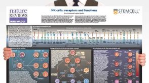 挂图Natural Killer Cells Overview of NK cell receptors, subsets, activation and function
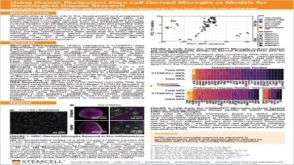 科学海报Using Human Pluripotent Stem Cell-Derived Microglia As Models For Neurological Disease Research
 沪公网安备31010102008431号