Sulforaphane targets pancreatic tumour-initiating cells by NF-kappaB-induced antiapoptotic signalling.
BACKGROUND AND AIMS: Emerging evidence suggests that highly treatment-resistant tumour-initiating cells (TICs) play a central role in the pathogenesis of pancreatic cancer. Tumour necrosis factor-related apoptosis-inducing ligand (TRAIL) is considered to be a novel anticancer agent; however,recent studies have shown that many pancreatic cancer cells are resistant to apoptosis induction by TRAIL due to TRAIL-activated nuclear factor-kappaB (NF-kappaB) signalling. Several chemopreventive agents are able to inhibit NF-kappaB,and favourable results have been obtained--for example,for the broccoli compound sulforaphane-in preventing metastasis in clinical studies. The aim of the study was to identify TICs in pancreatic carcinoma for analysis of resistance mechanisms and for definition of sensitising agents. METHODS: TICs were defined by expression patterns of a CD44(+)/CD24(-),CD44(+)/CD24(+) or CD44(+)/CD133(+) phenotype and correlation to growth in immunodeficient mice,differentiation grade,clonogenic growth,sphere formation,aldehyde dehydrogenase (ALDH) activity and therapy resistance. RESULTS: Mechanistically,specific binding of transcriptionally active cRel-containing NF-kappaB complexes in TICs was observed. Sulforaphane prevented NF-kappaB binding,downregulated apoptosis inhibitors and induced apoptosis,together with prevention of clonogenicity. Gemcitabine,the chemopreventive agents resveratrol and wogonin,and the death ligand TRAIL were less effective. In a xenograft model,sulforaphane strongly blocked tumour growth and angiogenesis,while combination with TRAIL had an additive effect without obvious cytotoxicity in normal cells. Freshly isolated patient tumour cells expressing markers for TICs could be sensitised by sulforaphane for TRAIL-induced cytotoxicity. CONCLUSION: The data provide new insights into resistance mechanisms of TICs and suggest the combination of sulforaphane with TRAIL as a promising strategy for targeting of pancreatic TICs.
View Publication
Bar EE et al. (OCT 2007)
Stem cells (Dayton,Ohio) 25 10 2524--33
Cyclopamine-mediated hedgehog pathway inhibition depletes stem-like cancer cells in glioblastoma.
Brain tumors can arise following deregulation of signaling pathways normally activated during brain development and may derive from neural stem cells. Given the requirement for Hedgehog in non-neoplastic stem cells,we investigated whether Hedgehog blockade could target the stem-like population in glioblastoma multiforme (GBM). We found that Gli1,a key Hedgehog pathway target,was highly expressed in 5 of 19 primary GBM and in 4 of 7 GBM cell lines. Shh ligand was expressed in some primary tumors,and in GBM-derived neurospheres,suggesting a potential mechanism for pathway activation. Hedgehog pathway blockade by cyclopamine caused a 40%-60% reduction in growth of adherent glioma lines highly expressing Gli1 but not in those lacking evidence of pathway activity. When GBM-derived neurospheres were treated with cyclopamine and then dissociated and seeded in media lacking the inhibitor,no new neurospheres formed,suggesting that the clonogenic cancer stem cells had been depleted. Consistent with this hypothesis,the stem-like fraction in gliomas marked by both aldehyde dehydrogenase activity and Hoechst dye excretion (side population) was significantly reduced or eliminated by cyclopamine. In contrast,we found that radiation treatment of our GBM neurospheres increased the percentage of these stem-like cells,suggesting that this standard therapy preferentially targets better-differentiated neoplastic cells. Most importantly,viable GBM cells injected intracranially following Hedgehog blockade were no longer able to form tumors in athymic mice,indicating that a cancer stem cell population critical for ongoing growth had been removed. Disclosure of potential conflicts of interest is found at the end of this article.
View Publication
产品类型:
产品号#:
01700
01705
01701
01702
72072
72074
产品名:
ALDEFLUOR™ 试剂盒
ALDEFLUOR™ DEAB试剂, 1.5 mM, 1 mL
ALDEFLUOR™检测缓冲液
环巴胺(Cyclopamine)
环巴胺(Cyclopamine)
Zhang M et al. (SEP 2014)
International journal of cancer 135 5 1132--41
Anti-β₂M monoclonal antibodies kill myeloma cells via cell- and complement-mediated cytotoxicity.
Our previous studies showed that anti-β2M monoclonal antibodies (mAbs) at high doses have direct apoptotic effects on myeloma cells,suggesting that anti-β2M mAbs might be developed as a novel therapeutic agent. In this study,we investigated the ability of the mAbs at much lower concentrations to indirectly kill myeloma cells by utilizing immune effector cells or molecules. Our results showed that anti-β2M mAbs effectively lysed MM cells via antibody-dependent cell-mediated cytotoxicity (ADCC) and complement-dependent cytotoxicity (CDC),which were correlated with and dependent on the surface expression of β2M on MM cells. The presence of MM bone marrow stromal cells or addition of IL-6 did not attenuate anti-β2M mAb-induced ADCC and CDC activities against MM cells. Furthermore,anti-β2M mAbs only showed limited cytotoxicity toward normal B cells and nontumorous mesenchymal stem cells,indicating that the ADCC and CDC activities of the anti-β2M mAbs were more prone to the tumor cells. Lenalidomide potentiated in vitro ADCC activity against MM cells and in vivo tumor inhibition capacity induced by the anti-β2M mAbs by enhancing the activity of NK cells. These results support clinical development of anti-β2M mAbs,both as a monotherapy and in combination with lenalidomide,to improve MM patient outcome.
View Publication
Li Y et al. (MAY 2010)
Clinical cancer research : an official journal of the American Association for Cancer Research 16 9 2580--90
Sulforaphane, a dietary component of broccoli/broccoli sprouts, inhibits breast cancer stem cells.
PURPOSE: The existence of cancer stem cells (CSCs) in breast cancer has profound implications for cancer prevention. In this study,we evaluated sulforaphane,a natural compound derived from broccoli/broccoli sprouts,for its efficacy to inhibit breast CSCs and its potential mechanism. EXPERIMENTAL DESIGN: Aldefluor assay and mammosphere formation assay were used to evaluate the effect of sulforaphane on breast CSCs in vitro. A nonobese diabetic/severe combined immunodeficient xenograft model was used to determine whether sulforaphane could target breast CSCs in vivo,as assessed by Aldefluor assay,and tumor growth upon cell reimplantation in secondary mice. The potential mechanism was investigated using Western blotting analysis and beta-catenin reporter assay. RESULTS: Sulforaphane (1-5 micromol/L) decreased aldehyde dehydrogenase-positive cell population by 65% to 80% in human breast cancer cells (P textless 0.01) and reduced the size and number of primary mammospheres by 8- to 125-fold and 45% to 75% (P textless 0.01),respectively. Daily injection with 50 mg/kg sulforaphane for 2 weeks reduced aldehyde dehydrogenase-positive cells by textgreater50% in nonobese diabetic/severe combined immunodeficient xenograft tumors (P = 0.003). Sulforaphane eliminated breast CSCs in vivo,thereby abrogating tumor growth after the reimplantation of primary tumor cells into the secondary mice (P textless 0.01). Western blotting analysis and beta-catenin reporter assay showed that sulforaphane downregulated the Wnt/beta-catenin self-renewal pathway. CONCLUSIONS: Sulforaphane inhibits breast CSCs and downregulates the Wnt/beta-catenin self-renewal pathway. These findings support the use of sulforaphane for the chemoprevention of breast cancer stem cells and warrant further clinical evaluation.
View Publication
The longevity of organisms is maintained by stem cells. If an organism loses the ability to maintain a balance between quiescence and differentiation in the stem/progenitor cell compartment due to aging and/or stress,this may result in death or age-associated diseases,including cancer. Ewing sarcoma is the most lethal bone tumor in young patients and arises from primitive stem cells. Here,we demonstrated that endogenous Ewing sarcoma gene (Ews) is indispensable for stem cell quiescence,and that the ablation of Ews promotes the early onset of senescence in hematopoietic stem progenitor cells. The phenotypic and functional changes in Ews-deficient stem cells were accompanied by an increase in senescence-associated β-galactosidase staining and a marked induction of p16(INK4a) compared with wild-type counterparts. With its relevance to cancer and possibly aging,EWS is likely to play a significant role in maintaining the functional capacity of stem cells and may provide further insight into the complexity of Ewing sarcoma in the context of stem cells.
View Publication
产品类型:
产品号#:
03434
03444
产品名:
MethoCult™ GF M3434
MethoCult™ GF M3434
Cremona CA and Lloyd AC (SEP 2009)
Journal of cell science 122 Pt 18 3272--81
Loss of anchorage in checkpoint-deficient cells increases genomic instability and promotes oncogenic transformation.
Mammalian cells generally require both mitogens and anchorage signals in order to proliferate. An important characteristic of many tumour cells is that they have lost this anchorage-dependent cell-cycle checkpoint,allowing them to proliferate without signals provided by their normal microenvironment. In the absence of anchorage signals from the extracellular matrix,many cell types arrest cell-cycle progression in G1 phase as a result of Rb-dependent checkpoints. However,despite inactivation of p53 and Rb proteins,SV40LT-expressing cells retain anchorage dependency,suggesting the presence of an uncharacterised cell-cycle checkpoint,which can be overridden by coexpression of oncogenic Ras. We report here that,although cyclin-CDK complexes persisted in suspension,proliferation was inhibited in LT-expressing cells by the CDK inhibitor p27(Kip1) (p27). Interestingly,this did not induce a stable arrest,but aberrant cell-cycle progression associated with stalled DNA replication,rereplication and chromosomal instability,which was sufficient to increase the frequency of oncogenic transformation. These results firstly indicate loss of anchorage in Rb- and p53-deficient cells as a novel mechanism for promotion of genomic instability; secondly suggest that anchorage checkpoints that protect normal cells from inappropriate proliferation act deleteriously in Rb- and p53-deficient cells to promote tumourigenesis; and thirdly indicate caution in the use of CDK inhibitors for cancer treatment.
View Publication
产品类型:
产品号#:
05401
05402
05411
产品名:
MesenCult™ MSC 基础培养基(人)
MesenCult™ MSC 刺激补充剂(人)
MesenCult™ 增殖试剂盒(人)
Lo J-F et al. (MAR 2011)
Cancer research 71 5 1912--23
The epithelial-mesenchymal transition mediator S100A4 maintains cancer-initiating cells in head and neck cancers.
Cancer-initiating cells (CIC) comprise a rare subpopulation of cells in tumors that are proposed to be responsible for tumor growth. Starting from CICs identified in head and neck squamous cell carcinomas (HNSCC),termed head and neck cancer-initiating cells (HN-CIC),we determined as a candidate stemness-maintaining molecule for HN-CICs the proinflammatory mediator S100A4,which is also known to be an inducer of epithelial-mesenchymal transition. S100A4 knockdown in HN-CICs reduced their self-renewal capability and their stemness and tumorigenic properties,both in vitro and in vivo. Conversely,S100A4 overexpression in HNSCC cells enhanced their stem cell properties. Mechanistic investigations indicated that attenuation of endogenous S100A4 levels in HNSCC cells caused downregulation of Notch2 and PI3K (phosphoinositide 3-kinase)/pAKT along with upregulation of PTEN,consistent with biological findings. Immunohistochemical analysis of HNSCC clinical specimens showed that S100A4 expression was positively correlated with clinical grading,stemness markers,and poorer patient survival. Together,our findings reveal a crucial role for S100A4 signaling pathways in maintaining the stemness properties and tumorigenicity of HN-CICs. Furthermore,our findings suggest that targeting S100A4 signaling may offer a new targeted strategy for HNSCC treatment by eliminating HN-CICs.
View Publication
产品类型:
产品号#:
01700
01705
01702
产品名:
ALDEFLUOR™ 试剂盒
ALDEFLUOR™ DEAB试剂, 1.5 mM, 1 mL
ALDEFLUOR™检测缓冲液
Lelaidier M et al. (OCT 2015)
Oncotarget 6 30 29440--55
TRAIL-mediated killing of acute lymphoblastic leukemia by plasmacytoid dendritic cell-activated natural killer cells.
Acute lymphoblastic leukemia (ALL) still frequently recurs after hematopoietic stem cell transplantation (HSCT),underscoring the need to improve the graft-versus-leukemia (GvL) effect. Natural killer (NK) cells reconstitute in the first months following HSCT when leukemia burden is at its lowest,but ALL cells have been shown to be resistant to NK cell-mediated killing. We show here that this resistance is overcome by NK cell stimulation with TLR-9-activated plasmacytoid dendritic cells (pDCs). NK cell priming with activated pDCs resulted in TRAIL and CD69 up-regulation on NK cells and IFN-γ production. NK cell activation was dependent on IFN-α produced by pDCs,but was not reproduced by IFN-α alone. ALL killing was further enhanced by inhibition of KIR engagement. We showed that ALL lysis was mainly mediated by TRAIL engagement,while the release of cytolytic granules was involved when ALL expressed NK cell activating receptor ligands. Finally,adoptive transfers of activated-pDCs in ALL-bearing humanized mice delayed the leukemia onset and cure 30% of mice. Our data therefore demonstrate that TLR-9 activated pDCs are a powerful tool to overcome ALL resistance to NK cell-mediated killing and to reinforce the GvL effect of HSCT. These results open new therapeutic avenues to prevent relapse in children with ALL.
View Publication
产品类型:
产品号#:
19062
19062RF
19055
19055RF
产品名:
EasySep™人浆细胞样DC富集试剂盒
RoboSep™ 人浆细胞样DC富集试剂盒含滤芯吸头
EasySep™人NK细胞富集试剂盒
RoboSep™ 人NK细胞富集试剂盒含滤芯吸头
Deng S et al. (JAN 2010)
PloS one 5 4 e10277
Distinct expression levels and patterns of stem cell marker, aldehyde dehydrogenase isoform 1 (ALDH1), in human epithelial cancers.
Aldehyde dehydrogenase isoform 1 (ALDH1) has been proved useful for the identification of cancer stem cells. However,our knowledge of the expression and activity of ALDH1 in common epithelial cancers and their corresponding normal tissues is still largely absent. Therefore,we characterized ALDH1 expression in 24 types of normal tissues and a large collection of epithelial tumor specimens (six cancer types,n = 792) by immunohistochemical staining. Using the ALDEFUOR assay,ALDH1 activity was also examined in 16 primary tumor specimens and 43 established epithelial cancer cell lines. In addition,an ovarian cancer transgenic mouse model and 7 murine ovarian cancer cell lines were analyzed. We found that the expression levels and patterns of ALDH1 in epithelial cancers are remarkably distinct,and they correlate with their corresponding normal tissues. ALDH1 protein expression levels are positively correlated with ALDH1 enzymatic activity measured by ALDEFLUOR assay. Long-term in vitro culture doesn't significantly affect ALDH1 activity in epithelial tumor cells. Consistent with research on other cancers,we found that high ALDH1 expression is significantly associated with poor clinical outcomes in serous ovarian cancer patients (n = 439,p = 0.0036). Finally,ALDH(br) tumor cells exhibit cancer stem cell properties and are resistant to chemotherapy. As a novel cancer stem cell marker,ALDH1 can be used for tumors whose corresponding normal tissues express ALDH1 in relatively restricted or limited levels such as breast,lung,ovarian or colon cancer.
View Publication
产品类型:
产品号#:
01700
01705
01701
01702
05620
产品名:
ALDEFLUOR™ 试剂盒
ALDEFLUOR™ DEAB试剂, 1.5 mM, 1 mL
ALDEFLUOR™检测缓冲液
MammoCult™ 人源培养基套装
Morrow M et al. (MAY 2004)
Blood 103 10 3890--6
TEL-AML1 promotes development of specific hematopoietic lineages consistent with preleukemic activity.
The t(12;21)(p13;q22) translocation is the most common chromosomal abnormality yet identified in any pediatric leukemia and gives rise to the TEL-AML1 fusion product. To investigate the effects of TEL-AML1 on hematopoiesis,fetal liver hematopoietic progenitor cells (HPCs) were transduced with retroviral vectors expressing this fusion protein. We show that TEL-AML1 dramatically alters differentiation of HPCs in vitro,preferentially promoting B-lymphocyte development,enhancing self-renewal of B-cell precursors,and leading to the establishment of long-term growth factor-dependent pre-B-cell lines. However,it had no effect on myeloid development in vitro. Further experiments were performed to determine whether TEL-AML1 also demonstrates lineage-specific activity in vivo. TEL-AML1-expressing HPCs displayed a competitive advantage in reconstituting both B-cell and myeloid lineages in vivo but had no effect on reconstitution of the T-cell lineage. Despite promoting these alterations in hematopoiesis,TEL-AML1 did not induce leukemia in transplanted mice. Our study provides a unique insight into the role of TEL-AML1 in leukemia predisposition and a potential model to study the mechanism of leukemogenesis associated with this fusion.
View Publication

 EasySep™小鼠TIL(CD45)正选试剂盒


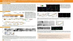 科学海报Robust Establishment and Expansion of Multilineage Human Fallopian Tube Organoids in Serum-Free Medium
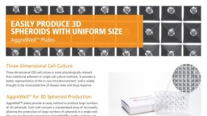 产品手册AggreWell™ for Spheroids
 沪公网安备31010102008431号