Lassailly F et al. (JUL 2010)
Blood 115 26 5347--54
Microenvironmental contaminations" induced by fluorescent lipophilic dyes used for noninvasive in vitro and in vivo cell tracking."
Determining how normal and leukemic stem cells behave in vivo,in a dynamic and noninvasive way,remains a major challenge. Most optical tracking technologies rely on the use of fluorescent or bioluminescent reporter genes,which need to be stably expressed in the cells of interest. Because gene transfer in primary leukemia samples represents a major risk to impair their capability to engraft in a xenogenic context,we evaluated the possibility to use gene transfer-free labeling technologies. The lipophilic dye 3,3,3',3' tetramethylindotricarbocyanine iodide (DiR) was selected among 4 near-infrared (NIR) staining technologies. Unfortunately we report here a massive transfer of the dye occurring toward the neighbor cells both in vivo and in vitro. We further demonstrate that all lipophilic dyes tested in this study (1,1'-dioctadecyl-3,3,3',3'-tetramethylindotricarbocyanine perchlorate [DiI],DiD,DiR,and PKH26) can give rise to microenvironmental contamination,including when used in suboptimal concentration,after extensive washing procedures and in the absence of phagocytosis or marked cell death. This was observed from all cell types tested. Eventually,we show that this microenvironmental contamination is mediated by both direct cell-cell contacts and diffusible microparticles. We conclude that tracking of labeled cells using non-genetically encoded markers should always be accompanied by drastic cross validation using multimodality approaches.
View Publication
产品类型:
产品号#:
09600
09650
19756
19756RF
产品名:
StemSpan™ SFEM
StemSpan™ SFEM
Du W et al. (APR 2011)
Blood 117 16 4243--52
Overexpression of IL-3Rα on CD34+CD38- stem cells defines leukemia-initiating cells in Fanconi anemia AML.
Patients with Fanconi anemia (FA) have a high risk of developing acute myeloid leukemia (AML). In this study,we attempted to identify cell-surface markers for leukemia-initiating cells in FA-AML patients. We found that the IL-3 receptor-α (IL-3Rα) is a promising candidate as an leukemia-initiating cell-specific antigen for FA-AML. Whereas IL-3Rα expression is undetectable on normal CD34(+)CD38(-) HSCs,it is overexpressed on CD34(+)CD38(-) cells from FA patients with AML. We examined the leukemia-initiating cell activity of IL-3Rα-positive FA-AML cells in a humanized" FA xenotransplant model in which we separated AML cells into IL-3Rα-positive and IL-3Rα-negative CD34 fractions and transplanted them into irradiated recipient mice. In all 3 FA-AML samples�
View Publication
Lindvall C et al. (NOV 2006)
The Journal of biological chemistry 281 46 35081--7
The Wnt signaling receptor Lrp5 is required for mammary ductal stem cell activity and Wnt1-induced tumorigenesis.
Canonical Wnt signaling has emerged as a critical regulatory pathway for stem cells. The association between ectopic activation of Wnt signaling and many different types of human cancer suggests that Wnt ligands can initiate tumor formation through altered regulation of stem cell populations. Here we have shown that mice deficient for the Wnt co-receptor Lrp5 are resistant to Wnt1-induced mammary tumors,which have been shown to be derived from the mammary stem/progenitor cell population. These mice exhibit a profound delay in tumorigenesis that is associated with reduced Wnt1-induced accumulation of mammary progenitor cells. In addition to the tumor resistance phenotype,loss of Lrp5 delays normal mammary development. The ductal trees of 5-week-old Lrp5-/- females have fewer terminal end buds,which are structures critical for juvenile ductal extension presumed to be rich in stem/progenitor cells. Consequently,the mature ductal tree is hypomorphic and does not completely fill the fat pad. Furthermore,Lrp5-/- ductal cells from mature females exhibit little to no stem cell activity in limiting dilution transplants. Finally,we have shown that Lrp5-/- embryos exhibit substantially impaired canonical Wnt signaling in the primitive stem cell compartment of the mammary placodes. These findings suggest that Lrp5-mediated canonical signaling is required for mammary ductal stem cell activity and for tumor development in response to oncogenic Wnt effectors.
View Publication
Adherent cells generated during long-term culture of human umbilical cord blood CD34+ cells have characteristics of endothelial cells and beneficial effect on cord blood ex vivo expansion.
Hematopoiesis depends on the association of hematopoietic stem cells with stromal cells that constitute the hematopoietic microenvironment. The in vitro development of the endothelial cell from umbilical cord blood (UCB) is not well established and has met very limited success. In this study,UCB CD34(+) cells were cultured for 5 weeks in a stroma-free liquid culture system using thrombopoietin,flt3 ligand,and granulocyte-colony stimulating factor. By week 4-5,we found that firmly adherent fibroblast-like cells were established. These cells showed characteristics of endothelial cells expressing von Willebrand factor,human vascular cell adhesion molecule-1,human intracellular adhesion molecule-1,human CD31,E-selectin,and human macrophage. Furthermore,when comparing an ex vivo system without an established endothelial monolayer to an ex vivo system with an established endothelial monolayer,better expansion of total nucleated cells,CD34(+) cells,and colony-forming units (CFUs)-granulocyte-macrophage and CFUs-granulocyte-erythroid-megakaryocyte-macrophage were found during culture. This phenomenon was in part due to the fact that a significant reduction of apoptotic fractions was found in the CD34(+) cells,which were cultured on the adherent monolayer for up to 5 weeks. To gather quantitative data on the number of endothelial cells derived from a given number of CD34 cells,we performed limiting dilution assay by using Poisson distribution: the number of tested cells (linear scale) producing a 37% negative culture (logarithmic scale) is the number of cells containing one endothelial cell. By this method,one endothelial cell may be found from 314 CD34(+) cells after 5 weeks of culture. These results suggest that the UCB CD34(+) cell fraction contains endothelial cell precursors,establishing the hematopoietic microenvironment and providing the beneficial effects through downregulating apoptosis on UCB expansion protocols. These observations may provide insight for future cellular therapy or graft engineering.
View Publication
产品类型:
产品号#:
04434
04444
产品名:
MethoCult™H4434经典
MethoCult™H4434经典
Petersen OW and Polyak K (MAY 2010)
Cold Spring Harbor perspectives in biology 2 5 a003160
Stem cells in the human breast.
The origins of the epithelial cells participating in the development,tissue homeostasis,and cancer of the human breast are poorly understood. However,emerging evidence suggests a role for adult tissue-specific stem cells in these processes. In a hierarchical manner,these generate the two main mammary cell lineages,producing an increasing number of cells with distinct properties. Understanding the biological characteristics of human breast stem cells and their progeny is crucial in attempts to compare the features of normal stem cells and cancer precursor cells and distinguish these from nonprecursor cells and cells from the bulk of a tumor. A historical overview of research on human breast stem cells in primary tissue and in culture reveals the progress that has been made in this area,whereas a focus on the cell-of-origin and reprogramming that occurs during neoplastic conversion provides insight into the enigmatic way in which human breast cancers are skewed toward the luminal epithelial lineage.
View Publication
De Giorgi U et al. (MAY 2011)
Cancer biology & therapy 11 9 812--5
Mesenchymal stem cells expressing GD2 and CD271 correlate with breast cancer-initiating cells in bone marrow.
Purpose: The bone marrow microenvironment is considered a critical component in the dissemination and fate of cancer cells in the metastatic process. We explored the possible correlation between bone marrow mesenchymal stem cells (BM-MSC) and disseminated breast cancer-initiating cells (BCIC) in primary breast cancer patients. Experimental design: Bone marrow mononuclear cells (BM-MNC) were collected at the time of primary surgery in 12 breast cancer patients. BM-MNC was immunophenotyped and BCIC was defined as epithelial cells (CD326+CD45-) with a stem-like" phenotype (CD44+CD24low/-�
View Publication
产品类型:
产品号#:
01700
01705
01702
产品名:
ALDEFLUOR™ 试剂盒
ALDEFLUOR™ DEAB试剂
ALDEFLUOR™测定缓冲液
Seiwert TY et al. ( 2009)
Cancer research 69 7 3021--3031
The MET receptor tyrosine kinase is a potential novel therapeutic target for head and neck squamous cell carcinoma.
Recurrent/metastatic head and neck cancer remains a devastating disease with insufficient treatment options. We investigated the MET receptor tyrosine kinase as a novel target for the treatment of head and neck squamous cell carcinoma (HNSCC). MET/phosphorylated MET and HGF expression was analyzed in 121 tissues (HNSCC/normal) by immunohistochemistry,and in 20 HNSCC cell lines by immunoblotting. The effects of MET inhibition using small interfering RNA/two small-molecule inhibitors (SU11274/PF-2341066) on signaling,migration,viability,and angiogenesis were determined. The complete MET gene was sequenced in 66 head and neck cancer tissue samples and eight cell lines. MET gene copy number was determined in 14 cell lines and 23 tumor tissues. Drug combinations of SU11274 with cisplatin or erlotinib were tested in SCC35/HN5 cell lines. Eighty-four percent of the HNSCC samples showed MET overexpression,whereas 18 of 20 HNSCC cell lines (90%) expressed MET. HGF overexpression was present in 45% of HNSCC. MET inhibition with SU11274/PF-2341066 abrogated MET signaling,cell viability,motility/migration in vitro,and tumor angiogenesis in vivo. Mutational analysis of 66 tumor tissues and 8 cell lines identified novel mutations in the semaphorin (T230M/E168D/N375S),juxtamembrane (T1010I/R988C),and tyrosine kinase (T1275I/V1333I) domains (incidence: 13.5%). Increased MET gene copy number was present with textgreater10 copies in 3 of 23 (13%) tumor tissues. A greater-than-additive inhibition of cell growth was observed when combining a MET inhibitor with cisplatin or erlotinib and synergy may be mediated via erbB3/AKT signaling. MET is functionally important in HNSCC with prominent overexpression,increased gene copy number,and mutations. MET inhibition abrogated MET functions,including proliferation,migration/motility,and angiogenesis. MET is a promising,novel target for HNSCC and combination approaches with cisplatin or EGFR inhibitors should be explored.
View Publication

 EasySep™小鼠TIL(CD45)正选试剂盒


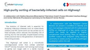 产品手册Highway1™: High-Purity Sorting of Bacterially-Infected Cells


 沪公网安备31010102008431号