Kurtzberg LS et al. (MAY 2011)
Clinical cancer research : an official journal of the American Association for Cancer Research 17 9 2777--87
Genz-644282, a novel non-camptothecin topoisomerase I inhibitor for cancer treatment.
PURPOSE: Genz-644282 [8,9-dimethoxy-5-(2-N-methylaminoethyl)-2,3-methylenedioxy-5H-dibenzo[c,h][1,6]naphthyridin-6-one] has emerged as a promising candidate for antitumor agents. This report describes the bone marrow colony-forming unit,granulocyte macrophage (CFU-GM) and tumor cell CFU activity of topoisomerase I (Top1) inhibitors,such as Genz-644282,topotecan,irinotecan/SN-38,and ARC-111,and examines their activity in several human tumor xenograft models. EXPERIMENTAL DESIGN: Colony-forming assays were conducted with mouse and human bone marrow and eight human tumor cell lines. In addition,29 human tumor cell lines representing a range of histology and potential resistance mechanisms were assayed for sensitivity to Genz-644282 in a 72-hour exposure assay. The efficacy of Genz-644282 was compared with standard anticancer drugs (i.e.,irinotecan,docetaxel,and dacarbazine) in human tumor xenografts of colon cancer,renal cell carcinoma,non-small cell lung cancer,and melanoma. RESULTS: Human bone marrow CFU-GM was more sensitive to the Top1 inhibitors than was mouse bone marrow CFU-GM. The ratio of mouse to human IC(90) values was more than 10 for the camptothecins and less than 10 for Genz-644282,which had more potency as a cytotoxic agent toward human tumor cells in culture than the camptothecins in the colony-forming and 72-hour proliferation assays. Genz-644282 has superior or equal antitumor activity in the human tumor xenografts than the standard drug comparators. CONCLUSIONS: On the basis of preclinical activity and safety,Genz-644282 was selected for development and is currently undergoing phase 1 clinical trial.
View Publication
Akcakanat A et al. ( 2009)
Molecular Cancer 8 1 75
The rapamycin-regulated gene expression signature determines prognosis for breast cancer
BACKGROUND: Mammalian target of rapamycin (mTOR) is a serine/threonine kinase involved in multiple intracellular signaling pathways promoting tumor growth. mTOR is aberrantly activated in a significant portion of breast cancers and is a promising target for treatment. Rapamycin and its analogues are in clinical trials for breast cancer treatment. Patterns of gene expression (metagenes) may also be used to simulate a biologic process or effects of a drug treatment. In this study,we tested the hypothesis that the gene-expression signature regulated by rapamycin could predict disease outcome for patients with breast cancer. RESULTS: Colony formation and sulforhodamine B (IC50 textless 1 nM) assays,and xenograft animals showed that MDA-MB-468 cells were sensitive to treatment with rapamycin. The comparison of in vitro and in vivo gene expression data identified a signature,termed rapamycin metagene index (RMI),of 31 genes upregulated by rapamycin treatment in vitro as well as in vivo (false discovery rate of 10%). In the Miller dataset,RMI did not correlate with tumor size or lymph node status. High (textgreater75th percentile) RMI was significantly associated with longer survival (P = 0.015). On multivariate analysis,RMI (P = 0.029),tumor size (P = 0.015) and lymph node status (P = 0.001) were prognostic. In van 't Veer study,RMI was not associated with the time to develop distant metastasis (P = 0.41). In the Wang dataset,RMI predicted time to disease relapse (P = 0.009). CONCLUSION: Rapamycin-regulated gene expression signature predicts clinical outcome in breast cancer. This supports the central role of mTOR signaling in breast cancer biology and provides further impetus to pursue mTOR-targeted therapies for breast cancer treatment.
View Publication
产品类型:
产品号#:
73362
73364
100-1050
产品名:
雷帕霉素
雷帕霉素
雷帕霉素
Niu C et al. (SEP 2009)
Blood 114 10 2087--96
c-Myc is a target of RNA-binding motif protein 15 in the regulation of adult hematopoietic stem cell and megakaryocyte development.
RNA-binding motif protein 15 (RBM15) is involved in the RBM15-megakaryoblastic leukemia 1 fusion in acute megakaryoblastic leukemia. Although Rbm15 has been reported to be required for B-cell differentiation and to inhibit myeloid and megakaryocytic expansion,it is not clear what the normal functions of Rbm15 are in the regulation of hematopoietic stem cell (HSC) and megakaryocyte development. In this study,we report that Rbm15 may function in part through regulation of expression of the proto-oncogene c-Myc. Similar to c-Myc knockout (c-Myc-KO) mice,long-term (LT) HSCs are significantly increased in Rbm15-KO mice due to an apparent LT-HSC to short-term HSC differentiation defect associated with abnormal HSC-niche interactions caused by increased N-cadherin and beta(1) integrin expression on mutant HSCs. Both serial transplantation and competitive reconstitution capabilities of Rbm15-KO LT-HSCs are greatly compromised. Rbm15-KO and c-Myc-KO mice also share related abnormalities in megakaryocyte development,with mutant progenitors producing increased,abnormally small low-ploidy megakaryocytes. Consistent with a possible functional interplay between Rbm15 and c-Myc,the megakaryocyte increase in Rbm15-KO mice could be partially reversed by ectopic c-Myc. Thus,Rbm15 appears to be required for normal HSC-niche interactions,for the ability of HSCs to contribute normally to adult hematopoiesis,and for normal megakaryocyte development; these effects of Rbm15 on hematopoiesis may be mediated at least in part by c-Myc.
View Publication
Ucar D et al. (MAR 2009)
Chemico-biological interactions 178 1-3 48--55
Aldehyde dehydrogenase activity as a functional marker for lung cancer.
Aldehyde dehydrogenase (ALDH) activity has been implicated in multiple biological and biochemical pathways and has been used to identify potential cancer stem cells. Our main hypothesis is that ALDH activity may be a lung cancer stem cell marker. Using flow cytometry,we sorted cells with bright (ALDH(br)) and dim (ALDH(lo)) ALDH activity found in H522 lung cancer cell line. We used in vitro proliferation and colony assays as well as a xenograft animal model to test our hypothesis. Cytogenetic analysis demonstrated that the ALDH(br) cells are indeed a different clone,but when left in normal culture conditions will give rise to ALDH(lo) cells. Furthermore,the ALDH(br) cells grow slower,have low clonal efficiency,and give rise to morphologically distinct colonies. The ability to form primary xenografts in NOD/SCID mice by ALDH(br) and ALDH(lo) cells was tested by injecting single cell suspension under the skin in each flank of same animal. Tumor size was calculated weekly. ALDH1A1 and ALDH3A1 immunohistochemistry (IHC) was performed on excised tumors. These tumors were also used to re-establish cell suspension,measure ALDH activity,and re-injection for secondary and tertiary transplants. The results indicate that both cell types can form tumors but the ones from ALDH(br) cells grew much slower in primary recipient mice. Histologically,there was no significant difference in the expression of ALDH in primary tumors originating from ALDH(br) or ALDH(lo) cells. Secondary and tertiary xenografts originating from ALDH(br) grew faster and bigger than those formed by ALDH(lo) cells. In conclusion,ALDH(br) cells may have some of the traditional features of stem cells in terms of being mostly dormant and slow to divide,but require support of other cells (ALDH(lo)) to sustain tumor growth. These observations and the known role of ALDH in drug resistance may have significant therapeutic implications in the treatment of lung cancer.
View Publication
Kakarala M and Wicha MS (JUN 2008)
Journal of clinical oncology : official journal of the American Society of Clinical Oncology 26 17 2813--20
Implications of the cancer stem-cell hypothesis for breast cancer prevention and therapy.
Recent research in breast biology has provided support for the cancer stem-cell hypothesis. Two important components of this hypothesis are that tumors originate in mammary stem or progenitor cells as a result of dysregulation of the normally tightly regulated process of self-renewal. As a result,tumors contain and are driven by a cellular subcomponent that retains key stem-cell properties including self-renewal,which drives tumorigenesis and differentiation that contributes to cellular heterogeneity. Advances in stem-cell technology have led to the identification of stem cells in normal and malignant breast tissue. The study of these stem cells has helped to elucidate the origin of the molecular complexity of human breast cancer. The cancer stem-cell hypothesis has important implications for early detection,prevention,and treatment of breast cancer. Both hereditary and sporadic breast cancers may develop through dysregulation of stem-cell self-renewal pathways. These aberrant stem cells may provide targets for the development of cancer prevention strategies. Furthermore,because breast cancer stem cells may be highly resistant to radiation and chemotherapy,the development of more effective therapies for this disease may require the effective targeting of this cell population.
View Publication

 EasySep™小鼠TIL(CD45)正选试剂盒


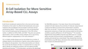 技术公告B Cell Isolation for More Sensitive Array-Based CLL Assays
 产品手册Highway1™: Fast, Gentle, and Automated Cell Sorting for Every Lab
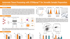 科学海报Automate Tissue Processing with STEMprep™ for Versatile Sample Preparation
 产品手册Highway1™: Fast, Gentle, and Automated Cell Sorting for Every Lab

 沪公网安备31010102008431号